




No CrossRef data available.
Article contents
- Extract
- The challenge
- Stress and burnout
- Strategies requiring innovative technology
- The preoperative phase
- The surgical phase
- The phase of care on the intensive care unit
- The post-operative phase
- Resonance of information
- Finding the Holy Grail
- The impact on our outcomes for first stage palliation
- Conclusions
- References
Achieving resonance in a programme for congenital cardiac surgery
Published online by Cambridge University Press: 21 September 2005
Article contents
- Extract
- The challenge
- Stress and burnout
- Strategies requiring innovative technology
- The preoperative phase
- The surgical phase
- The phase of care on the intensive care unit
- The post-operative phase
- Resonance of information
- Finding the Holy Grail
- The impact on our outcomes for first stage palliation
- Conclusions
- References
Extract
In 1995, Miami Children's Hospital recognized an institutional problem with its programme providing surgical treatment for congenital cardiac malformations. There was a high rate of mortality for neonatal surgery, and no patients had survived attempted first stage palliation for hypoplastic left heart syndrome. The hospital enlisted nationally recognized consultants in congenital cardiac surgery and cardiology to review the existing programme, and to make recommendations for improvement. Based on these recommendations, a new team was recruited. The recruits were a young attending surgeon, an interventional cardiologist, and a cardiac intensivist, attracted from recognized centers of excellence in Boston and Toronto.
Information
- Type
- Norwood Procedure and Staged Palliation
- Information
- Copyright
- © 2004 Cambridge University Press
The volume of patients is increasingly recognized as the foundation for clinical excellence in care. The relationship between volume and performance is well documented,1–4 and may be particularly true for the most complex procedures performed in neonates, such as first stage palliation for hypoplastic left heart syndrome. In deciding to rebuild a center in South Florida, the new recruits assessed the existing level of care to determine whether there was a need for, and the volume to support, a new programme. The best results for first stage palliation were known to be achieved in centers using an approach based on the existence of a dedicated cardiac team (Table 1).
Table 1. Published outcomes for first stage palliation of hypoplasia of the left heart.

At that time, in 1995, there were three competing programmes in South Florida, including one at the University of Miami, which held the certificate of need for pediatric cardiac transplantation. Miami Children's Hospital had no obstetrical service, so all neonatal patients were referred from these, and other outside, often competing, institutions. Despite this level of competition, and based on interviews with potential referring physicians from the region, such as pediatric cardiologists, adult cardiologists, neonatologists and pediatricians, we anticipated a minimum of 200 open heart referrals per year, and so the programme was initiated.
Extensive planning with the hospital administration resulted also in creation of a service for pediatric cardiology, with parallel financial incentives between the hospital and the physicians, and capital expenditure for development of the programme. The initial budget for personnel and capital was 5 million dollars. Based on the model of the centers in Boston and Toronto, we created dedicated teams for pediatric cardiac surgery, catheterization, and intensive care. Relationships were developed with regional obstetricians, pediatricians, neonatologists, and patients using a coordinated campaign for education and marketing based on the philosophy of the new cardiac teams. We determined also to introduce innovative technology focused on reducing the trauma of care for patients having congenital cardiac malformations. Incorporation of the referring physicians into the planned care was critical for initial stabilization and subsequent management of the patients, and innovative strategies for communication were evolved to streamline this flow of information.
The challenge
The “Status Quo”
When we arrived in 1995, we entered a hospital environment with open clinical privileges for any licensed physician. There had been numerous challenges to our proposed model for a dedicated cardiac programme. Historically, the patients with congenital cardiac malformations had been cared for in the pediatric and neonatal intensive care units. The staffs of these units were resistant to building a competing unit for cardiac intensive care. The hospital administrators had encouraged a policy of open privilege for cardiac surgery and interventional catheterization to increase the volume of patients. Our nursing administration resisted the creation of a specialized “cardiac” nursing team. Over the first year, nonetheless, we initiated and defended our model of the dedicated congenital heart team. We appointed a director of nursing for the dedicated cardiac intensive care unit. The dedicated cardiac team performed all diagnostic and therapeutic procedures within the hospital, intentionally avoiding competition with community based cardiologists for outpatient diagnostic procedures. Dedicated teams were built in interventional cardiology, the cardiac intensive care unit, and cardiac surgery. The last component of the team to become completely dedicated was anesthesia, which now consists of three full-time cardiac anesthesiologists, covering all catheterizations and surgeries. A wing of the hospital was built adjacent to the cardiac operating room and catheterization laboratory, on the same floor as the Heart station diagnostic laboratory.
Stress and burnout
Our previous experience, along with published evidence,5, 6 indicated that caring for patients undergoing staged palliation for hypoplastic left heart syndrome can be traumatic to the cardiac team, as well as for the patient. These experiences had shown that neonates undergoing first stage palliation were often the most stressful patients, presenting acute and chronic clinical challenges, emotional stress, and large volumes of data requiring integration. We responded by enhancing our lines of communication. The team participated in three weekly conferences, consisting of cardiac intensive care unit teaching rounds, combined cardiologic and cardiac surgical case presentations, and a review of morbidity and mortality. Discussions were focused on evidence-based medicine, with frequent web-based searches of the literature conducted during the conference discussions to identify and adopt successful strategies created in other cardiac programmes. Real time reviews of data from the programmes were used to assess performance, and to formulate plans for care. Reviews of clinical incidents emphasized what we did right, distinguishing it from what we did wrong, and reinforced our shared goals and philosophy. The leaders of the programme enlisted support for those suffering grief, and strongly suppressed any suggestions of bullying and blaming.
Nursing retention was high, and by 2002, the average experience among 27 full-time nurses on our pediatric cardiac intensive care unit was 8.7 years. We monitored carefully the volumes of patients so as to manage nursing ratios and extra shifts. We also anticipated the need for additional recruitment of clinicians, attempting to stay at least two physicians deep in every specialty. We were ready and willing to make rapid changes amongst our physicians to enhance performance, and there was significant turnover amongst our clinical personnel during the first 8 years. Prior to building the new program in 1995, the cardiac team at Miami Children's Hospital had consisted of two surgeons and three cardiologists. After the subsequent 8 years, only two of the original five physicians remained. In the meantime, nonetheless, the team had been joined by 21 new physicians, with 8 departing, leaving in 2003 a total of 18 dedicated pediatric cardiac physicians.
Early experiences demonstrated that patients undergoing first stage palliation were a sensitive test of the concept of the resonance of the dedicated cardiac team. As an example, we were initially unable to include respiratory care as a dedicated component of the team. One evening in 1996, a neonate 3 weeks from the first stage of palliation was given nebulizer treatment by a respiratory therapist. The alarms monitoring saturations of oxygen, and the electrocardiogram, were turned off, and the child was bagged with 100% oxygen for several minutes to deliver the nebulized therapy. The child arrested, and could not be resuscitated. A member of the respiratory therapy team was subsequently assigned to the cardiac team and educated on functionally univentricular physiology. Since then, there has been no recurrence of this type of event.
Strategies requiring innovative technology
Challenges arose during each phase of care for patients undergoing first stage palliation, so a series of technological and strategic innovations were developed or adopted to address them.
The preoperative phase
Because all patients with hypoplastic left heart syndrome were referred from outside hospitals, we were limited in our ability to manage the initial resuscitation of neonates with complex functionally univentricular physiology. We responded by developing a programme for fetal echocardiography and prenatal diagnosis, thus optimizing the logistics of delivery. A transport team was also created, so that members of the cardiac team would be available to diagnose, resuscitate, and transport neonates with complex cardiac malformations. We built a system for cardiopulmonary bypass that could be transported rapidly by air, thus providing complete support using bypass at remote sites in unstable newborns.7
In the more recent era, preoperative medical management has focused on optimizing systemic delivery of oxygen, guided by serial monitoring of lactate in the blood. Techniques to optimize delivery of oxygen have become standard for the management of patients with hypoplastic left heart syndrome, and include attempting to achieve an optimal ratio of pulmonary-to-systemic flows by manipulating inspired gases, or systemic vasodilation. If necessary, ventricular pump function was improved with modest doses of catecholamines. Milrinone was our drug of choice when initiating medical management of low cardiac output states.
The surgical phase
Early in the development of the programme, it became obvious that bleeding after surgery contributed to mortality. To combat this problem, we used fresher blood products, and routinely delayed closure of the sternum to decrease the risk of tamponade. So as to facilitate minimally invasive surgical procedures, we tasked the perfusion team with redesigning the pump to allow use of lower profile venous cannulas. The perfusion team responded by building a completely Heparin-bonded circuit dependent on venous pull.8, 9 In addition to permitting the low profile venous cannulas to be used through small incisions during minimally invasive procedures, the new approach had the added benefit of allowing bi-caval venous cannulation during neonatal procedures such as the first stage of palliation for the hypoplastic left heart syndrome. These smaller cannulas enabled us to perform intra-cardiac neonatal repairs without circulatory arrest. Efforts were made to reduce priming volumes, and to optimize concentration of blood, along with our chosen pH strategy. Aprotinin was routinely used to reduce the inflammatory response.10 Carbon dioxide was infused into the operative field to reduce air embolism. Hemostatic agents, such as fibrin glue, and smaller gauge sutures, were adopted to seal suture lines.
Patients undergoing the first stage of palliation were known to have the potential for rapid deterioration in the peri-operative period, and to be difficult to resuscitate.11 With these potential problems in mind, we designed a rapid cardiopulmonary support system to be used as a ventricular assist device in place of conventional extracorporeal membrane oxygenation. We considered the conventional technique of extracorporeal oxygenation to be a traumatic form of cardiopulmonary support in the setting of first stage palliation, since programmes using this approach had found that bleeding, along with the duration of cardiopulmonary resuscitation before support, were predictors of mortality. We designed our cardiopulmonary support system specifically to reduce bleeding, and to reduce the time needed for its assembly. Systems then used for extracorporeal oxygenation were large and immobile, relied on gravity drainage, were slow to assemble, had a large surface area in contact with blood, while additionally requiring large blood priming volumes. The need for systemic heparinization whilst using extracorporeal oxygenation was known to be associated with prolonged post-operative bleeding, and intracranial hemorrhage. In contrast, our novel streamlined cardiopulmonary support system was compact and portable, had a small crystalloid priming volume at 250 ml, was completely Heparin-bonded, and could be set up in 5 min. The system was made available in the cardiac intensive care unit, the operating room, and the catheterization laboratory to provide support within 15 min at any time. Use of the system allowed patients found to be unstable after first stage palliation to be transitioned from cardiopulmonary bypass in the operating room directly to the mobile support system, to have their heparin reversed to achieve hemostasis, and then to be transferred to the cardiac intensive care unit for recovery, or to the catheterization laboratory for diagnosis and treatment of correctable anatomic lesions.
Early mortality after first stage palliation was also known sometimes to be related to poor ventricular function, and poor perfusion of end organs. This was due to multiple factors, including but not limited to excessive flow of blood to the lungs, inadequate myocardial preservation, residual obstruction in the aortic arch, and tricuspid insufficiency. To combat all these difficulties, we made several modifications to our operative technique. At first, we used shunts of 3 mm diameter for patients weighing less than 2.5 kg in an effort to improve diastolic coronary arterial perfusion. Subsequently, in patients weighing less than 2.2 kg, we changed to using ventriculo-pulmonary conduits fashioned either from polytetrafluorethylene tubing of 6 mm diameter, or small homografts, thus hoping to eliminate the coronary arterial steal, and to improve systemic perfusion.
We were also concerned with the neurologic trauma produced by prolonged circulatory arrest. In 2001, therefore, we adopted the strategy, developed in Japan,12 to minimize or eliminate circulatory arrest during first stage palliation by using continuous perfusion through the brachiocephalic artery. This required modifications of our technique for bypass, using arterial cannulas of 6 mm diameter, and perfusing retrogradely through the modified Blalock-Taussig shunt. Continuous perfusion of the brain and body decreased the pressures placed on the surgeons by the limited time available for the critical reconstruction of the aortic arch, particularly when fashioning the proximal aortic suture line. Imprecise suturing at this stage can kink or obstruct the ascending aorta, producing lethal ischemia or resulting in significant bleeding.
We found that some patients were unstable immediately on arrival in the cardiac intensive care unit after the first stage of palliation. To deal with this difficulty, we enhanced communication within the team. The cardiac nurse practitioners routinely relayed information to the family of the patient, and to the team in the cardiac intensive care unit, throughout the operative procedure. The cardiac intensivists established a physical presence in the operating room as patients were separated from bypass, planning inotropic strategy, monitoring the lines, managing the state of coagulation, and assessing hemodynamics and operative events along with the surgical and anesthetic teams. This set the stage for smoother transitions to the cardiac intensive care unit. We also streamlined the exchange of equipment by using monitors of the same pattern in the operating room, catheterization laboratory, and cardiac intensive care unit.
The phase of care on the intensive care unit
In 2000, we adopted a strategy of anticipatory manipulation of systemic cardiac output and the ratio of pulmonary to systemic flows of blood so as to create optimal hemodynamics. This was accomplished with the adoption of tests carried out at the point of care so as to eliminate laboratory delay, ventilatory manipulation with nitric oxide and nitrogen, continuous monitoring of saturations of oxygen, and routine analysis of the lactate in the blood. This latter analysis was performed serially, and adjacent to the patient, using a hand-held device (i-Stat 1 Analyzer, i-Stat Corporation, East Windsor, NJ) that produced immediate results. We then used the levels of lactate in the blood as an indicator of the systemic delivery of oxygen. We devised a plan for treatment wherein systemic delivery of oxygen was increased until the measured levels of lactate showed an acceptable trend toward normal. Post-operative inotropic support consisted of a combination of milrinone and dopamine in all patients, adding epinephrine at doses of less than 0.1 μg/kg/min if needed. Cardiopulmonary support was used preemptively in patients with rising lactates, or in those with evidence of inadequate systemic perfusion, to avoid cardiac arrest. The attending surgical team, along with the dedicated teams from the cardiac operating room and those providing perfusion, were on continuous call for rapid cannulation, and a “Code Blue” beeping system was instituted simultaneously to alert the entire cardiac team in the event of hemodynamic deterioration.
The post-operative phase
Our patients often required complex interventions, with surgical and bypass support, after their first stage of palliation. To reduce the risk and trauma of these interventional catheterizations in unstable patients, we developed a series of hybrid procedures synthesizing technology and techniques from surgery and interventional catheterization. These procedures were performed in both the operating room and the catheterization laboratory, depending on the clinical situation. As an example, two infants with gradients across the aortic arch subsequent to the palliation have had stents placed in the arch at the time of their second stage bidirectional cavopulmonary anastomoses, one stent being inserted in the operating room, and one in the catheterization laboratory. To accomplish this, the surgeon placed the patient on bypass, with the heart warm and beating, and placed a sheath in the ascending aorta. Side by side with the surgical team, the interventional team then used angiographic guidance to position and place the stents, with the potential for future redilation to sizes fit for adults. The surgeons then completed the bi-directional cavopulmonary anastomosis. This strategy allowed placement of large stents into the infant aortic arch without trauma to the femoral vessels, and with total hemodynamic stability ensured by the use of the cardiopulmonary bypass. The surgical team routinely used the cardiopulmonary support system for patients needing urgent diagnostic or interventional catheterization. Such support was initiated both in the operating room and the cardiac intensive care unit, and proved successful in patients requiring stenting of shunts as an emergency because of early acute thrombosis, a situation that usually proves lethal.13
Resonance of information
Our systems used to manage information have evolved dramatically over the period of eight years. On our arrival in 1995, there were no computers in the cardiac surgical office, the operating room, the echocardiography station or the catheterization laboratory. We rapidly provided computers in these areas, supplementing the hardware with the necessary technology to acquire clinical data and measure performance (Table 2). We then designed software to reach the ultimate goal of having one hand on a patient, while having direct access to the clinical data concerning that patient in the other hand.
Table 2. Evolution of technology used to manage information at Miami Children's Hospital.

The management of staged palliation of patients with complex functionally univentricular anatomy single is a supreme test of the ability of the team to share and preserve clinical data. The challenges to effective management of the data accruing from these patients are legion. Our patients were frequently referred from remote locations, far from their providers of primary care, thus making communication before and after interventions difficult. The cardiac team consisted of a large number of personnel, acquiring massive volumes of three-dimensional data from remote sites. Indeed, the echocardiographic and catheterization laboratories, the operating room, and the intensive care unit essentially became remote islands of information. Decisions concerning management decisions were made continually, minute by minute in the cardiac intensive care unit, and year by year over the period of follow-up, and needed to be based on accurate data. Yet the manufacturers of the equipment used in these locations used proprietary and closed databases for the storage of information. Furthermore, the three-dimensional visual data acquired from catheterizations, echocardiograms, and operative imaging consumed large volumes of space for storage.
To keep pace with this staggering cascade of acquisition of data, we incorporated an expert on information technology into the cardiac team. This new member of the team rapidly came to know each other member, worked with them daily, and learned the types of data they generated and needed, the information systems they used, and their personal idiosyncrasies. He also shared the overall philosophies and goals established by the team, in particular the mission to reduce the cumulative trauma of caring for patients with congenital cardiac malformations, and developed technologies to achieve them. Despite diligent efforts to expand the systems we used for the management of information, the “holy grail”, namely to access all data from any location and at any time, remained elusive until the year 2002.
Finding the Holy Grail
Conventional forms of rounds share the common goal of exchanging information to improve the care of the patients. Cardiac teams typically achieve this goal using card rounds, work rounds, teaching rounds, and grand rounds. To consolidate the diverse information generated over time for patients with congenital heart disease, we designed a novel form of rounds at Miami Children's Hospital, which we labeled the internet rounds. Our system, now available commercially as “I-Rounds™” (Teges LLC, Austin, TX), is a web-based system that permits secure access to critical data at any time, in any place, and using any web-enabled device. We now use I-Rounds to collect and exchange data on every patient admitted to the cardiac program. With each hospitalization, a web-based record is created that accesses all relevant hospital demographic and diagnostic systems. A digital picture of each patient is taken daily and embedded in the web-based progress note (Fig. 1). This creates a dashboard allowing access to all intra-operative images, images taken before and after repair of each lesion, real time access to hospital laboratory data, the intensive care unit monitors, X-rays, echo reports, perfusion records, and progress notes. Laboratory trends are plotted, and critical values, such as increasing levels of lactate in the blood, generate alerts. All team members and referring physicians have password-protected access to the data, which is encrypted. Logins are monitored and recorded, creating a record of the time, duration, and frequency of internet rounds performed by the cardiac team. The volume of data stored in the system is continuously measured.

Figure 1. A single screen snapshot, for the 10th post-operative day, of a patient's current clinical data generated by the I-Rounds™ system on the day of discharge after first stage palliation.
The impact on our outcomes for first stage palliation
Over the past 8 years, patients have been admitted to our programme from 287 cities, in 45 countries and 25 of the United States of America. Our number of patients having open heart surgery now exceeds 200 cases per year, every year, with a total of 1810 open heart procedures being undertaken during the period of study. Prospective data concerning mortality have been collected on every patient undergoing cardiac surgery from June 15, 1995 to the end of the period using the CardioAccess© Congenital Heart Surgery Database, which was developed in part at our hospital. This data has been reported to the Pediatric Cardiac Care Consortium since 1997. Statistical analysis has included the use of Chi-square analysis of mortality data, and t-test analysis of length of stay, utilizing SigmaStat for Windows version 2.03 (SPSS Inc., Chicago, II).
During the period, we carried out first stage palliation in 83 neonates, this being part of the overall number of 2244 procedures for repair of congenital cardiac malformations (Table 3). We referred three patients with hypoplastic left heart syndrome to outside institutions for transplantation. Of those undergoing first stage palliation, 19 died (23% mortality), while 70 died after other procedures (3.1% mortality). The neonates undergoing first stage palliation had a median age of 7 days, with a range from 0 to 43 days, and weighed 3.1 kg, with a range from 1.4 to 4.5 kg. The cardiopulmonary support system was used peri-operatively in 12 of the patients undergoing first stage palliation (14%). The system was deployed in the operating room for 3, of whom 1 survived, in the catheterization laboratory for one, who survived, and 8 times in the cardiac intensive care unit, with 4 survivors. Thus, half of the patients survived after the system was used to support patients undergoing first stage palliation.
Table 3. Number of patients undergoing the first stage of palliation, and other operations for other congenital cardiac malformations, at Miami Children's Hospital during the 8 years of study between June, 1995 and July, 2003, as assessed in groups of 2 years.

The outcomes for first stage palliation have improved progressively over the period of our study (Fig. 2). The mortality has decreased from 47% in the first two years, to 8% in the last two years. A similar decrease in mortality has been achieved in the patients undergoing surgery for other congenital cardiac malformations (Fig. 3). The median period required for post-operative stay after first stage palliation also decreased over the period of study, from 28 days during the first four years to 20 days during the last 4 years (p = 0.04). The mean times required for circulatory arrest decreased with the advent of continuous perfusion through the brachiocephalic artery, from 71 min to 16 min (Fig. 4).

Figure 2. Mortality for the first stage of palliation (S1P) for patients with hypoplasia of the left heart (HLHS) as analysed over 8 years in increments of 2 years.

Figure 3. Mortality by year for first stage palliation compared with that for all other operations on patients with congenital cardiac malformations.

Figure 4. The time required for circulatory arrest in minutes for first stage palliation according to the sequence of operations.
Conclusions
Through a broad set of initiatives introduced over the past 8 years, incorporating team-building, adoption of novel strategies for surgery, interventions, and intensive care, and development of unique systems for the management of information, we have achieved remarkable improvements in the outcomes for the first stage palliation for hypoplasia of the left heart at Miami Children's Hospital. The results for our overall surgical procedures have paralleled those achieved for first stage palliation, supporting our hypothesis that such palliation is a good global indicator of the performance of the cardiac team. Unique strategies for management of information based on the use of the web have facilitated communication between members of our extended team, and have provided real-time access to data for three-dimensional imaging. We now intend to investigate further the impact of this system on long-term outcomes for patients with hypoplasia of the left heart. The data stored on our “I-Rounds” system will become the cumulative memory of our programme, in effect representing our shared clinical experience. This measurable shared memory may become an independent predictor of performance, and a basis for comparison of the level of experience with other programmes. As a team, we continue to strive to maintain optimal states of resonance for our clinical performance and collection of information, as we seek to fulfill our mission to reduce the cumulative trauma of caring for patients with congenital cardiac malformations.
References
Jenkins KJ, Newburger JW, Lock JE, Davis RB, Coffman GA, Iezzoni LI. In-hospital mortality for surgical repair of congenital heart defects: preliminary observations of variation by hospital caseload. Pediatrics 1995; 95: 323–330.Google Scholar
Jenkins KJ, Gauvreau K. Center-specific differences in mortality: preliminary analyses using the Risk Adjustment in Congenital Heart Surgery (RACHS-1) method. J Thorac Cardiovasc Surg 2002; 124: 97–104.Google Scholar
Sollano JA, Gelijns AC, Moskowitz AJ, Heitjan DF, Cullinane S, Saha T, Chen JM, Roohan PJ, Reemtsma K, Shields EP. Volume–outcome relationships in cardiovascular operations: New York State, 1990–1995. J Thorac Cardiovasc Surg 1999; 117: 419–428.Google Scholar
Hannan EL, Racz M, Kavey RE, Quaegebeur JM, Williams R. Pediatric cardiac surgery: the effect of hospital and surgeon volume on in-hospital mortality. Pediatrics 1998; 101: 963–969.Google Scholar
Donchin Y, Seagull FJ. The hostile environment of the intensive care unit. Curr Opin Crit Care 2002; 8: 316–320.Google Scholar
Fischer JE, Calame A, Dettling AC, Zeier H, Fanconi S. Experience and endocrine stress responses in neonatal and pediatric critical care nurses and physicians. Crit Care Med 2000; 28: 3281–3288.Google Scholar
Jacobs JP, Ojito JW, McConaghey TW, Boden BD, Chang AC, Aldousany A, Zahn EM, Burke RP. Rapid cardiopulmonary support for children with complex congenital heart disease. Ann Thorac Surg 2000; 70: 742–749.Google Scholar
Ojito J, Hannan RL, Miyaji K, White JA, McConaghey TW, Jacobs JP, Burke RP. Assisted venous drainage cardiopulmonary bypass in congenital heart surgery. Ann Thorac Surg 2001; 71: 1267–1271.Google Scholar
Miyaji K, Hannan RL, Ojito J, Jacobs JP, White JA, Burke RP. Heparin-coated cardiopulmonary bypass circuit: clinical effects in pediatric cardiac surgery. J Card Surg 2000; 15: 194–198.Google Scholar
Ohuchi H, Okabe H, Nagata N, Kaneko Y. Aprotinin reduces homologous blood transfusions when pediatric cardiac surgery must be redone. Nippon Kyobu Geka Gakkai Zasshi 1996; 44: 1980–1985.Google Scholar
Parra DA, Totapally BR, Zahn E, Jacobs J, Aldousany A, Burke RP, Chang AC. Outcome of cardiopulmonary resuscitation in a pediatric cardiac intensive care unit. Crit Care Med 2000; 28: 3296–3300.Google Scholar
Imoto Y, Kado H, Shiokawa Y, Fukae K, Yasui H. Norwood procedure without circulatory arrest. Ann Thorac Surg 1999; 68: 559–561.Google Scholar
Zahn EM, Chang AC, Aldousany A, Burke RP. Emergent stent placement for acute Blalock-Taussig shunt obstruction after stage 1 Norwood surgery. Cathet Cardiovasc Diagn 1997; 42: 191–194.Google Scholar
Ishino K, Stumper O, De Giovanni JJ, Silove ED, Wright JG, Sethia B, Brawn WJ, de Leval M. The modified Norwood procedure for hypoplastic left heart syndrome: early to intermediate results of 120 patients with particular reference to aortic arch repair. J Thorac Cardiovasc Surg 1999; 117: 920–930.Google Scholar
Clancy RR, McGaurn SA, Wernovsky G, Spray TL, Norwood WI, Jacobs ML, Murphy JD, Gaynor JW, Goin JE. Preoperative risk-of-death prediction model in heart surgery with deep hypothermic circulatory arrest in the neonate. J Thorac Cardiovasc Surg 2000; 119: 347–357.Google Scholar
Malec E, Januszewska K, Kolz J, Pajak J. Factors influencing early outcome of Norwood procedure for hypoplastic left heart syndrome. Eur J Cardiothorac Surg 2000; 18: 202–206.Google Scholar
Drinkwater DC Jr, Aharon AS, Quisling SV, Dodd D, Reddy VS, Kavanaugh-McHugh A, Doyle T, Patel NR, Barr FE, Kambam JK, Graham TP, Chang PA. Modified Norwood operation for hypoplastic left heart syndrome. Ann Thorac Surg 2001; 72: 2081–2086.Google Scholar
Gaynor JW, Mahle WT, Cohen MI, Ittenbach RF, DeCampli WM, Steven JM, Nicolson SC, Spray TL. Risk factors for mortality after the Norwood procedure. Eur J Cardiothorac Surg 2002; 22: 82–89.Google Scholar
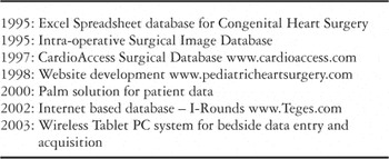
Table 2.

A single screen snapshot, for the 10th post-operative day, of a patient's current clinical data generated by the I-Rounds™ system on the day of discharge after first stage palliation.

Table 3.

Mortality for the first stage of palliation (S1P) for patients with hypoplasia of the left heart (HLHS) as analysed over 8 years in increments of 2 years.

Mortality by year for first stage palliation compared with that for all other operations on patients with congenital cardiac malformations.

The time required for circulatory arrest in minutes for first stage palliation according to the sequence of operations.
 You have Access