Introduction
One of the challenges facing reconstructive surgeons is to provide reconstructive options that provide good cosmetic and functional results whilst minimising morbidity and peri-operative risk. This is particularly true in reconstruction of the nose where there are limits to what can be achieved with local flaps and skin grafts. Pedicled flaps require a longer, more complex procedures and are multistage, which can be problematic in elderly patients with significant co-morbidities.
Whilst skin grafts are often easier to execute and have a lower burden on the patient, their sunken nature means that they are cosmetically unsatisfactory to some patients and their survival can be reduced when used over the cartilage and bone of the nasal skeleton.
There are several established pieces of workReference Nguyen, Washington and Soon1, Reference von Lindern, Niederhagen, Appel and Bergé2 detailing the use of biologic dressings to promote granulation at the defect site, improving the tissue depth and vascularity before playing host to a skin graft in a second procedure after a period of time (often a few weeks). However, we describe a novel one-stage procedure using the oxidised regenerated cellulose and collagen matrix, Promogran™ (3M), to fill a defect and allow placement of a full-thickness skin graft (FTSG) at the time of primary surgery, resulting in improved contour and improved survival over bone and cartilage.
Surgical technique
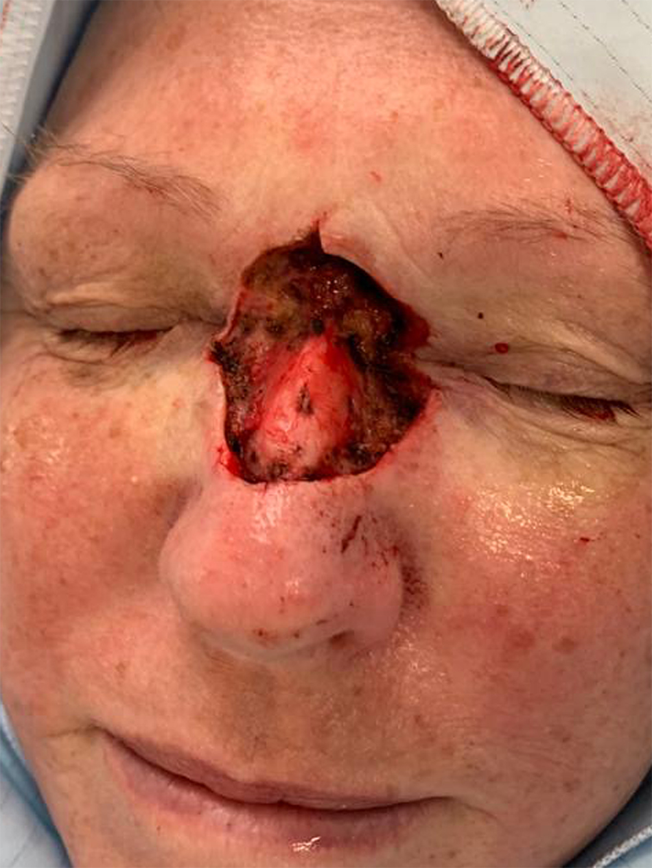
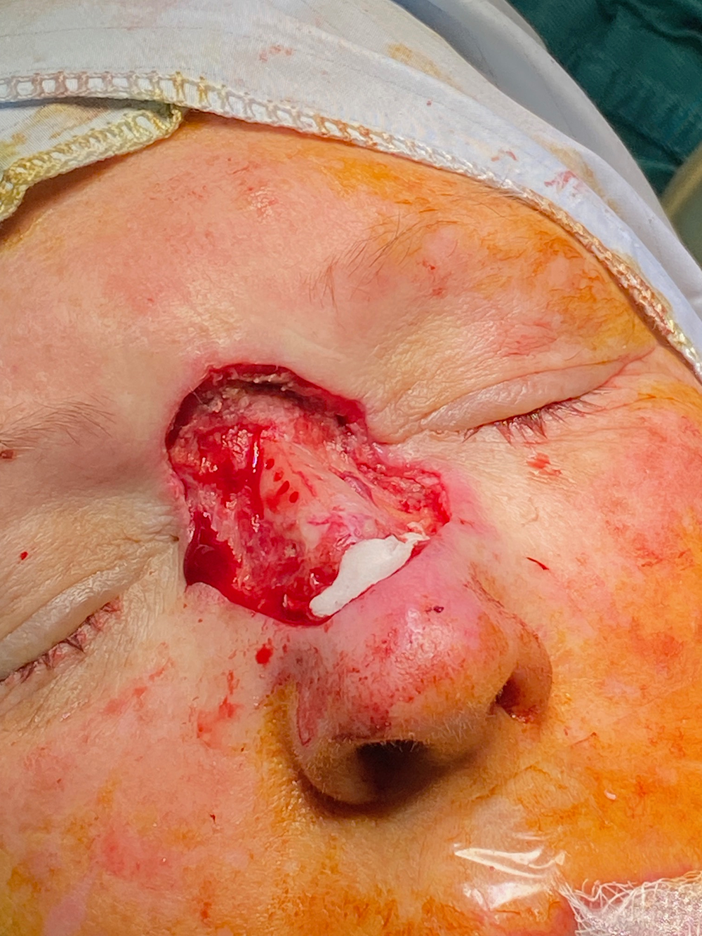
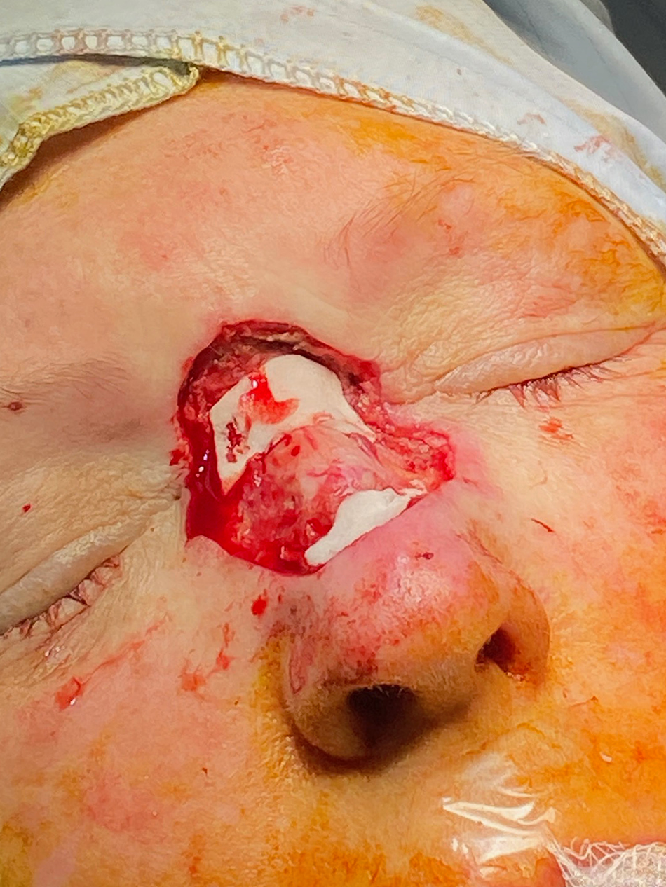
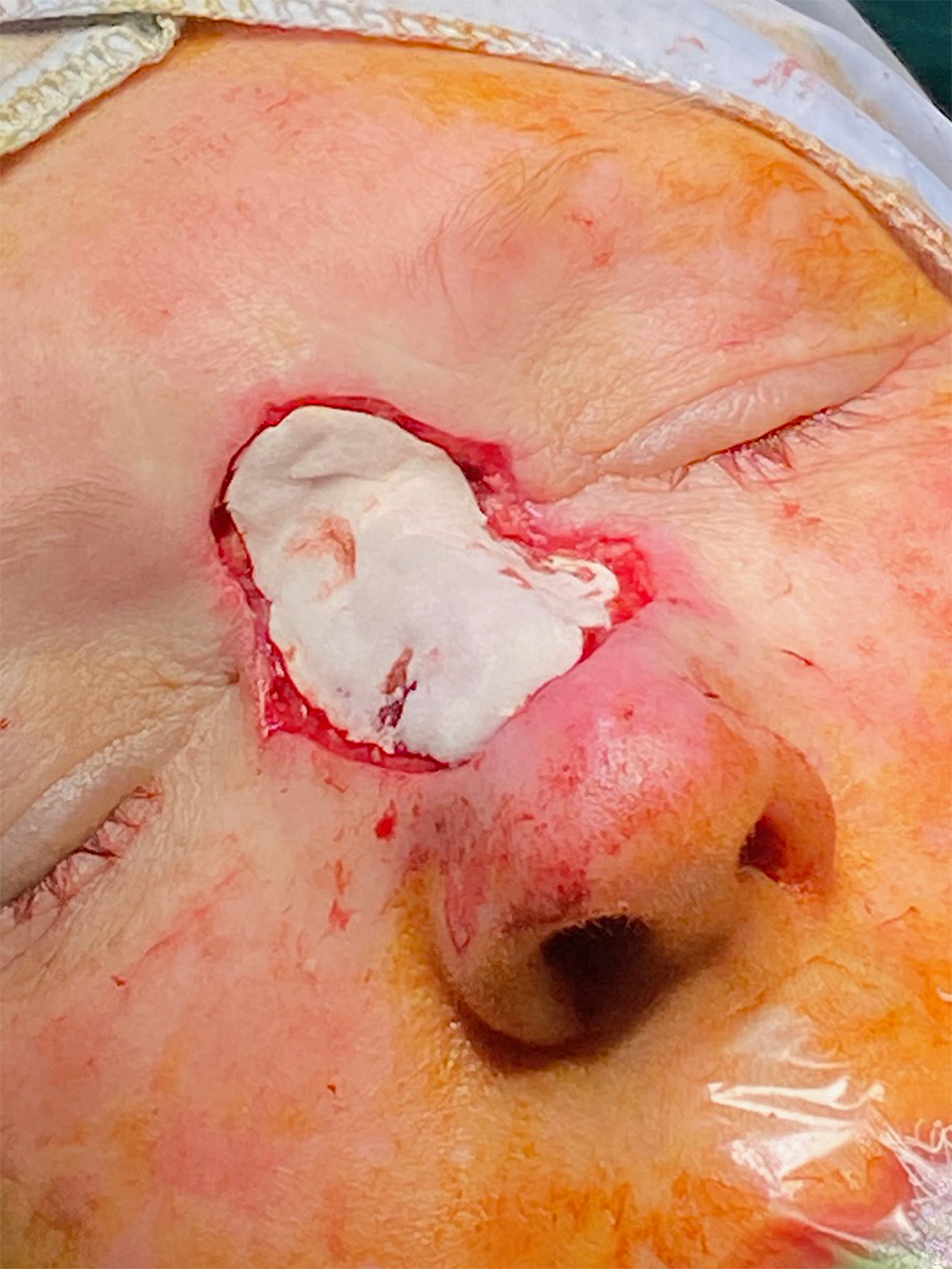
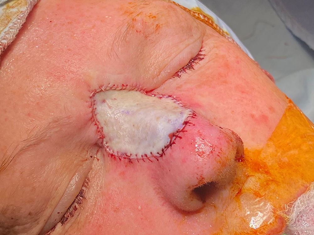
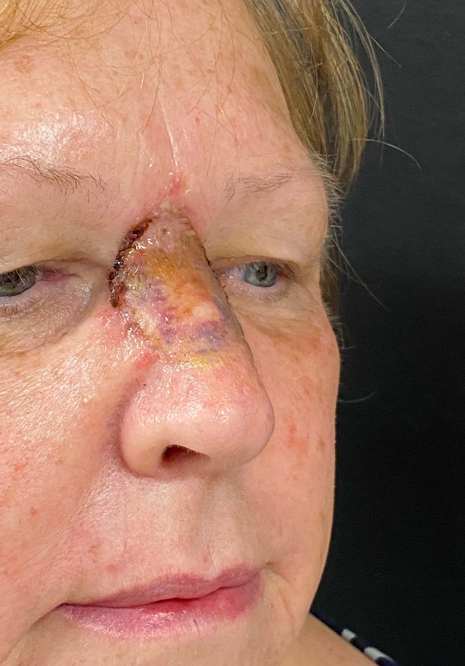
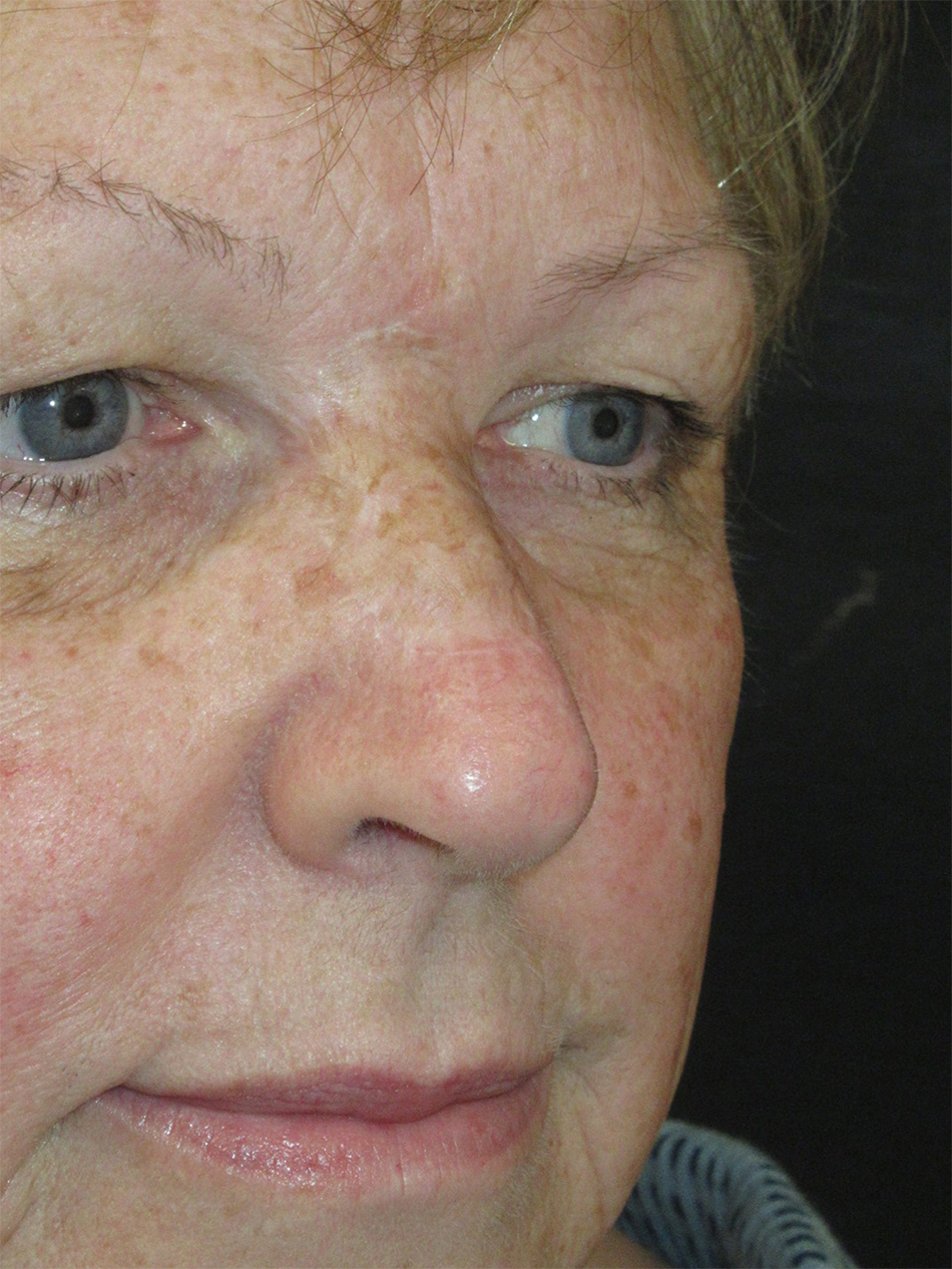
After the skin lesion has been removed, the wound bed is prepared by encouraging a gentle capillary ooze (i.e., not an entirely dry wound). If this is over an area of bone or cartilage then small, partial-thickness defects can be made with a drill, curette or a biopsy punch to stimulate a small amount of bleeding. The FTSG is harvested from the supraclavicular area, thinned to be free of fat and then kept in saline until the wound bed is ready to receive it. The Promogran™ is then cut to size and a single layer placed in the wound bed. It is important to avoid any excess Promogran™ to ensure universal growth of the granulation tissue in the underbed. It may also be desirable to only insert a small segment into the wound to try and recreate a natural contour, such as lesions involving the nasal ala. Alternatively, a double layer could be used in some areas for the same purpose; in these circumstances, adequate ooze of blood to seep through to both layers is ensured as dry matrix will lead to skin graft failure. If the inserted matrix is not saturated with the blood seeped through the bed and the wound bed is dry, a small amount of blood can be drawn through venepuncture and applied to the matrix before inserting the full thickness graft over the matrix. The senior author (S.A.) favours a rapidly absorbable braided suture (e.g., 5-0 Vicryl Rapide) to secure the skin graft in place. The graft is coated in an antibiotic ointment (e.g., mupirocin) and typically a paraffin gauze dressing is sutured over the site and impregnated with antibiotic ointment. Patients are then advised to regularly apply ointment to the dressing (or directly onto the graft if no dressing has been applied) for 3 weeks before they are reviewed in the outpatient clinic. If a dressing has been sutured in, it will be removed at this juncture (Figures 1 to 7).
Defect following Mohs’ resection.

Nasal bones drilled to promote bleeding.

Deep layer of Promogran™.

Superficial layer of Promogran™.

Full thickness skin graft secured with Vicryl Rapide.

2 weeks post operative appearance.

12 months post operative appearance.

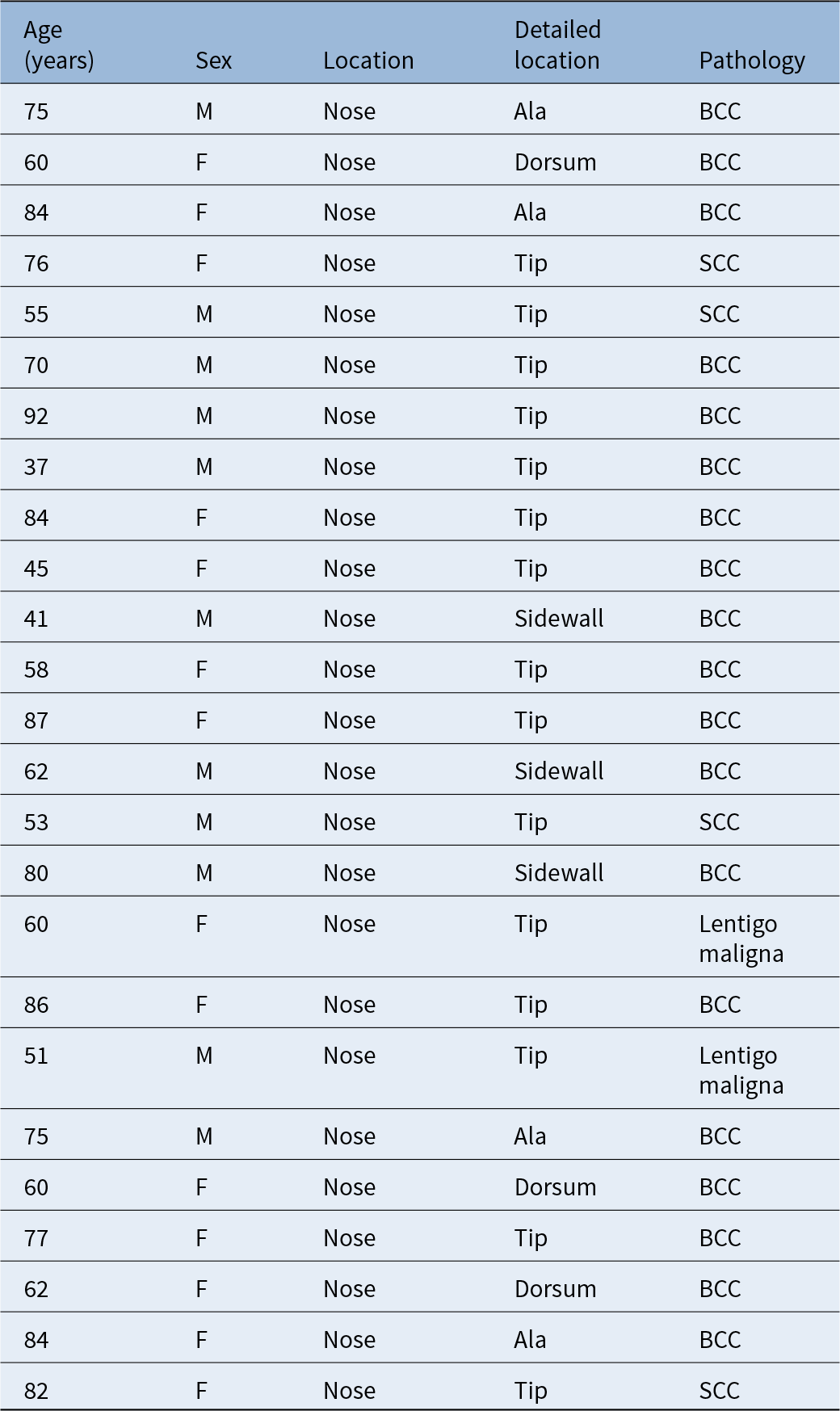
Application of this technique has resulted in a subjective improvement in appearance compared to traditional FTSG reconstruction, as judged by the surgeon and patients themselves (no formal or objective measurement was undertaken). The senior author (S.A.) has employed this technique on 25 patients from 2021 to 2024 (Table 1). All patients were followed up for at least 3 months after surgery (the cases presented in this manuscript were followed up by 1 year). All FTSGs survived except one of the early cases where the centre of the graft failed owing to the Promogran™ staying dry and free of blood during the insertion (hence, we ensure the Promogran™ is saturated with blood before placing the skin graft).
Additional patients treated with technique

BCC = basal cell carcinoma; F = female; M = male; SCC = squamous cell carcinoma.
Discussion
Skin grafting is a type of free tissue transfer without a native blood supply and has existed in western literature since the early 1800s (and has likely been used in practice for over 3000 years).Reference Kok, White, Watkinson and Clarke3 FTSGs are composed of both the whole epidermis and dermis layers, accompanied sometimes by a small volume of subcutaneous tissue. Split-thickness skin grafts (STSGs) contain the whole epidermis and some dermis (depending on the thickness of the graft).Reference Adams and Ramsey4 During the first 24–48 hours after placement, the skin graft receives transudate from the bed and becomes oedematous in a process known as plasmatic imbibition.Reference Kok, White, Watkinson and Clarke3–Reference Ramsey, Walker and Patel5 It is helped to adhere to the underlying bed of tissue by fibrin produced by the recipient site. After 48 hours revascularisation of the graft occurs, initially with the cut ends of the vessels in the recipient bed and graft joining up and subsequently with the formation of new vessels with their growth penetrating the graft. After 3 weeks, revascularisation is complete, and graft remodelling occurs for up to a year.Reference Kok, White, Watkinson and Clarke3 Lymphatic circulation begins within a week and reinnervation begins 2–4 weeks after implantation but can continue throughout the remodelling period.Reference Adams and Ramsey4 The above process is reliant on a well-vascularised recipient bed; however, some surfaces, such as exposed bone, prove to be unfavourable for graft survival. Through a process known as “bridging”, grafts can revascularise from the edges of healthy skin around the donor site up to 5 mm; meaning that typically only grafts of 1 cm or less can survive over an avascular bed, provided there is healthy surrounding tissue.Reference Adams and Ramsey4
Several physiological processes occur in a wound bed covered with Promogran™ that potentially play a role in the effects described on FTSGs. In the context of chronic wounds, Promogran™ has been shown to inactivate key matrix metalloproteases (MMPs) which are responsible for impairment of wound healing.Reference Cullen, Smith, McCulloch, Silcock and Morrison6 MMPs tend to be elevated in chronic, non-healing wounds and have been shown to play a key role in several factors that influence wound healing, such as the regulation of growth factors and cytokines.Reference Guarnera and Restuccia7 Promogran™ has also been shown to act as a free radical scavengerReference Barrett and Moore8, Reference Cullen, Watt, Lundqvist, Silcock, Schmidt and Bogan9 which may be relevant in the context of skin graft survival due to the proposed role of free radicals in graft necrosis.Reference Angel, Zhang, Rogers, Caplan, Dorsett-Martin and Blain10 In vitro studies of Promogran™ have shown increased fibroblast proliferation and cell migration, Reference Hart, Silcock, Gunnigle, Cullen, Light and Watt11 which may help FTSGs adhere to the wound bed and shorten the revascularisation period. Fibroblast collagen deposition may also partially explain increased tissue bulk. Additionally, rat models of acute wounds showed significantly increased levels of platelet-derived growth factor, keratinocyte growth factor, insulin-like growth factor-I and insulin-like growth factor binding protein-3 in wounds treated with Promogran™.Reference Jeschke, Sandmann, Schubert and Klein12
The use of Promogran™ can have several benefits in certain situations for nasal skin reconstruction with skin grafts. The additional granulation under the FTSG adds bulk to the tissue to reduce the typical sunken appearance caused by excision of the subcutaneous tissues and, with careful placement, it can help recreate the natural contours of structures like the nasal ala. Whilst the aesthetic results may be inferior to flap reconstruction, a one-stage procedure (often performed under local anaesthesia) and the ability to place a skin graft over exposed cartilage and bone adds to the appeal of Promogran™ application, which may be preferable for many patients, especially those with multiple co-morbidities. This may also prove helpful in cases with a risk of positive oncological margins, lest a complex reconstruction option be wasted. The technique of using Promogran™ adds a vital tool to the facial plastic surgeon’s arsenal of reconstructive options in a field where creative problem solving is often required.
What is already known on the subject
• Full-thickness skin grafts (FTSGs) often have a lack of bulk and sunken appearance, leading to a poor cosmetic outcome
• FTSGs placed over avascular structures (i.e., cartilage/bone) can have reduced chances of survival
• Two-stage procedures using biologic dressings can enhance graft success by promoting granulation tissue but are time-consuming and burdensome
• Promogran™ (oxidised regenerated cellulose and collagen matrix) is used in chronic wounds to modulate matrix metalloproteinases and support tissue regeneration
What this paper adds
• Achieves improved graft survival, contour and appearance, even over bone or cartilage, with careful matrix saturation and surgical technique.
• Demonstrates clinical applicability in 25 patients, with only one failure due to a dry matrix—emphasising the importance of blood-soaked preparation.
• Offers a minimally invasive, one-stage alternative for nasal reconstruction, particularly suitable for elderly or high-risk patients.
Acknowledgements
The authors declare that they have no relevant financial interests, relationships or conflicts of interest to disclose. No funding or material support was received for the preparation of this manuscript.
Ethical standards
This work describes a surgical technique and retrospective review of cases and was reviewed by the authors’ institutional governance process and deemed exempt from formal research ethics committee approval. All procedures contributing to this work comply with the ethical standards of the relevant national and institutional guidelines on human experimentation and with the Helsinki Declaration of 1975, as revised in 2013. Written informed consent was obtained from all patients for inclusion in this study and for the use of their clinical photographs.