Introduction
The formation, growth and maturation process of ovarian follicles is known as folliculogenesis and is influenced by various substances, including angiotensin II (ANG II) (Ferreira et al., Reference Ferreira, Gasperin, Santos, Rovani, Santos, Gutierrez, Oliveira, Reis and Gonçalves2011a). This substance and its receptors are part of the renin–angiotensin system (RAS) (Yoshimura, Reference Yoshimura1997), which plays important roles in regulation of ovarian follicle development, atresia and maturation (Ferreira et al., Reference Ferreira, Oliveira, Fernandes, Moraes and Gonçalves2007; Barreta et al., Reference Barreta, Oliveira, Ferreira, Antoniazzi, Gasperin, Sandri and Gonçalves2008; Portela et al., Reference Portela, Gonçalves, Veiga, Nicola, Buratini and Price2008).
ANG II actions are mediated through interaction with two main types of membrane receptors, AGTR1 and AGTR2, which have been detected in the granulosa and inner theca cells of rat antral follicles (Husain et al., Reference Husain, Bumpus, Silva and Speth1987). Moreover, rabbit granulosa cells express high levels of AGTR2 in preovulatory follicles (Yoshimura et al., Reference Yoshimura, Karube, Aoki, Oda, Koyama, Nagai, Akimoto, Hirano and Nakamura1996). In bovine, mRNA expression for AGTR1 and AGTR2 was localized in both theca and granulosa cells of antral follicles (Portela et al., Reference Portela, Gonçalves, Veiga, Nicola, Buratini and Price2008).
The activity of ANG II has been related to oocyte maturation, ovulation, steroidogenesis (swine: Li et al., Reference Li, Jiao, Liu, Chen, Wang and Wang2004; rabbit: Yoshimura et al., Reference Yoshimura, Karubc, Koyama, Shiokawa, Nanno and Nakamura1992, Reference Yoshimura, Karube, Oda, Koyama, Shiokawa, Akiba, Yoshinaga and Nakamura1993; Feral et al., Reference Feral, Le Gall and Leymarie1995; bovine: Acosta et al., Reference Acosta, Berisha, Ozawa, Sato, Schams and Miyamoto1999; Giometti et al., Reference Giometti, Bertagnolli, Ornes, Da Costa, Carambula, Reis, De Oliveira, Emanuelli and Gonçalves2005; Stefanello et al., Reference Stefanello, Barreta, Porciuncula, Arruda, Oliveira, Oliveira and Gonçalves2006) and follicular growth in vivo (bovine: Ferreira et al., Reference Ferreira, Gasperin, Rovani, Santos, Barreta, Bohrer, Price and Gonçalves2011b). Giometti et al. (Reference Giometti, Bertagnolli, Ornes, Da Costa, Carambula, Reis, De Oliveira, Emanuelli and Gonçalves2005) found that bovine oocytes cultured for 12 h in medium conditioned with follicular cells had inhibited nuclear maturation, and the majority of oocytes remained at the germinal vesicle stage. However, this effect was reversed after the addition of ANG II to the medium. In addition, a recent study demonstrated that ANG II is an essential cofactor for luteinising hormone (LH) in the early increase of metalloprotease expression/activity that induces the cascade of events leading to ovulation (Portela et al., Reference Portela, Zamberlam, Gonçalves, de Oliveira and Price2011). To our knowledge, only one study has evaluated the influence of ANG II on isolated preantral follicles (<300 µm in diameter), obtaining no significant effect on follicular diameter or antrum formation after 16 or 30 days of in vitro culture (Shuttleworth et al., Reference Shuttleworth, Broughton Pipkin and Hunter2002).
Although the presence of ANG II receptors in ovaries from different species suggests its importance in follicular development, there are no data about the quantification of AGTR1 and AGTR2 mRNA in goat ovarian follicles. Therefore, we hypothesized that these receptors are present in goat ovaries and angiotensin would influence the in vitro development of early preantral follicles (primordial, intermediate and primary follicles). The ability to activate dormant ovarian follicles may play a pivotal role in establishing a successful in vitro culture model for maturing primordial follicles (Desai et al., Reference Desai, Alex, Abdel Hafez, Calabro, Goldfarb, Fleischman and Falcone2010). Therefore, a much deeper understanding of factors regulating caprine preantral follicle development is still needed to improve their capacity to grow, to be fertilized and to produce embryos.
Thus, the present study was designed to determine the steady-state level of AGTR1 and AGTR2 mRNA in different follicular stages and to investigate the influence of different concentrations of ANG II (0, 1, 5, 10, 50 or 100 ng/ml) on the viability, activation and growth of preantral follicles enclosed in caprine ovarian tissue cultured for 1 or 7 days.
Materials and methods
Unless otherwise stated, the culture media, synthetic human ANG II, and other chemicals used in the present study were purchased from Sigma Chemical Co. (St Louis, MO, USA).
Quantification of mRNA levels for AGTR1 and AGTR2 in goat ovarian follicles
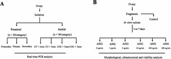
To quantify levels of AGTR1 and AGTR2 mRNA, 30 ovaries from 15 goats (Capra hircus) were collected at a local slaughterhouse from adult (1–3 years old) mixed-breed goats. Immediately postmortem, the ovaries were washed once in 70% alcohol for 10 s and twice in MEM supplemented by HEPES and antibiotics (100 µg/ml penicillin and 100 µg/ml streptomycin). After this preparation, 10 ovaries were used to isolate primordial, primary and secondary follicles. The remaining ovaries were used to collect cumulus–oocyte complexes (COCs), mural granulosa cells, and theca cells from small and large antral follicles (Fig. 1 A).
Experimental design.

Primordial, primary, and secondary follicles were isolated using a previously described mechanical procedure (Lucci et al. Reference Lucci, Amorim, Báo, Figueiredo, Rodrigues, Silva and Gonçalves1999). After isolation, these follicles were washed several times to completely remove the stromal cells and were then placed by category into separate Eppendorf tubes in three groups of 10. This procedure was completed within 2 h, and all samples were stored at −80°C until the RNA was extracted. From a second group of ovaries (n = 15), COCs aspirated from small (1–3 mm) and large (3–6 mm) antral follicles were recovered. Compact COCs were selected from the follicle content as previously described (van Tol and Bevers, Reference van Tol and Bevers1998). Thereafter, three groups of 10 COCs were stored at −80°C until RNA extraction. To collect the mural granulosa and theca-cell complex (GT), small (n = 30) and large antral follicles (n = 30) were isolated from the ovaries (n = 5) and dissected free from the stromal tissue with forceps as previously described (van Tol and Bevers, Reference van Tol and Bevers1998). The follicles were then bisected, and the GT were collected and stored at −80°C.
Total RNA isolation was performed using a Trizol Plus RNA Purification Kit (Invitrogen, São Paulo, Brazil). According to the manufacturer's instructions, 1 ml of Trizol solution was added to each frozen sample, and the lysate was aspirated through a 20-gauge needle before centrifugation at 10,000 g for 3 min at room temperature. All lysates were then diluted 1:1 with 70% ethanol and subjected to a mini-column. After the binding of the RNA to the column, DNA digestion was performed using RNase-free DNase (340 Kunitz units/ml) for 15 min at room temperature. After washing the column three times, the RNA was eluted with 30 µl RNase-free water.
Prior to reverse transcription, the eluted RNA samples were incubated for 5 min at 70°C and chilled on ice. Reverse transcription was then performed in a total volume of 20 µl, which comprised 1 µg of sample RNA, 4 µl 5× reverse transcriptase buffer (Invitrogen), 8 U RNaseOUT, 150 U Superscript III reverse transcriptase, 0.036 U random primers (Invitrogen), 10 mM DTT, and 0.5 mM of each dNTP, distilled water completing the final volume to 20 µl. The mixture was incubated for 1 h at 42°C, followed by 5 min at 80°C, then it was stored at −20°C. Negative controls were prepared under the same conditions but without the inclusion of the reverse transcriptase.
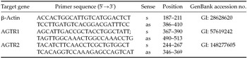
Quantification of the AGTR1 and AGTR2 mRNAs was performed using SYBR Green. PCR reactions were composed of 1 µL cDNA as a template in 7.5 µl of SYBR Green Master Mix (PE Applied Biosystems, Foster City, CA, USA), 5.5 µL of ultra-pure water, and 0.5 µM of each primer. The primers were designed to amplify AGTR1 and AGTR2 mRNA. β-Actin was selected as endogenous control for normalization of gene expression in all samples, as a previous study had demonstrated that this reference gene is the most stable in goat preantral follicles (Frota et al., Reference Frota, Leitão, Costa, Brito, van den Hurk and Silva2011). (Table 1). Primer specificity and amplification efficiency was verified for each gene. The thermal cycling profile for the first round of PCR was as follows: initial denaturation and activation of the polymerase for 15 min at 94°C, followed by 40 cycles of 15 s at 94°C, 30 s at 60°C, and 45 s at 72°C. The final extension was for 10 min at 72°C. All reactions were performed in a real-time PCR Mastercycler (Eppendorf, Germany). The ΔΔCT method was used to transform CT values into normalized relative steady-state mRNA levels (Livak & Schmittgen, Reference Livak and Schmittgen2001).
Primer pairs used for real-time PCR analyses

as, antisense; s, sense.
Experimental design for in vitro culture of caprine ovarian tissue
For in vitro culture, additional pairs of caprine ovaries (n = 8 ovaries) were collected and washed as described above. The ovaries were transported within one hour to the laboratory in tubes containing MEM-HEPES and antibiotics (100 µg/ml penicillin and 100 µg/ml streptomycin) at 4°C (Chaves et al., Reference Chaves, Martins, Saraiva, Celestino, Lopes, Correia, Lima-Verde, Matos, Báo, Name, Campello, Silva and Figueiredo2008).
The organ culture system used herein was previously described in detail (Chaves et al., Reference Chaves, Martins, Saraiva, Celestino, Lopes, Correia, Lima-Verde, Matos, Báo, Name, Campello, Silva and Figueiredo2008). In the laboratory, ovarian cortex tissue samples from each ovarian pair were cut into 13 slices (approximately 3 × 3 mm with a 1-mm thickness) using a needle and scalpel under sterile conditions. The tissue slices were then either directly fixed for histological and ultrastructural analysis (fresh control) or placed in culture for 1 or 7 days. Ovarian slices were incubated in 1 ml of culture media consisting of MEM supplemented with ITS (10 µg/ml insulin, 5.5 µg/ml transferrin and 5 ng/ml selenium), 2 mM glutamine, 2 mM hypoxanthine and 1.25 mg/ml bovine serum albumin (BSA) in the presence of 0, 1, 5, 10, 50 or 100 ng/ml ANG II (Fig. 1 B) in 24-well plates at 39°C in 5% CO2 in air. Each treatment was repeated four times, and the ovaries of four different animals were consequently used. The culture media were replenished every other day.
Morphological analysis and assessment of in vitro follicular growth
Before culture (fresh control) and after 1 or 7 days in culture, all of the pieces cultured with or without ANG II were fixed in Carnoy's solution for 12 h and then dehydrated in increasing concentrations of ethanol (Dinâmica, São Paulo, Brazil). After paraffin embedding (Synth, São Paulo, Brazil), the caprine tissue slices were cut into 7-μm sections, and each section was mounted on a glass slide and stained by periodic acid-Schiff and hematoxylin (Dinâmica). Follicle stage and survival were assessed microscopically on serial sections. Coded slides were blindly examined using a microscope (Nikon, Japan) under ×400 magnification.
The developmental stages of follicles have been defined previously (Silva et al., Reference Silva, van den Hurk, Costa, Andrade, Nunes, Ferreira, Lôbo and Figueiredo2004) as primordial (one layer of flattened granulosa cells around the oocyte) or growing (intermediate: one layer of flattened to cuboidal granulosa cells; primary: one layer of cuboidal granulosa cells; secondary: two or more layers of cuboidal granulosa cells around the oocyte). Individual follicles were further classified as histologically normal when an intact oocyte surrounded by well-organized granulosa cells in one or more layers without a pyknotic nucleus was observed. Atretic follicles were defined as those with a retracted oocyte, pyknotic nucleus, and/or disorganized granulosa cells that were detached from the basement membrane. Overall, 120 follicles were evaluated for each treatment (30 follicles per treatment for one repetition × four repetitions = 120 follicles).
To evaluate follicular activation, the percentages of healthy primordial and growing follicles were calculated before (fresh control) and after culture in each medium. In addition, follicle and oocyte diameters were measured only in morphologically normal follicles. The follicle diameter was recorded as the length from edge to edge of the basement membrane or from the outside edge of the theca-cell layer when present. Oocyte diameter was recorded as the length from edge to edge of the oocyte membrane. Two perpendicular diameters were recorded for each parameter, and the average of these two values was reported. Furthermore, each follicle was examined in every section in which the nucleus appeared. This ensured that each follicle was only counted once, regardless of its size.
Viability assessment of follicles cultured in vitro
Based on the morphological analysis, the viability of the follicles cultured with the ANG II concentrations that promoted the highest percentages of normal follicle growth was further analyzed using fluorescent probes. For this end, additional pairs of goat ovaries (n = 4 ovaries) were collected, washed and transported to the laboratory as described above. Then, ovarian tissue was cut into fragments and one of these fragments was immediately processed for follicle isolation, and the remaining fragments were cultured for 7 days with ANG II (10 or 50 ng/ml) as described above. After the culture period, fragments were processed for mechanical isolation using the methods described by Lucci et al. (Reference Lucci, Amorim, Báo, Figueiredo, Rodrigues, Silva and Gonçalves1999).
Thereafter, the viability of isolated preantral follicles was assessed through a two-color fluorescence cell viability assay based on the simultaneous determination of live and dead cells using calcein-AM and ethidium homodimer-1, respectively. The first probe detected the intracellular esterase activity of viable cells, and the second labeled nucleic acids of non-viable cells that had plasma membrane disruption. The test was performed by adding 4 μM calcein-AM and 2 μM ethidium homodimer-1 (Molecular Probes, Invitrogen, Karlsruhe, Germany) to the suspension of isolated follicles and incubating at 37°C for 15 min. After labeling, follicles were washed once by centrifugation at 100 g for 5 min, resuspended in MEM and mounted on a glass microscope slide in 5 ml anti-fading medium (DABCO, Sigma, Deisenhofen, Germany) to prevent photobleaching. Finally, follicles were examined using a Axioskop 40 fluorescence microscope (Zeiss, Germany). The emitted fluorescence signals of calcein-AM and ethidium homodimer-1 were collected at 488 and 568 nm, respectively. Oocytes and granulosa cells were considered alive if the cytoplasm stained positively with calcein-AM (green) and the chromatin was not labelled with ethidium homodimer-1 (red).
Ultrastructural analysis of caprine preantral follicles
For a more detailed evaluation of follicular morphology after histological analysis, ultrastructural studies were performed in non-cultured follicles (fresh control) and in follicles that were cultured for 7 days in the lowest concentration of ANG II (10 ng/ml), which showed the best results for follicular survival and viability. A section with a maximum dimension of 1 mm3 was cut from each fragment of the ovarian tissue and fixed in Karnovsky solution (4% paraformaldehyde and 2.5% glutaraldehyde in 0.1 M sodium cacodylate buffer at pH 7.2) for 3 h at room temperature (approximately 25°C). After three washes in sodium cacodylate buffer, specimens were post-fixed in 1% osmium tetroxide, 0.8% potassium ferricyanide and 5 mM calcium chloride in 0.1 M sodium cacodylate buffer for 1 h at room temperature. The samples were then dehydrated using a gradient of acetone solutions and then embedded in SPIN-PON resin (Sigma Company, St Louis, MO). Subsequently, semi-thin sections (3 µm) were cut, stained with toluidine blue and analyzed using light microscopy at ×400 magnification. Ultra-thin sections (60–70 nm) were obtained from preantral follicles, which were classified as morphologically normal in semi-thin sections according to the criteria adopted in histology. Subsequently, the ultra-thin sections were contrasted with uranyl acetate and lead citrate and examined under a Morgani-FEI transmission electron microscope operating at 80 kV. The following parameters were evaluated based on a previous study (Lucci et al., Reference Lucci, Silva, Carvalho, Figueiredo and Báo2001): the density and the integrity of ooplasmic and granulosa cell organelles, vacuolization and the integrity of the basement and nuclear membranes.
Statistical analysis
Data of mRNA expression from primordial, primary and secondary follicles and mRNA expression in small and large antral follicles were compared by Tukey's multiple comparisons test (P < 0.05). Percentages of the surviving follicles at both stages (i.e., primordial and developing) and follicle and oocyte diameters were initially evaluated for homoscedasticity and normal distribution of the residues, by Bartlett's and Shapiro–Wilk tests, respectively. Confirmed both requirements underlying analysis of variance, the effects of treatment (ANG II concentrations), time and treatment by time interaction were analyzed using PROC MIXED of SAS (2002). The model was Y ijk =µ + C i + F j + T k + (CT) ik + e ijk , where Y ijk is the observation of the jth ovarian fragment in the ith treatment at the kth time of culture, µ is the overall mean, C i is the ith treatment, F j is the random effect of the jth ovarian fragment within the ith treatment, T k is the kth time of culture, (CT) ik is the treatment by time interaction term and e ijk is the random residual effect. Dunnett's test was applied to compare ANG II-treated groups with the fresh control (Steel et al., Reference Steel, Torrie and Dickey1997). Comparisons amongst treatments or between times were further analyzed by Student–Newman–Keuls (SNK) test. A probability of P < 0.05 indicated a significant difference and results were expressed as mean ± standard error of the mean.
Results
Levels of AGTR1 and AGTR2 mRNA in goat ovarian follicles
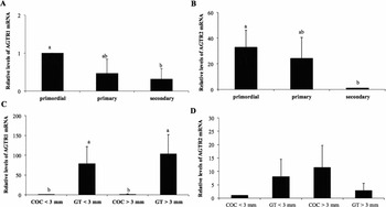
Primordial follicles had significantly higher levels of AGTR1 and AGTR2 receptor mRNA than the secondary follicle stages (P < 0.05), but the levels did not differ from those of primary follicles (P > 0.05; Fig. 2 A, B).
Steady-state levels of AGTR1 and AGTR2 mRNAs in goat ovarian follicles (means ± SEM). (A, B) Primordial, primary and secondary follicles. (C, D) Cumulus–oocyte complexes (COCs) and granulosa and theca cells (GT) from small and large antral follicles. a,b P < 0.05.

No differences were observed in the AGTR1 and AGTR2 receptor mRNA levels for COCs or granulosa/theca cells collected from small and large antral follicles (P > 0.05; Fig. 2 C–F). Granulosa/theca cells from either small or large antral follicles had significantly higher levels of AGTR1 mRNA than their respective COCs (P < 0.05; Fig. 2 G, H).
Effect of ANG II on follicular survival after in vitro culture
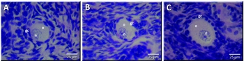
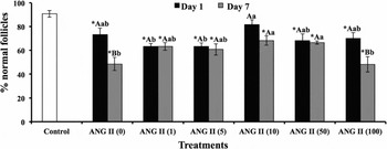
The present study analyzed a total of 1560 caprine preantral follicles. Figure 3 shows histological sections of primordial (A), intermediate (B) and primary (C) morphologically normal follicles after 7 days of culture in 10 ng/ml ANG II. Statistical analysis revealed that there was a significant effect of the concentration of ANG II (P < 0.0129), the culture time (P < 0.0002) and the interaction between concentration ANG II and culture time (P < 0.0234). As shown in Fig. 4, a significant reduction (P < 0.05) in the percentage of morphologically normal follicles was observed in all treatments when compared to the fresh control (90.83%), except when the fragments were cultured for 1 day in 10 ng/ml ANG II (81.67%; P > 0.05). Moreover, after 7 days, a significantly higher percentage of normal follicles (P < 0.05) was observed in tissues that were cultured with 10 or 50 ng/ml ANG II than those cultured with 0 or 100 ng/ml ANG II.
Histological sections, stained with periodic acid–Schiff hematoxylin, showing primordial (A), intermediate (B) and primary (C) normal follicles after culture in 10 ng/ml ANG II for 7 days. o: oocyte; n: oocyte nucleus; gc: granulosa cells. Magnification was at ×400.

Percentage (mean ± SEM) of morphologically normal caprine preantral follicles in the fresh control and after in vitro culture for 1 or 7 days in the absence or presence of ANG II. *Differs significantly from fresh control follicles; A,Bdifferent uppercase letters represent significant differences between columns (days of culture); a,bdifferent lowercase letters represent significant differences between lines (ANG II concentrations).

Assessment of preantral follicle viability by fluorescence
Based on the results of the morphological analysis, a viability trial was performed using follicles (n = 30) from the fresh control and from the treatments that maintained the highest percentage of morphologically normal follicles after 7 days of culture (αMEM+ supplemented with 10 or 50 ng/ml ANG II). The results showed that the percentage of viable follicles was similar in the fresh control group (85%) and those cultured in 10 ng/ml or 50 ng/ml ANG II (93.33 or 90.0%, respectively) (P > 0.05).
Ultrastructural analysis of caprine preantral follicles
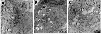
The ultrastructural characteristics of follicles from the fresh control (Fig. 5 A) and those cultured with 10 ng/ml ANG II were similar (Fig. 5 B). The follicles showed intact basement and nuclear membranes and a large oocyte nucleus. In addition, vesicles and organelles were uniformly distributed in the ooplasm, especially the mitochondria. Granulosa cells were ultrastructurally normal and showed an elongated and large nucleus with an irregular membrane (Fig. 5 C).
Ultrastructural analysis of caprine non-cultured follicles (fresh control) (A, C) and follicle cultured for 7 days in medium containing 10 ng/ml ANG II (B). gc, granulosa cells; m, mitochondria; arrow, oocyte membrane. (A, B: bar = 2 µm; C: bar = 1 µm). Three to five follicles per group were examined, and the photomicrographs are representative examples.

Follicular activation and growth after in vitro culture
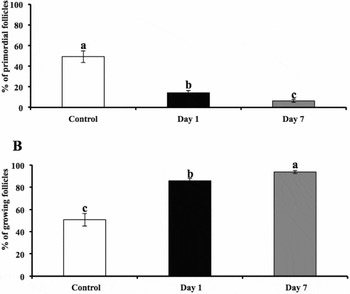
Analysis of follicular activation showed no significant effect of concentration of ANG II (P < 0.1574), but only culture time (P < 0.0054). There was no significant interaction between concentration of ANG II and culture time (P < 0.4757). Figure 6(A, B) shows the percentage of primordial and growing follicles, respectively, in ovarian cortical tissue before and after in vitro culture. After 1 or 7 days of culture, a significant reduction in the percentage of primordial follicles, reflecting an increase in the percentage of growing ones, was observed in all treatments compared to fresh control group. Furthermore, an increase in activation was observed at day 7 compared to day 1 of culture.
Percentage (mean ± SEM) of primordial (A) and growing (B) follicles in the fresh control and after in vitro culture for 1 or 7 days in the absence or presence of ANG II. a,b,cDifferent lowercase letters represent significant differences between columns (days of culture).

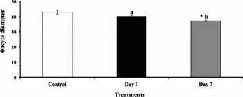
Regarding to follicular diameter, analysis showed no significant effect of concentration of ANG II (P < 0.1039), the culture time (P < 0.5443), or the interaction between concentration ANG II and culture time (P < 0.2207). Concerning specifically to oocytes, a significant decrease in their diameter (P < 0.0120) was observed with the progression of culture period from 1 to 7 days (Fig. 7). Conversely, ANGII concentration did not affect significantly this variable (P < 0,0621), neither the interaction between ANGII and time culture (P < 0,3501).
Oocyte diameter (mean ± SEM) in the fresh control and after in vitro culture for 1 or 7 days in the absence or presence of ANG II. *Differs significantly from fresh control follicles; a,bdifferent lowercase letters represent significant differences between columns (days of culture).

Discussion
This study showed the AGTR1 and AGTR2 mRNA levels in goat follicles and examined the effects of ANG II on the survival and development of preantral follicles after in vitro culture of caprine ovarian tissue for 7 days. Primordial follicles had higher AGTR1 and AGTR2 mRNA levels than secondary follicles. Contrary to our results, in mice, Yoon et al. (Reference Yoon, Kim, Chung, Choi, Lee, Cha and Lee2006) observed an increase in the AGTR2 mRNA expression using microarray analysis during the transition from primary to secondary follicle. The discrepancy may reflect a physiological difference between species, such as the length of folliculogenesis, which is shorter in mice (17–21 days) than in goats (about 6 months). Moreover, in the present study, the AGTR1 mRNA levels were higher in granulosa/theca cells from small and large antral follicles when compared to their respective COCs. The effects of ANG II on blood pressure regulation are mediated by AGTR1, whereas the physiological roles of the AGTR2 receptor are still uncertain (Paul et al., Reference Paul, Mehr and Kreutz2006). It is likely that AGTR1 regulates blood flow while AGTR2 controls oocyte growth. Some studies using RT-PCR have also demonstrated the presence of AGTR1 and AGTR2 mRNA in both the granulosa and theca cells of bovine antral follicles (Berisha et al., Reference Berisha, Schams and Miyamoto2002; Portela et al., Reference Portela, Gonçalves, Veiga, Nicola, Buratini and Price2008).
Higher rates of morphologically normal follicles were observed in tissues cultured for 7 days in medium supplemented with 10 or 50 ng/ml ANG II when compared with 0 ng/ml ANG II, and fluorescence microscopy confirmed these results. Moreover, the ultrastructural integrity of follicles cultured in the presence of 10 ng/ml ANG II (the lowest concentration that maintained morphology) was preserved. As the ovarian fragments consisted mostly of primordial follicles and the mRNA levels of angiotensin receptors were higher in caprine primordial than in secondary follicles, it is possible that ANG II maintained follicular viability through its binding to its receptors. A recent study showed that AGTR2 expression was higher in healthy follicles compared to atretic follicles, suggesting that AGTR2 is related to the maintenance of follicular viability (Portela et al., Reference Portela, Gonçalves, Veiga, Nicola, Buratini and Price2008). In addition, ANG II appears to regulate the induction of other autocrine growth factors, such as insulin-like growth factor-1 (IGF-1; Stefanello et al., (Reference Stefanello, Barreta, Porciuncula, Arruda, Oliveira, Oliveira and Gonçalves2006) and fibroblast growth factor-2 (FGF-2; Itoh et al., Reference Itoh, Mukoyama, Pratt, Gibbons and Dzau1993), which may have functioned as a survival factor for the follicles in the current study (Garor et al., Reference Garor, Abir, Erman, Felz, Nitke and Fisch2009).
After 7 days of culture, a high percentage of growing follicles was observed in all treatments when compared to the fresh control and one day of culture. However, no difference between treated follicles with or without ANG II was found, which indicates that ANG II did not affect caprine follicular activation. This can be due to the fact that αMEM+ is an enriched medium, containing non-essential amino acids, carbohydrates, inorganic salts, sodium pyruvate, lipoic acid, biotin, vitamins and DNA precursors, all of which promote cell division (Hartshorne, Reference Hartshorne1997). Other authors have also shown high developing follicle counts in human ovarian tissue cultured for 6 days with basic medium alone (α-MEM) (Lerer-Serfaty et al., Reference Lerer-Serfaty, Samara, Fisch, Shachar, Kossover, Seliktar, Ben-Haroush and Abir2013).
Treatment with ANG II did not influence follicular or oocyte diameter after 7 days of culture. Shuttleworth et al. (Reference Shuttleworth, Broughton Pipkin and Hunter2002) also found no significant effect of ANG II (10−10 mol/l) on the diameter of swine isolated preantral follicles after 16 or 30 days of culture. Some authors have reported that the addition of ANG II to bovine granulosa cells in culture did not affect cell proliferation or estradiol secretion, but inhibited mRNA that encoded serine protease inhibitor E2, a protein that is involved in tissue remodeling (Portela et al., Reference Portela, Gonçalves, Veiga, Nicola, Buratini and Price2008). The decrease in oocyte diameter observed after 7 days of in vitro culture could be an indicative that only the follicles with lower oocyte diameter survived.
This study demonstrated a high expression of AGTR1 and AGTR2 in primordial follicles. In antral follicles, granulosa/theca cells from either small or large antral follicles had higher levels of AGTR1 mRNA. In addition, 10 ng/ml ANG II maintained the viability of caprine preantral follicles after 7 days of in vitro culture, although did not influence follicular or oocyte diameter. In conclusion, these findings suggest that ANG II could have a significant application in improving the quality of follicles that are used for in vitro culture.
Acknowledgements
This work was supported by CNPQ (RENORBIO: grant number 554812/2006-1). Jamily Bezerra Bruno is a recipient of a grant from FUNCAP/CAPES (Brazil). The authors thank José Leandro da Silva Neto for his technical support in classical histology.
Disclosures
The authors declare that there is no potential conflict of interest that can be perceived as prejudicing the impartiality of the research reported.
Supplementary materials
Supplementary materials are available with this paper. Please visit http://dx.doi.org/10.1017/S0967199415000544