Introduction
Fascioliasis is an important food-borne helminthic disease caused by the two trematode species, Fasciola hepatica and F. gigantica (Mas-Coma, Reference Mas-Coma2005; Ashrafi et al., Reference Ashrafi, Valero, Massoud, Sobhani, Solaymani-Mohammadi, Conde, Khoubbane, Bargues and Mas-Coma2006a). This disease inflicts deleterious economic losses in ruminants due to reduction in weight, milk yield, fertility rate and condemned livers (Schweizer et al., Reference Schweizer, Braun, Deplazes and Torgerson2005; Elitok et al., Reference Elitok, Elitok and Kabu2006; Charlier et al., Reference Charlier, Duchateau, Claerebout, Williams and Vercruysse2007). Furthermore, WHO has recently included fascioliasis as an important human disease among neglected diseases, with human cases reported from countries in Europe, America, Asia, Africa and Oceania (Mas-Coma et al., Reference Mas-Coma, Valero and Bargues2009).
The definitive host range of both fasciolid species is wide, including mainly livestock species (Mas-Coma et al., Reference Mas-Coma, Bargues and Valero2005), while intermediate hosts or vectors are freshwater snails of the family Lymnaeidae (Bargues & Mas-Coma, Reference Bargues and Mas-Coma2005). The geographical distribution of both aetiological agents overlaps in many African and Asian countries (Mas-Coma et al., Reference Mas-Coma, Bargues and Valero2005, Reference Mas-Coma, Valero and Bargues2009). Despite the importance of differentiating between infection by each fasciolid species, due to their distinct epidemiological, pathological and control characteristics, there is, unfortunately, neither a direct coproantigen test nor an indirect immunological test available for their differential diagnosis (Valero et al., Reference Valero, Ubeira, Khoubbane, Artigas, Muiño, Mezo, Pérez-Crespo, Periago and Mas-Coma2009b, Reference Valero, Periago, Pérez-Crespo, Rodríguez, Perteguer, Gárate, González-Barberá and Mas-Coma2012a, Reference Valero, Periago, Perez-Crespo, Angles, Villegas, Aguirre, Strauss, Espinoza, Herrera, Terashima, Tamayo, Engels, Gabrielli and Mas-Comab). Diagnostic tools available at present are only useful to differentiate fascioliasis from other diseases. Hitherto, the specific differentiation can only be made by either a morphometrical study of adult flukes and eggs (Periago et al., Reference Periago, Valero, Panova and Mas-Coma2006; Valero et al., Reference Valero, Perez-Crespo, Periago, Khoubbane and Mas-Coma2009a, Reference Valero, Perez-Crespo, Khoubbane, Artigas, Panova, Ortiz, Maço, Espinoza and Mas-Coma2012c) or by molecular tools (Marcilla et al., Reference Marcilla, Bargues and Mas-Coma2002; Mas-Coma et al., Reference Mas-Coma, Valero and Bargues2009).
The overlapping distribution of F. hepatica and F. gigantica has also led to a long-ranging controversy over the taxonomic identity of the Fasciola species found in countries of the Far East, in which some resemble F. hepatica, whereas others resemble F. gigantica, with intermediate forms also being present and involving phenomena such as abnormal gametogenesis, diploidy, triploidy and mixoploidy, parthenogenesis and hybridization events between different genotypes (see reviews in Mas-Coma & Bargues, Reference Mas-Coma and Bargues1997; Mas-Coma et al., Reference Mas-Coma, Valero and Bargues2009). A comprehensive multidisciplinary study has, however, demonstrated that F. hepatica and F. gigantica should be considered valid species, despite their capacity to hybridize and give rise to intermediate forms in overlapping areas (Mas-Coma et al., Reference Mas-Coma, Valero and Bargues2009).
Pakistan is one of the Asian countries where both fasciolid species overlap, and fascioliasis is highly prevalent in livestock, including cattle, buffaloes, sheep and goats (Khan et al., Reference Khan, Sajid, Khan, Iqbal and Hussain2010), and the two fasciolid species are frequently reported (Chaudhry & Niaz, Reference Chaudhry and Niaz1984; Masud & Majid, Reference Masud and Majid1984; Siddiqi & Shah, Reference Siddiqi and Shah1984; Maqbool et al., Reference Maqbool, Arshad, Mahmood and Hussain1994, Reference Maqbool, Hayat, Akhtar and Hashmi2002; Khan et al., Reference Khan, Sajid, Khan, Iqbal and Iqbal2009). In the Punjab province of Pakistan, fascioliasis is of special concern in bovines. Fascioliasis has been found to be endemic in various districts, with prevalences ranging from 10.48 to 40.31% (Chaudhry & Niaz, Reference Chaudhry and Niaz1984; Masud & Majid, Reference Masud and Majid1984; Maqbool et al., Reference Maqbool, Hayat, Akhtar and Hashmi2002; Khan et al., Reference Khan, Sajid, Khan, Iqbal and Iqbal2009), and results indicate the presence of F. gigantica to be more widespread than F. hepatica in five Punjabi districts (Khan et al., Reference Khan, Sajid, Khan, Iqbal and Iqbal2009). Furthermore, human cases have also been described in rural areas of Lahore, Central Punjab, Pakistan, thus suggesting fascioliasis to be an even greater public heath problem than previously estimated (Qureshi et al., Reference Qureshi, Tanveer, Qureshi, Maqbool, Gill and Ali2005).
Faced with the present situation, it is evident that further studies about the epidemiology and transmission of the disease to humans and animals are required to obtain the baseline on which to establish appropriate control measures in that endemic area of Pakistan. This study represents a step further in this endeavour, by analysing the morphometric characteristics of fasciolid adults infecting the buffalo, one of the main livestock species present.
Studies were performed on the basis of standardized measurements known to be useful for the differentiation of both fasciolid species (Periago et al., Reference Periago, Valero, Panova and Mas-Coma2006). Therefore, a computer image analysis system (CIAS) method (Valero et al., Reference Valero, Panova and Mas-Coma2005) was applied to flukes collected from buffaloes. Since it is the first time that such a study has been performed in Pakistan, the results are compared to pure fasciolid populations, namely (a) F. hepatica from the European Mediterranean area; and (b) F. gigantica from Burkina Faso; i.e. geographical areas where both species do not co-exist, according to previously obtained results (Periago et al., Reference Periago, Valero, Panova and Mas-Coma2006). Although metacercarial infectivity does not appear to differ in isolates from different livestock species (Valero & Mas-Coma, Reference Valero and Mas-Coma2000), the definitive animal host species is known to pronouncedly influence the phenotype of both adult stage and eggs of the liver fluke, mainly due to the different size of the liver duct microhabitat (Valero et al., Reference Valero, Darce, Panova and Mas-Coma2001a, Reference Valero, Panova, Comes, Fons and Mas-Coma2002, Reference Valero, Perez-Crespo, Periago, Khoubbane and Mas-Coma2009a). For this reason, only parasites obtained from bovines were used in this study.
Materials and methods
Collection and examination of flukes
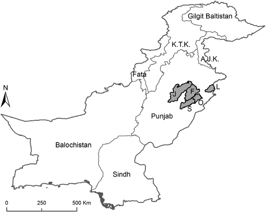
The study area comprises irrigated land with a well-established water canal system of the Indus river basin, commonly categorized as Central Punjab in Pakistan. The buffaloes originated from different districts of the Central Punjab area, namely the districts of Lahore, Faisalabad, Okara, Sahiwal and Jhang (fig. 1). The existence of rivers and canals provides excellent breeding grounds for the development and survival of freshwater snails serving as potential intermediate hosts for a variety of digenetic trematode parasites.
Map of Pakistan to show the origin of infected buffaloes in five districts (grey) of Central Punjab. J, Jhang; F, Faisalabad; S, Sahiwal; O, Okara; and L, Lahore.

In September 2011, the post-mortem examinations of 19 buffaloes were conducted immediately after slaughtering to ascertain the presence of adult liver flukes. The adult worms found in bile ducts were collected with the help of rubber-coated forceps in order to avoid any structural damage. Only adult flukes having gravid uteri (n= 81) were included in the study. Fasciolid specimens providing the largest worm variability in their size, maturity and gravid uteri were used for characterization. Previously published data on F. hepatica from Spain (Europe) and F. gigantica from Burkina Faso (Africa) were used as adult standard or genetically pure populations, corresponding to liver flukes from naturally infected bovines (Periago et al., Reference Periago, Valero, Panova and Mas-Coma2006; Mas-Coma et al., Reference Mas-Coma, Valero and Bargues2009).
Fasciolid specimens were fixed in Bouin's solution between two slides with a little pressure, depending on the thickness of the flukes. The fasciolids were stained with Grenacher's borax carmine and subsequently differentiated, dehydrated and mounted in Canada balsam. The eggs were collected from the distal part of the uteri of fully mature liver flukes by applying a little pressure on the acetabulum to force out mature eggs through the genital pore. In total, 345 fully mature eggs were included, being brownish dark and having well-formed shell walls. The comparison was made with previously published data for egg standard populations of F. hepatica from Spain and F. gigantica from Burkina Faso (Periago et al., Reference Periago, Valero, Panova and Mas-Coma2006).
Morphometrics
An accurate morphometric study was conducted to discriminate phenotypically between F. hepatica and F. gigantica. Only adult flukes found in naturally infected bovines were used for the phenotypic comparison. All measurements of adult worms and eggs were made according to a previously described standardized methodology (Valero et al., Reference Valero, Panova and Mas-Coma2005, Reference Valero, Perez-Crespo, Periago, Khoubbane and Mas-Coma2009a; Periago et al., Reference Periago, Valero, Panova and Mas-Coma2006, Reference Periago, Valero, El Sayed, Ashrafi, El Wakeel, Mohamed, Desquesnes, Curtale and Mas-Coma2008). After egg collection from the uteri of flukes, standardized measurements were taken using a microscope and images captured by a digital camera (Nikon Coolpix), which were then analysed by image analysis software (ImagePro plus version 5.0 for Windows, Media Cybernetics, Silver Spring, Maryland, USA).
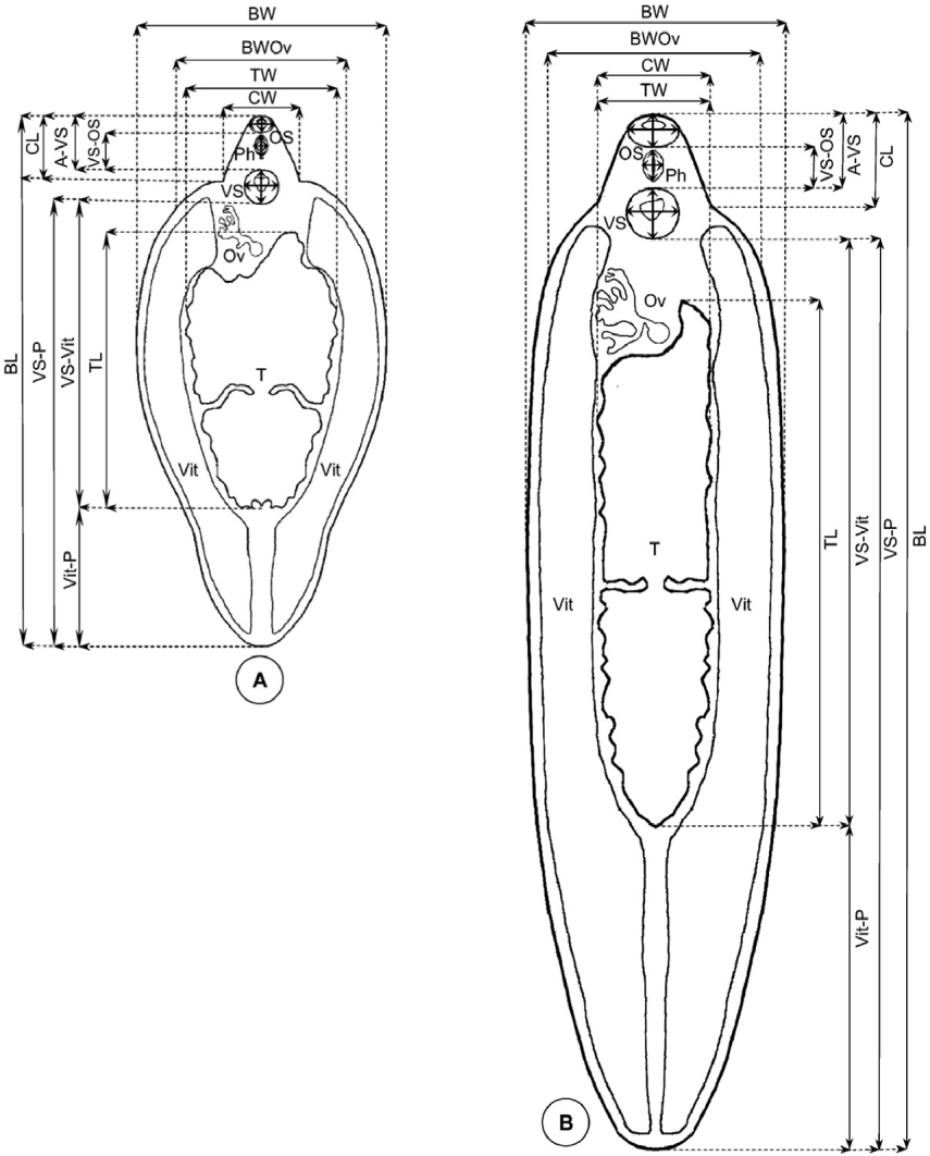
For adult fasciolids, the following standardized measurements were taken (fig. 2):
● Lineal biometric characters (mm): body length (BL), maximum body width (BW), body width at ovary level (BWOv), body perimeter (BP), body roundness (BR), cone length (CL), cone width (CW), maximum diameter of oral sucker (OS max), minimum diameter of oral sucker (OS min), maximum diameter of ventral sucker (VS max), minimum diameter of ventral sucker (VS min), distance between the anterior end of the body and ventral sucker (A–VS), distance between the oral sucker and ventral sucker (OS–VS), distance between the oral sucker and the union of the vitelline glands (VS–Vit), distance between the union of the vitelline glands and the posterior end of the body (Vit–P), distance between the ventral sucker and the posterior end of the body (VS–P), pharynx length (PhL), pharynx width (PhW), testicular space (taking both testes together) length (TL), testicular space width (TW), testicular space perimeter (TP).
● Areas (mm2): body area (BA), oral sucker area (OSA), ventral sucker area (VSA), pharynx area (PhA), testicular space area (TA).
● Ratios: body length over body width (BL/BW), body width at ovary level over cone width (BWOv/CW), oral sucker area over ventral sucker area (OSA/VSA), and body length over the distance between the ventral sucker and the posterior end of the body (BL/VS–P).
Standardized measurements of fasciolid adults described by Valero et al. (Reference Valero, Panova and Mas-Coma2005) and Periago et al. (Reference Periago, Valero, Panova and Mas-Coma2006, Reference Periago, Valero, El Sayed, Ashrafi, El Wakeel, Mohamed, Desquesnes, Curtale and Mas-Coma2008): (A) Fasciola hepatica; (B) Fasciola gigantica. The key to abbreviations for all measurements is shown in table 1.
For fasciolid eggs, the following standardized measurements were taken:
● Lineal biometric characters (μm): egg length (EL), egg width (EW), egg perimeter (EP), egg roundness (ER).
● Area (μm2): egg area (EA).
● Ratios: egg length over egg width (EL/EW).
Liver fluke body roundness (BR = BP2/4πBA) and egg roundness (ER = EP2/4πEA) were used to measure the body and egg shapes, respectively. A circular object will always have a roundness of 1.00, while more irregular objects will have larger values, indicating how circular an object is (Anonymous, 2001). Morphometric measurements used for F. hepatica adults follow a logistic growth model with respect to time (Valero et al., Reference Valero, Darce, Panova and Mas-Coma2001a, Reference Valero, Panova and Mas-Comab, Reference Valero, Panova and Mas-Coma2005). This implies that the morphometric development of the fasciolid adult is not limited but ‘damped’ and does not exceed certain characteristic maxima (Valero et al., Reference Valero, Marcos, Fons and Mas-Coma1998). Since the morphometric maximum values are characteristic for each population, they are considered the comparative base of this study (table 1).
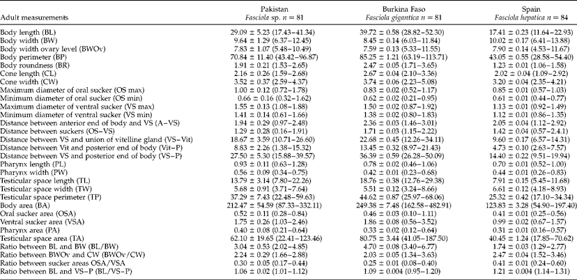
Comparative morphometrics with mean values ± SD (range in size) of 81 adult liver flukes in buffaloes from Pakistan, 81 Fasciola gigantica from Burkina Faso and 84 F. hepatica from Spain (after Periago et al., Reference Periago, Valero, Panova and Mas-Coma2006); n=number of adult liver flukes measured; lineal biometric characters in mm, areas in mm2 and ratios without units.
Data analysis
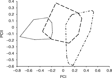
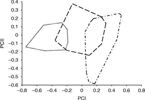
Morphological variation is quantified by geometrical morphometrics (Rohlf & Marcus, Reference Rohlf and Marcus1993), a technique offering an estimate of size by which different axes of growth are integrated into a single variable (the ‘centroid size’; Bookstein, Reference Bookstein1989). The estimate of size is contained in a single variable reflecting variation in many directions, as many as there are landmarks under study, and shape is defined as their relative positions after correction for size, position and orientation. With these informative data, and the corresponding software freely available to conduct complex analyses, significant biological and epidemiological features can be quantified more accurately (Dujardin, Reference Dujardin2008). Current statistical techniques in morphometrics make it possible to test the null hypothesis of conspecific populations being simply the allometric extension of each other, provided a common allometric trend is identifiable (Rohlf & Marcus, Reference Rohlf and Marcus1993; Klingenberg, Reference Klingenberg, Marcus, Corti, Loy, Naylor and Slice1996; Dujardin & Le Pont, Reference Dujardin and Le Pont2004). Multivariate analyses were applied to calculate the phenotypic variations among fasciolid adults, using size-free canonical discriminant analysis on the covariance of log-transformed measurements to assess phenotypic variations between the samples. These analyses are applied to exclude the effect of within-group ontogenetic variations by reducing the effect of each character on the first pooled within-group principal component (a multivariate size estimator) (Dos Reis et al., Reference Dos Reis, Pessoa and Strauss1990). The principal component analysis is used to summarize most of the variations in a multivariate dataset in few dimensions (Dujardin & Le Pont, Reference Dujardin and Le Pont2004). Phenotypic analysis of fasciolid adults was conducted by using various modules of the CLIC package (by J.P. Dujardin, http://momedujardin.wordpress.com). The results were statistically significant when P< 0.05. The following non-redundant measurements (one measurement is not included in another) used for fasciolid adults were BL, BW, BP, OS max, OS min, VS max, VS min, A-VS, VS-Vit, Vit-P, PhL, PhW and TL, where at least one dimension was measured among the most important morphological characters. The remaining variables were all significantly correlated with the first principal component (PCI), contributing 72% to overall variations. PCI could therefore be accepted as a general indicator of size (Bookstein, Reference Bookstein1989), so that the resulting factor maps (fig. 3) clearly illustrate global size differences in the populations analysed.
Principal component analysis of adult fasciolids from natural infections in buffaloes in Pakistan (dashed line) compared with F. hepatica from Spain (dotted line) and F. gigantica from Burkina Faso (dashed and dotted line) (Periago et al., Reference Periago, Valero, Panova and Mas-Coma2006); samples are projected on to the first (PCI, 72%) and second (PCII, 14%) principal components.
Results
Fasciolid populations from Pakistan were grouped according to the maximum and minimum values of given differentiating morphological measurements previously proposed for F. hepatica, F. gigantica or Fasciola sp. ( = intermediate forms) (Periago et al., Reference Periago, Valero, Panova and Mas-Coma2006): BR: 1.06–1.58 in F. hepatica, 1.71–3.65 in F. gigantica; BL/BW: 1.29–2.80 in F. hepatica, 3.40–6.78 in F. gigantica; and VS–P: 8.86–25.08 mm in F. hepatica, 26.28–50.09 mm in F. gigantica. The specimens from Pakistan were first grouped according to BR and BL/BW, and, secondarily, according to VS–P. Using these criteria, adult specimens from Pakistan were grouped into F. hepatica-like (3.7%), F. gigantica-like (22.2%) or Fasciola sp.-like (74.1%).
Secondly, integration of morphological data from all adult specimens from Pakistan (without the above-mentioned previous classification) was made through principal component analysis. Adults from Spain and Burkina Faso were included in the same analysis, as F. hepatica and F. gigantica standard populations, respectively. Fasciolid variables all significantly correlated with PC1.The resulting factor maps (fig. 3) clearly illustrate global size differences in the populations analysed, where each group is represented by its perimeter. Two independent zones can be distinguished: one zone is made up of samples from Spain, while the other zone consists only of samples from Burkina Faso. These zones overlap with the samples from Pakistan. The multivariate analysis used to measure the changes in size of fasciolid adults from Pakistan and compared with the above-mentioned standard populations for each pure fasciolid species showed that the size of most fasciolids from Pakistan is situated between F. hepatica and F. gigantica standard populations. These results confirm that intermediate forms of fasciolids exist in Central Punjab. Nevertheless, it is worth mentioning that the samples from Pakistan overlap with F. hepatica and F. gigantica standard populations, but in the absence of specimens of standard F. hepatica and F. gigantica from Pakistan it is also possible that the forms identified as F. hepatica-like and F. gigantica-like could be the extreme values of the morphometric distribution of the Fasciola sp.-like specimens.
Discussion
In Pakistan, fascioliasis in humans from Lahore, Central Punjab, was diagnosed in the chronic stage of the disease, i.e. through the coprological detection of fasciolid eggs (Qureshi et al., Reference Qureshi, Tanveer, Qureshi, Maqbool, Gill and Ali2005). Liver fluke disease has recently proved to be highly pathogenic when chronic, and not only during the acute phase as previously thought. This aspect is of great importance in human endemic areas of developing countries where the majority of infected subjects, mainly children, are detected in the chronic stage (Valero et al., Reference Valero, Santana, Morales, Hernandez and Mas-Coma2003, Reference Valero, Navarro, Garcia-Bodelon, Marcilla, Morales, Garcia, Hernandez and Mas-Coma2006, Reference Valero, Girones, Garcia-Bodelon, Periago, Chico-Calero, Khoubbane, Fresno and Mas-Coma2008). Additionally, appropriate surveys in these human endemic areas have shown that liver fluke infected patients are usually concomitantly infected by other parasites, related to an immune suppression caused by Fasciola infection in chronic fascioliasis (Girones et al., Reference Girones, Valero, Garcia-Bodelon, Chico-Calero, Punzon, Fresno and Mas-Coma2007). This pathological scenario of great concern may be aggravated when considering that results of recent studies indicate that the occurrence of F. gigantica is more widespread than that of F. hepatica in five Punjabi districts (Khan et al., Reference Khan, Sajid, Khan, Iqbal and Iqbal2009), similarly to northern Iran (Ashrafi et al., Reference Ashrafi, Massoud, Holakouei, Mahmoodi, Joafshani, Valero, Fuentes, Khoubbane, Artigas, Bargues and Mas-Coma2004). The considerably larger size of F. gigantica adult worms when compared to F. hepatica, and consequently its higher pathogenicity, must be taken into account in Pakistan. Moreover, fascioliasis is a trematode disease that is highly susceptible to climatic factors (Fuentes et al., Reference Fuentes, Valero, Bargues, Esteban, Angles and Mas-Coma1999, Reference Fuentes, Malone and Mas-Coma2001), which explains the impact of climate change on transmission of this disease (Mas-Coma et al., Reference Mas-Coma, Valero and Bargues2008). Pakistan's latitudinal situation is prone to be affected by climate change, which has indeed already been detected, showing a rise in the mean temperature in coastal areas, a 10–15% decrease of precipitation in the coastal belt, the presence of hyper-arid plains, and an increase in summer and winter precipitation in the northern part of the country (Farooq & Kahn, Reference Farooq and Khan2004; Cruz et al., Reference Cruz, Harasawa, Lal, Wu, Anokhin, Punsalmaa, Honda, Jafari, Li, Huu Ninh, Parry, Canziani, Palutikof, van der Linden and Hanson2007). When considering all the aforementioned facts, it becomes evident that assessing the present situation of F. hepatica and F. gigantica is a priority for Pakistan.
The present phenotypic study on fasciolids infecting buffaloes from Central Punjab demonstrates the presence of intermediate forms (Fasciola sp.) (table 1, fig. 3). Likewise, previous studies showed the presence of morphologically intermediate forms in geographically sympatric areas of F. hepatica and F. gigantica in Africa and Asia. In Africa, the presence of intermediate forms has been morphometrically evidenced by means of the CIAS methodology in Egypt (Periago et al., Reference Periago, Valero, El Sayed, Ashrafi, El Wakeel, Mohamed, Desquesnes, Curtale and Mas-Coma2008). In Asia, a varied spectrum of morphological forms of fasciolids has been described in several countries, such as India (Varma, Reference Varma1953), Japan (Watanabe & Iwata, Reference Watanabe and Iwata1954; Watanabe, Reference Watanabe1962; Oshima et al., Reference Oshima, Akahane and Shimazu1968a, Reference Oshima, Akahane, Koyama, Shimazu and Haradab; Terasaki et al., Reference Terasaki, Noda, Shibahara and Itagaki2000), Korea (Chu & Kim, Reference Chu and Kim1967), the Philippines (Kimura et al., Reference Kimura, Shimizu and Kawano1984), Thailand (Srimuzipo et al., Reference Srimuzipo, Komalamisra, Choochote, Jitpakdi, Vanichthanakorn, Keha, Riyong, Sukontasan, Komalamisra, Sukontasan and Tippawangkosol2000), Iran (Moghaddam et al., Reference Moghaddam, Massoud, Mahmoodi, Mahvi, Periago, Artigas, Fuentes, Bargues and Mas-Coma2004), Vietnam (Itagaki et al., Reference Itagaki, Sakaguchi, Terasaki, Sasaki, Yoshihara and Van Dung2009) and China (Peng et al., Reference Peng, Ichinomiya, Ohtori, Ichikawa, Shibahara and Itagaki2009), based on traditional microscopic measurements. In Iran, however, the presence of intermediate forms was verified by the same methodology as the one applied in this study (Periago et al., Reference Periago, Valero, El Sayed, Ashrafi, El Wakeel, Mohamed, Desquesnes, Curtale and Mas-Coma2008).
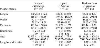
The size measurements of eggs of fasciolid intermediate forms in the current study, of 115.07–186.68/79.12–114.09 μm (table 2), show intermediate values when compared to the standard fasciolid populations of both species previously described (Periago et al., Reference Periago, Valero, Panova and Mas-Coma2006) and which were proved by molecular methods to be genetically pure F. hepatica and pure F. gigantica (Mas-Coma et al., Reference Mas-Coma, Valero and Bargues2009). Fasciolid egg size ranges depend upon livestock species and geographical areas, respectively, as previously described by applying the CIAS methodology (Valero et al., Reference Valero, Perez-Crespo, Periago, Khoubbane and Mas-Coma2009a). Egg length minimum values obtained in the present study approach minimum values found in F. hepatica from Bolivian cattle, F. hepatica from Egyptian cattle and those from Georgia. Egg length maximum values approach maxima obtained in F. gigantica from Egyptian cattle and F. gigantica from Vietnamese cattle, but are smaller than the maxima described in those from Burkina Faso. The results obtained from fasciolids encountered in the Central Punjab area agree with results from previous studies carried out in other Asian countries, where a large egg measurement overlap was detected (Watanabe, Reference Watanabe1962; Akahane et al., Reference Akahane, Harada and Oshima1970; Sahba et al., Reference Sahba, Arfaa, Faramandian and Jalali1972; Kimura et al., Reference Kimura, Shimizu and Kawano1984; Srimuzipo et al., Reference Srimuzipo, Komalamisra, Choochote, Jitpakdi, Vanichthanakorn, Keha, Riyong, Sukontasan, Komalamisra, Sukontasan and Tippawangkosol2000).
Comparative morphometrics with mean values ± SD and range in size of mature eggs of fasciolids from Pakistan with data previously published by Periago et al. (Reference Periago, Valero, Panova and Mas-Coma2006) for standard populations from Spain and Burkina Faso; n=number of eggs measured; lineal biometric characters in μm, areas in μm2, ratios without units.
According to Mas-Coma et al. (Reference Mas-Coma, Valero and Bargues2009), Iran and Pakistan are examples of Asian countries presenting a zonal overlap of the two fasciolid species. The zonal overlap refers to areas with different altitudes, where the highlands offer the necessary cold–mild weather conditions for Galba/Fossaria lymnaeid species and F. hepatica, and where the lowlands offer the warm–hot climate necessary for Radix lymnaeid species and F. gigantica. Thus, definitive hosts become co-infected by both fasciolids when moving from lowlands to neighbouring highlands and vice versa. In this manner, animals and humans may become co-infected due to interzonal transhumance, nomadic migrations, transportation, trade and/or other reasons (Mas-Coma et al., Reference Mas-Coma, Valero and Bargues2009). Previous studies in Pakistan (Kendall, Reference Kendall1954; Kendall & Parfitt, Reference Kendall and Parfitt1959) indicated that F. hepatica is confined to the highlands, related to the presence of its intermediate snail host Galba truncatula, while F. gigantica is found in lowland areas, given the distribution of Radix auricularia.
In the case of Iran, in Gilan province, G. truncatula is frequent in the Talesh Mountains, where it has been found to transmit F. hepatica (Ashrafi et al., Reference Ashrafi, Massoud, HolakoueiNainei, Jo-Afshani, Mahmoodi, Ebedati, Rezvani, Artigas, Bargues and Mas-Coma2007), whereas R. gedrosiana is restricted to the lowlands and does not appear to have the capacity to colonize highland regions (Ashrafi et al., Reference Ashrafi, Massoud, Holakouei, Mahmoodi, Joafshani, Valero, Fuentes, Khoubbane, Artigas, Bargues and Mas-Coma2004). This explains why F. gigantica is the most prevalent species (91.1%) in the human endemic area around the cities of Rasht and Bandar-Anzali, whereas the scarcity of F. hepatica in animals in these lowlands (8.9%) might be the consequence of F. hepatica-infected animal transportation from the highlands to the lowlands (Ashrafi et al., Reference Ashrafi, Valero, Panova, Massoud and Mas-Coma2006b). Neither can the possibility of hybrid-intermediate flukes, resulting from long-term introgression, with an F. hepatica-like form be ruled out (Ashrafi et al., Reference Ashrafi, Valero, Panova, Massoud and Mas-Coma2006b). Although altitudinal livestock transhumance does not appear to take place in Gilan nowadays, it is likely to have taken place in the past (Mas-Coma et al., Reference Mas-Coma, Valero and Bargues2009).
The intermediate forms, as well as the F. hepatica-like and F. gigantica-like adult worms, found in buffaloes from the Central Punjab area in the present study may also be explained by zonal overlap (Mas-Coma et al., Reference Mas-Coma, Valero and Bargues2009). In fact, nomads move their animals from the highlands to the lowlands in the winter season, looking for grazing lands. Similarly, animals are moved from the lowlands to the cooler highlands when warmer conditions return. Further work on this very complex orographic and transhumance scenario is required to understand the epidemiology of the disease in each locality, to assess how transmission of fascioliasis occurs throughout the Central Punjab area and thus establish appropriate control measures for each endemic subzone in the future.
Acknowledgements
Studies were funded by Projects No. SAF2010–20805 of the Ministry of Science and Innovation, Madrid, Spain; by the Red de Investigación de Centros de Enfermedades Tropicales – RICET (Project No. ISCIII-RETIC RD06/0021/0017 of the Programa de Redes Temáticas de Investigación Cooperativa RETICS/ FEDER), Fondo de Investigación Sanitaria (FIS), Ministry of Health, Madrid; and by Project No. 2012/042 of the PROMETEO Programme, Generalitat Valenciana, Valencia, Spain. The study was conducted during a 6-month pre-doctorate fellowship stay of the first author (K.A.) at the Department of Parasitology of the University of Valencia, Spain, funded by the International Research Support Initiative Programme (IRSIP) of the Higher Education Commission (HEC) of Pakistan and Indigenous HEC Fellowship Programme, Pakistan. The English stylistic help by Mr R. Wilk (Valencia, Spain) is greatly acknowledged.