Introduction
Oral tongue squamous cell carcinoma (SCC) is usually treated with initial surgical resection with or without post-operative chemo- and radiotherapy.Reference Robertson, Soutar, Paul, Webster, Leonard and Moore1 Loco-regional recurrences occur in approximately one in four patients with early (i.e. tumour (T) stage T1 or T2) oral tongue SCC, justifying aggressive treatment.Reference Brennan, Corry, Kleid, Porceddu, Yuen and Rischin2 There are a number of histological factors that have an impact on the prognosis for early oral cancer, most importantly the presence of lymph node metastases, extracapsular extension, and close or involved surgical margins.Reference Bernier, Cooper, Pajak, van Glabbeke, Bourhis and Forastiere3 Although the former two factors have an impact on adjuvant treatment, the latter is the only factor that may be improved by the surgeon.Reference Gross, Patel, Carvalho, Chu, Kowalski and Boyle4
Traditionally, a 1-cm margin is taken in all planes around a macroscopic or palpable oral tongue SCC.Reference Bradley, MacLennan, Brakenhoff and Leemans5 Pathologists and clinicians have agreed to define involved margins as less than 1 mm and close margins as 5 mm or less, while margins greater than 5 mm are designated as clear.Reference Meier, Oliver and Varvares6 However, mucosal margins shrink by approximately 30–50 per cent with formalin fixation and slide preparation.Reference Johnson, Sigman, Funk, Robinson and Hoffman7 This results in a final pathological margin of approximately 5 mm (below which a clear margin is termed a close margin) where the surgeon measured 1 cm, leaving little room for error. Wider margins, however, may have an impact on functional outcomes and significantly affect quality of life.
Our study hypothesis was that wider mucosal margins for oral SCC may be justified in order to reduce the number of cases with close or involved surgical margins. The functional impact of this approach might be minimised if wider margins could be taken at only one point e.g. on the deep surface of the surgical specimen. This study aimed to determine the site of closest margins for previously untreated early oral SCC cases managed over the 10-year period ending in January 2009 at Royal Melbourne Hospital, Melbourne, Australia.
Materials and methods
Institutional review board approval was obtained.
All previously untreated T1 and T2 oral tongue SCC cases managed at the Royal Melbourne Hospital between January 1999 and January 2009 were identified (n = 81) from the prospectively maintained head and neck oncology database and the medical records (using International Classification of Diseases Coding 9 and 10). Retrospective staging was based on the sixth edition of the American Joint Committee on Cancer Staging Manual.Reference Greene, Page, Fleming, Fritz, Balch and Haller8 Retrospective chart review identified 68 T1 tumours and 13 T2 tumours, with a median follow up of 38 months (range 12–136 months). No patients had distant metastases at presentation.
Cases were discussed at weekly multidisciplinary head and neck team meetings, where the initial surgical plan was decided based on surgical, radiation oncologist and patient preference, patient age, comorbidities, and the presence of other poor prognostic factors. Tumour depth was assessed using examination under anaesthetic, computed tomography, magnetic resonance imaging and, increasingly over time, intra-oral ultrasound.
As in our previously published protocol (Brennan et al.), at the time of study, a primary tumour size of more than 7 mm depth was used as the threshold for post-operative radiotherapy of the neck, with tumours of lesser depth receiving neck dissection.Reference Brennan, Corry, Kleid, Porceddu, Yuen and Rischin2 This practice has since been abandoned, as explained in our previous study.Reference Brennan, Corry, Kleid, Porceddu, Yuen and Rischin2
The site of the closest histological margin was taken from pathology reports. Where doubt existed, slides were re-evaluated by a senior head and neck pathologist (AT) and categorised as follows: clear if margin more than 5.0 mm (n = 24), close if 1.1–5.0 mm (n = 44) and involved if less than 1.0 mm (n = 10). Note that these three groups were separate (cases with involved margins were not included in the close group as we felt they may behave differently). The site of the closest margin in all three groups was also noted.
The practice at our institution was to mark the anterior margin as ‘12 o'clock’; previous sections were then marked as being taken relative to this. Thus, it was generally possible for the pathologist to work out the orientation of the close margin relative to this marker point.
If frozen sections or re-resections were undertaken, these were recorded but were not taken as changing the category of margin status listed above. The site of close and involved margins was classified according to its quadrant, i.e. medial, lateral, anterior or posterior. Close and involved margins were also designated as deep (i.e. more than 3 mm from the cut mucosal edge) or mucosal (i.e. less than 3 mm from the mucosal edge, or on the actual mucosa).
Inadequate information was available for margin assessment in three cases, and so these cases were excluded from analysis. Patients with margins considered involved after frozen section were considered by the multidisciplinary clinic for re-resection (preferred), post-operative radiotherapy or observation.
Other prognostic variables were recorded, including depth of invasion, tumour differentiation, maximum diameter, lymphovascular invasion, perineural invasion, pathological cervical nodes and extracapsular extension.
Statistical analysis was performed using the Statistical Package for the Social Sciences version 13.0 software program (SPSS Inc, Chicago, Illinois, USA). Survival was estimated using the Kaplan–Meier method from the date of surgery to the date of recurrence or status (alive/dead) at last follow up.
Results
Patient characteristics
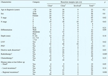
The median age at diagnosis was 63 years (range 21–90 years). Clear margins occurred in 24 patients (30.8 per cent), close margins in 44 (56.4 per cent) and involved margins in 10 (12.8 per cent) (Table I). There was a non-significant trend towards deeper tumours having more involved margins (p = 0.18). Clear or close margins were just as likely to be achieved in T1 cases (90 per cent) as in T2 cases (82 per cent) and N0 (100 per cent) and N+ cases (82 per cent). Perineural or lymphovascular invasion was seen equally in cases with involved margins (20 per cent) and cases with clear or close margins (21 per cent).
T1 & T2 Oral tongue SCC: patient characteristics

*n = 24; †n = 44; ‡n = 10; **n = 78. T = tumour stage; SCC = squamous cell carcinoma; pts = patients; N = node; ECE = extracapsular extension; LVI = lymphovascular invasion; PNI = perineural invasion
Primary radiotherapy was preferred in patients with close/ involved margins for treatment of the N0 neck (46 per cent) whereas prophylactic neck dissection was favoured for the N0 neck when margins were clear (58 per cent) under a previously published research protocol.Reference Brennan, Corry, Kleid, Porceddu, Yuen and Rischin2
Patients with deeper tumours were more likely to undergo neck dissection for the N0 neck than patients with thin tumours (p = 0.02). Four patients (22 per cent) with a tumour depth of 2.1–4.0 mm had a prophylactic neck dissection, as did 15 patients (58 per cent) with a tumour depth of 4.1–7.0 mm.
Post-operative radiotherapy was used in 30 patients (38 per cent) with doses of 50 to 66 Gy delivered to the primary site and involved cervical nodes. Concurrent single agent chemotherapy with cisplatin was used in six patients. Two of these six patients receiving chemoradiotherapy (33 per cent) had neck dissections. Use of radiotherapy (p < 0.01) and chemotherapy (p = 0.01) increased with primary tumour depth.
Site of closest margin
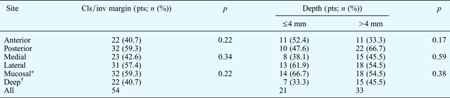
In cases with a close or involved margin, the closest margin was more likely to be posterior (59 per cent) than anterior (41 per cent, p = 0.22) and this trend was more pronounced with deeper tumours (67 vs 33 per cent, p = 0.17), although still not reaching significance (Table II).
T1 & T2 Oral tongue SCC: site of close or involved margin
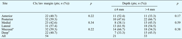
Total n = 78. *Including deep resections <3 mm. † >3 mm. T = tumour stage; SCC = squamous cell carcinoma; Cls/inv = close or involved; pts = patients
There was a non-significant trend for the closest margin to be on the lateral aspect (57 per cent) rather than the medial (43 per cent, p = 0.34). A majority had the closest margin at or near the mucosal edge (59 per cent) rather than on the deep surface (41 per cent, p = 0.22). However, none of these trends showed a clear, statistically significant difference, with close and involved margins occurring in all directions.
Frozen sections were taken in 55 patients (70.5 per cent), including 37 (84 per cent) with close margins and seven (70 per cent) with involved margins. Frozen sections were positive for dysplasia in seven (12.7 per cent) and positive for cancer in two (3.6 per cent), and were repeated in these patients until negative. Re-resection was undertaken in a further two individuals (20 per cent) with involved margins, although no tumour was found in either case.
Recurrence and survival
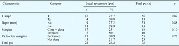
Local recurrence occurred in 22 patients (28 per cent) at a median of 12 months (Table III). Five (50 per cent) of the 10 patients with involved margins developed local recurrence. Recurrence occurred in one patient with involved margins despite re-resection (representing 50 per cent of those re-resected), and in three (42.3 per cent) who had further frozen sections taken. Nine (21 per cent) of the 43 patients with close margins had local recurrence, compared with eight (33 per cent) of the 24 patients with clear margins (p = 0.10).
T1 & T2 Oral tongue SCC: local recurrence

T = tumour stage; SCC = squamous cell carcinoma; pts = patients; FS = frozen section
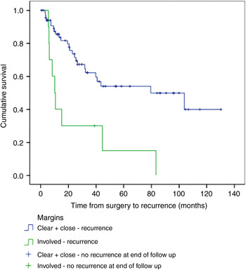
Disease-free survival was worse for patients with tumours with involved margins, compared with those with clear or close margins (Figure 1, p = 0.002). There was a trend to worse disease- free survival for patients with tumours of more than 7.1 mm invasion depth, compared with those with tumours of less than 7 mm depth (p = 0.08).
Kaplan–Meier disease-free survival curve for tumour stage 1 and 2 oral tongue squamous cell carcinoma, by surgical margin status (n = 78, p = 0.002).

Discussion
Surgery remains the initial management for most T1 and T2 oral tongue SCC tumours, yet local recurrence remains a significant issue (28 per cent in this series). This study found that involved surgical margins had a trend to worse local recurrence (p = 0.10) and significantly worse survival (p = 0.002). Over the last 10 years at our institution, 14 per cent of patients had involved margins and 55 per cent had close margins. Consequently, we feel that wider surgical margins may be justified, being the only prognostic factor that surgeons have the ability to improve. This study found that close or involved margins occurred in all directions (medial, lateral, anterior, posterior, mucosal and deep) and were equally likely in all tumour types (T1, T2, N0, N+, deep, superficial, with perineural invasion and without). These findings suggest that a wider margin should be taken in all directions and for all early oral tongue SCC tumour types.
The 1 cm rule for surgical margins around a macroscopic or palpable tumour is based on historic use and has not been scientifically challenged.Reference DiNardo, Lin, Karageorge and Powers9 Note that a 1-cm macroscopic margin does not result in a 1-cm histological margin. In our series, the incidence of tumours with involved (14 per cent) or close (55 per cent) margins was worryingly high.
Centres of excellence have published rates of histological close and involved margins of approximately 20 per cent for oral tongue SCC (Gross et al.).Reference Gross, Patel, Carvalho, Chu, Kowalski and Boyle4 However, the overall rate of involved margins in the USA has been estimated as 16 per cent, similar to that in our series.Reference Jacobs, Ahmad, Cassiano, Schuller, Scott and Laramore10 In a review of 301 tumours, Woolgar and Triantafyllou found that 13 per cent had involved margins and a further 43 per cent had close margins, similar to our findings.Reference Woolgar and Triantafyllou11 Most centres audit their incidence of close and involved margins; however, surprisingly, the recommended excision margins have not been challenged.
Given the incidence of involved surgical margins in our series (14 per cent), clearly the visible tumour edge did not reflect the microscopic tumour edge in all cases. This may have been due to tumour microsatellites or direct infiltration by the tumour, both of which have been shown to be associated with a worse prognosis.Reference Yan, Ko and Change12
The concept of field cancerisation was first observed in oral tongue tumours, describing multicentric, microscopic foci of concurrent tumour growth.Reference Slaughter, Southwick and Smejkal13 These concurrent, microscopic tumours could have an impact on surgical margins as ‘involved margin microsatellites’, and may also explain the high tumour recurrence rates observed even in cases with clear margins.
Nevertheless, the overall effectiveness of surgical excision for early tongue SCCs argues for predominant local treatment, and also implies that field cancerisation is a local effect. Although adjuvant therapy may have improved over the time of our trial (e.g. with the introduction of intensity modulated radiotherapy and chemoradiotherapy), and there was a higher incidence of adjuvant therapy use in patients with involved margins (Table I), local control was still clearly worse in this group. We believe that wider surgical margins should improve local control.
In the current series, our finding of improved local control (p = 0.10) and survival (p = 0.002) in patients with non-involved margins is well supported in the literature.Reference Bernier, Cooper, Pajak, van Glabbeke, Bourhis and Forastiere3
The strength of the conclusions of this paper is limited by the number of patients enrolled, which was reduced by strict exclusion of all patients with previously treated dysplastic or malignant oral lesions (who would clearly have a higher rate of recurrence). Further subgroup analysis with a larger series would be desirable to exclude a potential confounding effect of other risk factors, such as perineural spread.
This series showed that involved margins were no more likely in T1 versus T2 tumours, deep versus superficial tumours, and tumours with perineural invasion versus those without. Thus, it would seem more likely that the surgical practice of taking a 1-cm margin around macroscopic or palpable tumours is at fault, rather than that certain tumours (e.g. those which are T2, deep or with perineural invasion) are more likely to have impalpable tendrils that result in involved margins.
Close and involved margins occurred in all directions, indicating that if wider margins are considered, they should be taken in all directions. We found that the closest margin was more commonly posterior (59 per cent) than anterior, lateral (57 per cent) than medial and mucosal (59 per cent) than deep.
Readers should note that, unlike involved margins, patients with close resection margins did not have a significantly worse prognosis than those with clear margins. Nevertheless, one would expect that a higher incidence of close margins would be associated with a higher incidence of involved margins, since the histological tumour edge does not occur exactly at the macroscopic, palpable tumour edge but significantly beyond. Clearly, wider margins would be expected to have an impact on functional outcomes, especially if vital structures (e.g. the contralateral lingual artery, the major branch of the lingual nerve or the hypoglossal nerve) were sacrificed in order to take the additional margin. Nevertheless, the recommendation might be that wider margins be taken at every point except that involving the vital structure; in this way, a lower incidence of involved margins could still be expected.
• Early oral tongue squamous cell carcinomas (SCCs) have historically been treated with a 1cm macroscopic resection margin
• In this study of early oral tongue SCCs, involved and close margins were common, occurring in all histological types and on all resection surfaces
• In this study of tumour stage one and two SCCs, involved and close margins were common, occurring in all histological types and on all resection surfaces
• Local recurrence occurred in 28 per cent (median 12 months) and was more likely with involved (50 per cent) than clear or close margins (25 per cent)
• Histological margin is one of the few prognostic factors the surgeon can influence
• A surgical margin wider than 1 cm may be justified
Patel and colleagues have demonstrated the use of frozen sections to improve margin control in oral SCC, resulting in survival rates similar to those of patients with clear margins.Reference Patel, Goldstein, Guillemaud, Bruch, Brown and Gilbert14 Due to the small numbers involved (two patients), it was difficult to assess the impact of re-resection following discovery of an involved margin in this series.Reference Patel, Goldstein, Guillemaud, Bruch, Brown and Gilbert14 Frozen sections were used in the majority of cases in this series (70 per cent), but an accurate retrospective assessment of true margin status is impossible without an en bloc resection.Reference Patel, Goldstein, Guillemaud, Bruch, Brown and Gilbert14 Certainly, frozen section still had utility, changing the extent of resection in 12 per cent of cases.
However, the presumption of frozen section use is that the surgeon can predict the closest margin by palpation or inspection after resection. Our study findings would suggest that this is not reliable (since close and involved margins occur in all tumour types and all directions) unless frozen sections are taken at all margins. We feel that wider, formal histological margins are more likely to result in accurate analysis of margins than frozen section analysis, and would probably result in the same functional outcomes and final defect size as standard excision with frozen section analysis in all planes.
Patients with involved margins were noted to have a high rate of regional recurrence (80 per cent, compared with 22 per cent in those with close or clear margins, p = 0.0001). One would expect a higher rate of regional recurrence associated with local recurrence, and this was often the case in our series. This emphasises the importance of local control and adequate local treatment in such cases. Other authors have found margin status to be less important than primary site and histological features (e.g. T stage, and perineural and lymphovascular invasion).Reference Brandwein-Gensler, Teixeira, Lewis, Lee, Rolnitzky and Hill15 This does not correspond with the findings of larger, prospective series such as that by Bernier et al., nor does it explain the primary role of surgery.Reference Bernier, Cooper, Pajak, van Glabbeke, Bourhis and Forastiere3 Surgery is a local treatment that acts only on the operated site. Histological clear margins may be one of the few prognostic factors on which good surgery can have an impact.
Conclusions
This study's findings support the hypothesis that wider surgical margins may be appropriate for most T1and T2 oral tongue SCC tumours, with the aim of reducing local recurrence and thereby improving survival. The historically accepted 1-cm margin around macroscopic and palpable oral tumours seems to provide inadequate pathological results. This study found that close and involved margins occurred in all directions (medial, lateral, anterior, posterior, mucosal and deep) and were equally likely in all tumour types (i.e. T1, T2, N0, N+, deep, superficial, and those with and without perineural invasion). These findings suggest that a wider margin should be considered in all directions and for all tumour types. Outcomes might still be improved even if vital structures are exempted from the wider margins.