Introduction
Individuals exposed to childhood maltreatment, even in the absence of presenting with psychiatric disorder, show altered functioning in a number of neurocognitive domains implicated in mental health vulnerability (McLaughlin et al., Reference McLaughlin, Sheridan, Tibu, Fox, Zeanah and Nelson2015; Puetz et al., Reference Puetz, Viding, Palmer, Kelly, Lickley, Koutoufa, Sebastian and McCrory2016a, Reference Puetz, Zweerings, Dahmen, Ruf, Scharke, Herpertz-Dahlmann and Konrad2016b; McCrory et al., Reference McCrory, Gerin and Viding2017a, Reference McCrory, Puetz, Maguire, Mechelli, Palmer, Gerin, Kelly, Koutoufa and Viding2017b). According to the theory of latent vulnerability, proximal neurocognitive changes may occur in response to maltreatment conferring a degree of adaptive value for the child in an abusive or neglectful environment, while increasing risk of poor psychiatric outcome in the longer term (McCrory and Viding, Reference McCrory and Viding2015). A major gap in our understanding relates to the potentially differential impact of childhood abuse v. childhood neglect. It has been persuasively argued, in part based on animal data, that the neurocognitive impact of abuse-related experiences (such as physical, emotional and sexual abuse) should differ from that of deprivation-related experiences (such as physical and emotional neglect) (McLaughlin et al., Reference McLaughlin, Sheridan and Lambert2014, Reference McLaughlin, Sheridan and Nelson2016; Sheridan and McLaughlin, Reference Sheridan and McLaughlin2014).
Psychological and neurobiological alterations in threat processing following maltreatment experience have received sustained research interest in recent years (McCrory et al., Reference McCrory, De Brito, Sebastian, Mechelli, Bird, Kelly and Viding2011, Reference McCrory, De Brito, Kelly, Bird, Sebastian, Mechelli, Samuel and Viding2013; Tottenham et al., Reference Tottenham, Hare, Millner, Gilhooly, Zevin and Casey2011). It has been shown that experiences of physical abuse are associated with preferential attention to threatening information and enhanced perceptual ability for threat related-cues, such as angry faces (Pollak and Tolley-Schell, Reference Pollak and Tolley-Schell2003). Experiences of physical abuse and family violence in children have also been associated with significantly increased amygdala response to threatening compared to neutral facial expressions (McCrory et al., Reference McCrory, De Brito, Sebastian, Mechelli, Bird, Kelly and Viding2011, Reference McCrory, De Brito, Kelly, Bird, Sebastian, Mechelli, Samuel and Viding2013). It remains unclear, however, whether heightened neural responsiveness to threat is associated with abuse experiences but not with deprivation-related experiences (Sheridan and McLaughlin, Reference Sheridan and McLaughlin2014; McLaughlin et al., Reference McLaughlin, Sheridan and Nelson2016). Participants in prior child and adult studies report poly-victimisation and experiences of both abuse and neglect (McCrory et al., Reference McCrory, De Brito, Sebastian, Mechelli, Bird, Kelly and Viding2011; Dannlowski et al., Reference Dannlowski, Stuhrmann, Beutelmann, Zwanzger, Lenzen, Grotegerd, Domschke, Hohoff, Ohrmann, Bauer, Lindner, Postert, Konrad, Arolt, Heindel, Suslow and Kugel2012; Puetz et al., Reference Puetz, Viding, Palmer, Kelly, Lickley, Koutoufa, Sebastian and McCrory2016a, Reference Puetz, Zweerings, Dahmen, Ruf, Scharke, Herpertz-Dahlmann and Konrad2016b) making it impossible to draw any strong conclusions regarding the differential impact of these experiences at the neurocognitive level.
Sheridan and McLaughlin (McLaughlin et al., Reference McLaughlin, Sheridan and Lambert2014) argue that these different forms of maltreatment should, at least in part, differentially impact the functioning of a number of brain regions. Specifically, they propose that maltreatment experiences characterised by abuse should primarily lead to changes in neural circuits that underlie emotion and emotional learning, including (but not limited to) the amygdala. By contrast, maltreatment experiences characterised by deprivation (such as neglect) are argued to have a broader impact. In particular, the absence of appropriate cognitive stimulation and sensory, motor, linguistic, and social inputs typically provided by a caregiver are hypothesised to constrain early forms of learning, producing long-term deficits in complex cognitive function and associative learning (McLaughlin et al., Reference McLaughlin, Sheridan and Lambert2014, Reference McLaughlin, Sheridan and Nelson2016). At the neural level, such neglect-related changes are thought to be reflected in the brain's stress pathways, and in areas involved in the processing of complex cognitive and social inputs, including fronto-parietal networks (McLaughlin et al., Reference McLaughlin, Sheridan and Lambert2014, Reference McLaughlin, Sheridan and Nelson2016; Sheridan and McLaughlin, Reference Sheridan and McLaughlin2014). A large body of evidence from institutionalised samples reported poorer emotion regulation ability, atypical stress reactivity and deficits in complex cognitive domains such as language, executive functioning and memory – not readily explained by hyperactive amygdala activity alone (Bos et al., Reference Bos, Fox, Zeanah and Nelson2009). A more parsimonious explanation is that the absence of age-appropriate experience compromises the neural substrates underlying associative and implicit learning. The neural basis for such experience-expectant learning is synaptic pruning, in which connections that are used frequently are selectively strengthened whilst those who co-activate infrequently are eliminated (Hebb, Reference Hebb1949). McLaughlin and Sheridan (McLaughlin et al., Reference McLaughlin, Sheridan and Lambert2014, Reference McLaughlin, Sheridan and Nelson2016; Sheridan and McLaughlin, Reference Sheridan and McLaughlin2014) propose that deprivation in the form of neglect results in accelerated and extreme synapse elimination as well as reduced myelination of axons, both known and established consequences of global deprivation in animal studies (e.g. O'Kusky, Reference O'Kusky1985).
A number of studies investigating neural responses to threat-related cues in children exposed to institutional settings or primarily experiences of neglect have reported heightened amygdala reactivity (e.g. Tottenham et al., Reference Tottenham, Hare, Millner, Gilhooly, Zevin and Casey2011) and global alterations in brain networks (McLaughlin et al., Reference McLaughlin, Sheridan and Lambert2014; Puetz et al., Reference Puetz, Parker, Kohn, Dahmen, Verma and Konrad2017). However, in addition to experiences of pronounced deprivation, children in these settings will likely have been exposed to multiple adversities in the course of their institutional care, and therefore these studies cannot easily arbitrate on the question of a differential impact of abuse v. neglect on amygdala response (Tottenham et al., Reference Tottenham, Hare, Millner, Gilhooly, Zevin and Casey2011). Consequently, there are no extant studies that can inform regional alterations across multiple brain areas that may be specifically associated with experiences of neglect. It is, however, plausible that in an institutional setting the absence of a stable caregiver who monitors the environment for safety may lead the child to become more vigilant to threat-related cues (with an associated increase in amygdala response) (McCrory and Viding, Reference McCrory and Viding2015).
The present study aimed to investigate whether different forms of maltreatment experience exert differential effects on emotion processing, specifically neural reactivity to interpersonal threat cues as indexed by angry facial expressions. Using a large cohort study (n = 1144), we were able to identify four groups of young adults self-reporting different profiles of childhood maltreatment: (i) maltreatment experiences characterised by abuse only (defined as physical, sexual and emotional abuse); (ii) maltreatment experiences characterised by neglect only (defined as physical and emotional neglect); and (iii) combined abuse and neglect experiences; and a non-maltreated group with no abuse/neglect experiences. Based on previous research (McCrory et al., Reference McCrory, De Brito, Sebastian, Mechelli, Bird, Kelly and Viding2011; Tottenham et al., Reference Tottenham, Hare, Millner, Gilhooly, Zevin and Casey2011; White et al., Reference White, Bogdan, Fisher, Muñoz, Williamson and Hariri2012), and in line with the framework set out by Sheridan and McLaughlin (Sheridan and McLaughlin, Reference Sheridan and McLaughlin2014) we first predicted that experiences of abuse-only would be associated with heightened amygdala reactivity as compared to the non-maltreated control group. Second, experiences of neglect-only would be associated with increased amygdala reactivity to threat in line with prior studies of institutionalised and neglect samples (Tottenham et al., Reference Tottenham, Hare, Millner, Gilhooly, Zevin and Casey2011). In addition, and based on the predictions made by Sheridan and McLaughlin (Sheridan and McLaughlin, Reference Sheridan and McLaughlin2014) we expected that this group would show significant differences in neural reactivity in a broad distributed set of fronto-parietal regions including the prefrontal, somatosensory and association cortices (Sheridan and McLaughlin, Reference Sheridan and McLaughlin2014; McLaughlin et al., Reference McLaughlin, Sheridan and Nelson2016). Given the lack of extant studies investigating threat processing in samples specifically characterised by neglect, no specific regional peaks have been documented that could inform regions of interest approach; consequently, we adopted a whole brain exploratory approach. Third, we predicted that the combined impact of abuse and neglect would be additive, such that individuals with abuse experiences across these domains would show both greater amygdala reactivity, alongside the broad differences in fronto-parietal networks including the prefrontal, somatosensory, and association cortices hypothesised for the neglect-only group.
Methods
Participants
Data were available for 1144 participants (collected between 2010 and 2016) from the Duke Neurogenetics study (DNS), which assessed a wide range of behavioural and biological traits among non-patient, young adult university student volunteers. In order to screen for maltreatment experiences, all participants in the study completed the Childhood Trauma Questionnaire (CTQ) (Bernstein and Fink, Reference Bernstein and Fink1998), a self-report measure measuring (1) emotional, (2) and physical neglect and (3) emotional, (4) physical and (5) sexual abuse, yielding separate scores for each domain and a composite overall total score.
The authors assert that all procedures contributing to this work comply with the ethical standards of the relevant national and institutional committees on human experimentation and with the Helsinki Declaration of 1975, as revised in 2008.
Group categorisation
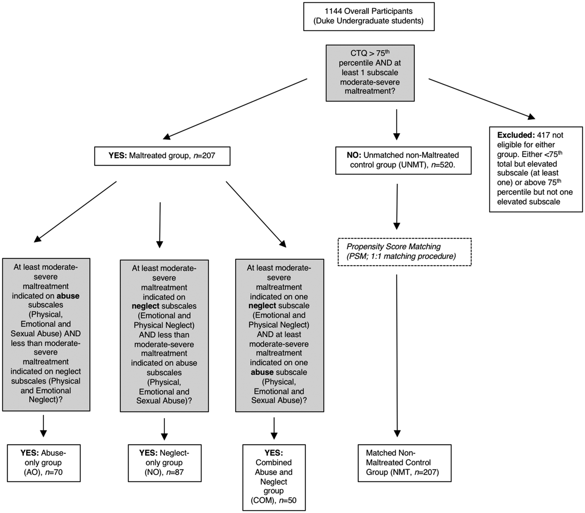
Please see Fig. 1 for a description of how the Maltreated groups were created using the CTQ (Bernstein and Fink, Reference Bernstein and Fink1998) as well as the matching procedure for the Non-Maltreated Control Participants. The creation of maltreatment groups allowed propensity score matching (PSM) of participants which ensured maximum comparability/reduced confounds between groups. Furthermore, our approach created groups that were characterised by clinically relevant maltreatment experiences. In this way, we could investigate whether different types of maltreatment related to different neural outcomes. Note, that there was a significant gender difference in the abuse only (AO) group, so the analyses of valence by group interactions for the AO group v. NMT group were subsequently controlled for gender.
Group categorisation process.

Propensity score matching
The R software package MatchIT (Ho et al., Reference Ho, Imai, King and Stuart2011) was used to implement two PSM methods to match participants. See online Supplementary data for additional information on PSM procedure.
Results of PSM
The best PSM matching method was the nearest neighbours (with a ratio of 1:1), as it kept all 207 maltreated individuals and matched them to 207 non-maltreated controls with a post-matching standardised overall mean difference of 0.04 and standardised mean difference for each variable ranging between 0.01 and 0.09 (see online Supplementary Table S1 in Appendix). The PSM procedure yielded a total sample size of n = 207 matched non-maltreated control participants with respect to age, gender, handedness, socio-economic status and psychopathology (see online Supplement).
Study-wide exclusion criteria were: (i) medical diagnoses of cancer, stroke, diabetes requiring insulin treatment, chronic kidney or liver disease, or lifetime history of psychotic symptoms, (ii) use of psychotropic, glucocorticoid, or hypolipidemic medication; and; (iii) conditions affecting cerebral blood flow and metabolism (e.g. hypertension).
All participants provided written informed consent in accord with Duke University guidelines.
Paradigm
Participants underwent a well-established perceptual face-matching task developed by Hariri and colleagues, which elicits robust amygdala reactivity in typically developing individuals and patient groups (Hariri et al., Reference Hariri, Mattay, Tessitore, Kolachana, Fera, Goldman, Egan and Weinberger2002, Reference Hariri, Drabant, Munoz, Kolachana, Mattay, Egan and Weinberger2005). Participants select one of two faces (shown on bottom) that matches the target face (shown on the top). Facial stimuli displaying neutral, angry, fearful and surprised expressions were taken from the standard picture set (Ekman and Friesen, Reference Ekman and Friesen1976). Blocks of each of the four face valence categories were presented in pseudorandom order within one run and interleaved with a sensorimotor control condition. A detailed description of the paradigm can be found elsewhere (see https://www.haririlab.com/paradigms/ and Hariri et al., Reference Hariri, Mattay, Tessitore, Kolachana, Fera, Goldman, Egan and Weinberger2002, Reference Hariri, Drabant, Munoz, Kolachana, Mattay, Egan and Weinberger2005). In light of previous findings suggesting a relationship between childhood maltreatment and hypervigilance to interpersonal threat, we restricted our analyses to the contrast of angry v. neutral faces. This decision has been informed by the findings of previous studies of children exposed to maltreatment, which indicate that the strongest difference compared to peer is in relation to the processing of angry faces, likely reflecting the increased experience-dependent salience of threatening facial expressions in this population (Pollak and Tolley-Shell, Reference Pollak and Tolley-Schell2003; McCrory et al., Reference McCrory, De Brito, Sebastian, Mechelli, Bird, Kelly and Viding2011). For completeness, we ran ROI analyses on the amygdala for the contrast fearful v. neutral faces for all group comparisons. In line with the prior neuro-cognitive literature (see above), we found no differential activation in the amygdala in response to fearful faces for any group comparison (NMT v. AO; NMT v. NO and NMT v. COM).
Finally, in order to rule out that differential brain activity to angry v. neutral face could be due to secondary differences in behavioural indices such as reaction time and accuracy we ran additional analyses on the behavioural data, which yielded no significant differences between the groups in relation to accuracy or reaction time.
fMRI protocol
Participant's brains were scanned using one of two identical 3T General Electric MR750 scanners at the Duke-UNC Brain Imaging and Analysis Center. Whole brain data were collected with the following sequence using an inverse-spiral pulse sequence to reduce susceptibility artefacts: TR = 2000 ms, TE = 30 ms, flip angle = 60; FOV = 240 mm; 3.75 × 3.75 × 4 mm voxels; inter-slice skip = 0. High-resolution three-dimensional structural images were acquired with the following parameters: TR = 7.7 s; TE = 3.0 ms; flip angle = 12; voxel size = 0.9 × 0.9 × 4 mm; FOV = 240 mm, inter-slice skip = 0.
fMRI data processing
Pre-processing followed a standard analyses pipeline as reported in Carré et al. (Reference Carré, Fisher, Manuck and Hariri2012) using SPM8 (also see online Supplementary data in appendix).
Fixed-effect statistics were then calculated for each individual by convolving box car functions modelling the conditions (i) angry faces and (ii) neutral faces with canonical haemodynamic response functions (i.e. angry > neutral faces). These individual contrast images were then entered into a series of pairwise t tests to assess for the presence of hypothesised group differences between groups. For the contrasts for which we had clear regional hypotheses (i.e. amygdala for AO v. NMT and NO v. NMT) we performed region-of-interest (ROI) analyses using small-volume corrections at p = 0.05 FWE. For those contrasts for which there was insufficient extant data to justify an ROI approach (i.e. NO v. NMT and COM v. NMT as well as the within MT group comparisons) we conducted whole brain analyses at a threshold of p = 0.001, ke = 10. Pairwise t tests were chosen over an omnibus F test as this is likely to increase sensitivity in cases where we do not expect perfectly co-localised differences between all groups.
ROI analyses
Region-of-interest (ROI) analyses were performed on the left and right amygdala. Because of the structural and functional heterogeneity of the amygdala (Davis and Whalen, Reference Davis and Whalen2001), we examined the ventral and dorsal amygdala independently to determine if, relative to the NMT group the AO, NO and COM groups showed differential activation in the amygdala's principal input and output regions, respectively. This approach is justified based on previous imaging research indicating that individual difference factors map onto specific regions of the amygdala (Etkin et al., Reference Etkin, Klemenhagen, Dudman, Rogan, Hen, Kandel and Hirsch2004; Manuck et al., Reference Manuck, Marsland, Flory, Gorka, Ferrell and Hariri2010; Hyde et al., Reference Hyde, Gorka, Manuck and Hariri2011; Carré et al., Reference Carré, Fisher, Manuck and Hariri2012). In line with the coordinates reported and used for the ventral and dorsal amygdala by Carré et al. (Reference Carré, Hyde, Neumann, Viding and Hariri2013), the ventral amygdala ROIs were anchored by the MNI coordinates x = ±21, y = −3, and z = −23 and the dorsal amygdala ROIs were anchored by the MNI coordinates x = ±21, y = −4, and z = −13 with a 6 mm sphere. Small volume corrections (SVC) were performed on the left and right amygdala, with a threshold of p = 0.05 FWE.
Results
Childhood Trauma Questionnaire
Between-group comparison of total Childhood Trauma Questionnaire [CTQ (Bernstein and Fink, Reference Bernstein and Fink1998)] scores are reported in the online Supplementary data.
Valence main effect in the non-maltreated (NMT) control group
Processing angry faces as compared to neutral faces elicited left ventral (ROI, p = 0.03, SVC-corrected) and right dorsal (p = 0.018, SVC-corrected) amygdala reactivity in the NMT control group, in line with prior research on non-maltreated individuals (Hariri et al., Reference Hariri, Mattay, Tessitore, Kolachana, Fera, Goldman, Egan and Weinberger2002, Reference Hariri, Drabant, Munoz, Kolachana, Mattay, Egan and Weinberger2005; Carré et al., Reference Carré, Hyde, Neumann, Viding and Hariri2013).
Valence by group interactions
(a) Abuse only (AO) group v. non-maltreated (NMT) control group
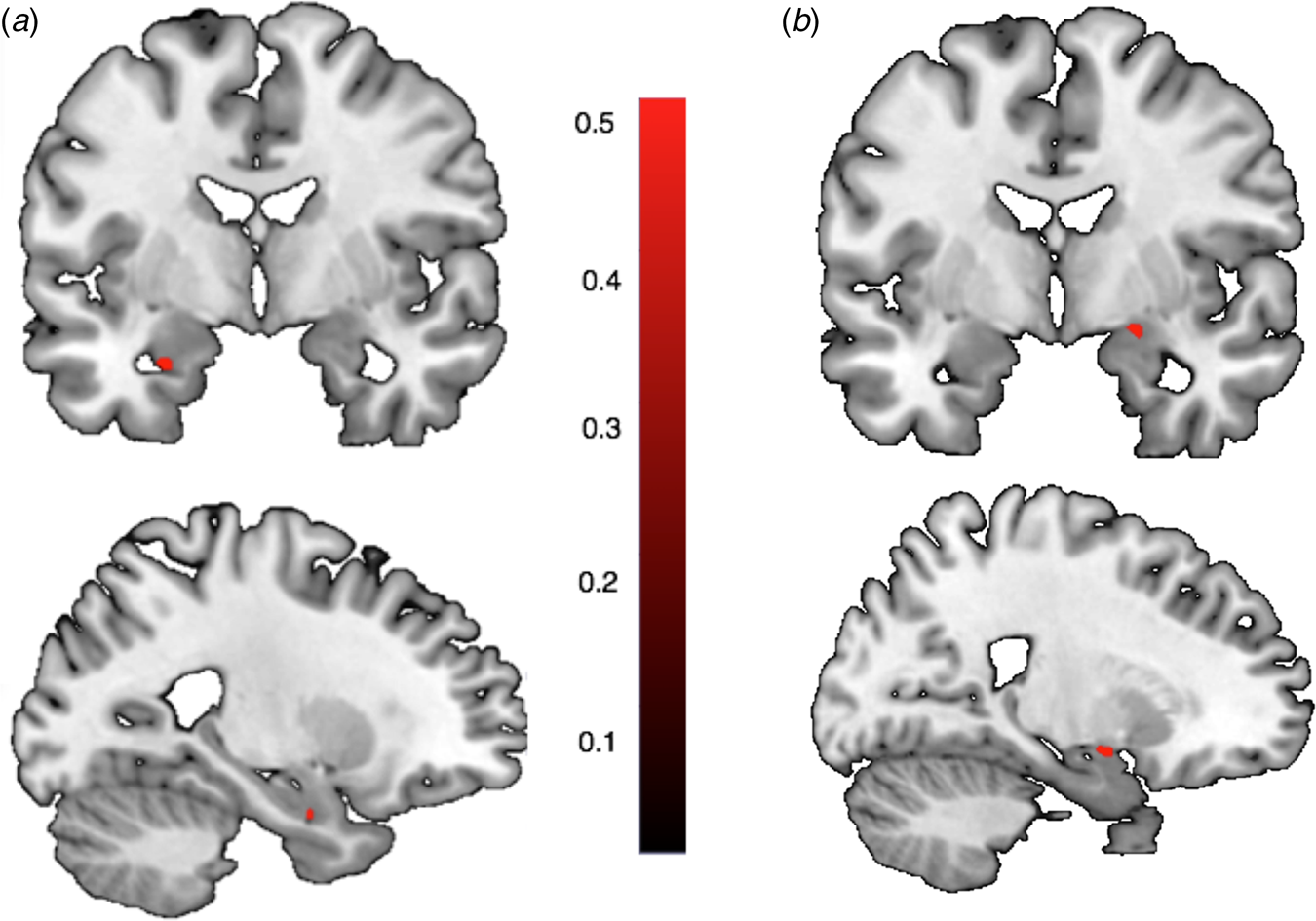
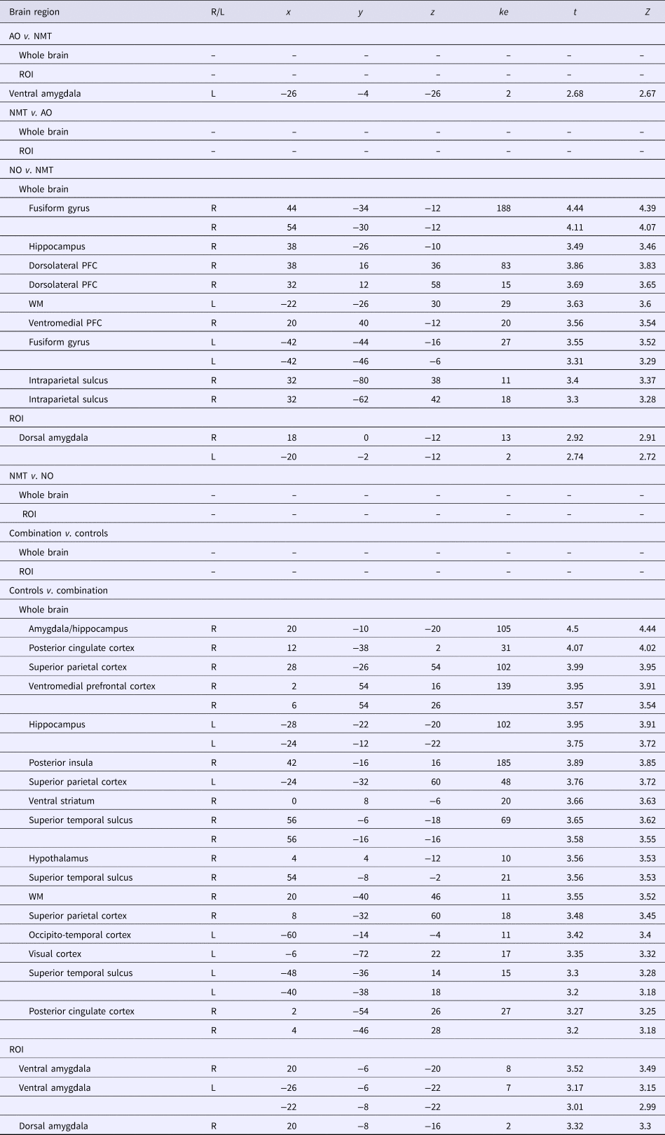
A significant valence × group interaction was found indicating that the AO group relative to the NMT control group showed heightened activation in the left ventral amygdala during the perceptual processing of angry faces compared to neutral faces (ROI, left: p = 0.043, SVC-corrected, see Fig. 2a). Exploratory whole brain analyses of regions more active in the AO relative to the NMT group or the reverse contrast between NMT group v. AO group did not yield any significant results (see Table 1).
(b) Neglect only group v. non-maltreated control group
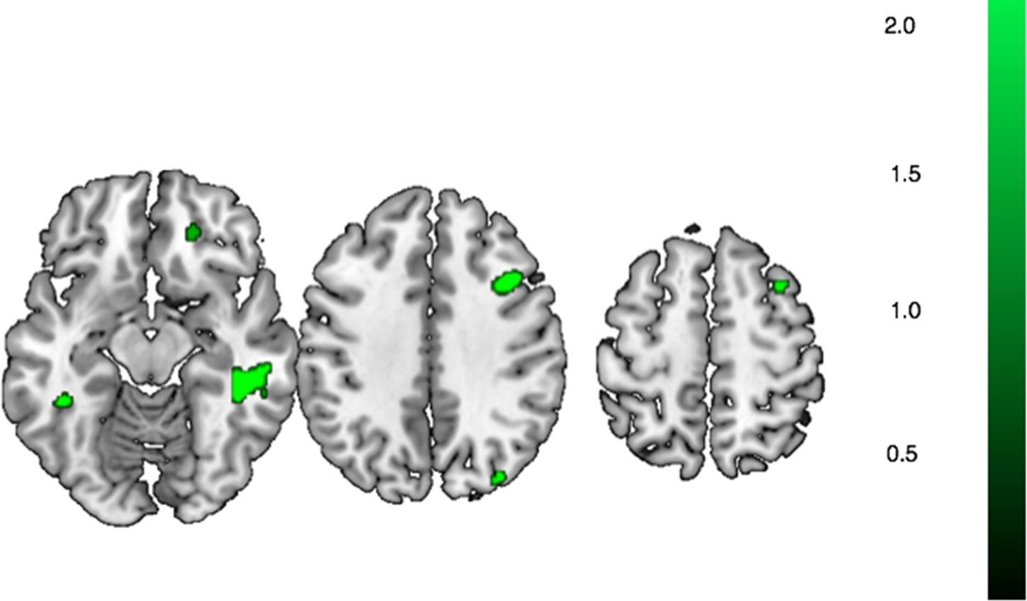
A significant valence × group interaction was found indicating that relative to the NMT group, the NO group showed heightened activation in the bilateral dorsal amygdala during the perceptual processing of angry faces compared to neutral faces (ROI, right: p = 0.024, left: p = 0.038; SVC-corrected; see Fig. 2b). Exploratory whole brain analyses of regions more active in the NO relative to the NMT group during the perceptual processing of angry faces v. neutral also included a range of brain areas including the dorsolateral and ventromedial PFC, bilateral fusiform gyrus as well as the intraparietal sulcus (see Fig. 3 and Table 1).
(c) Combined abuse and neglect (COM) group v. non-maltreated (NMT) control group
No brain areas were found to be more active in the COM group as compared to the NMT control group at ROI or exploratory whole brain level. The reverse contrast however, revealed significantly reduced activations in the bilateral ventral (right: p = 0.005, left: p = 0.013) and right dorsal amygdala (p = 0.008; all SVC-corrected at p = 0.05 FWE) in the COM group relative to the NMT control group. That is, individuals in the COM group tended to show hypo-activation of the amygdala compared to their non-maltreated peers. Exploratory whole brain analyses revealed significantly reduced activity in the COM group relative to the NMT group in several brain regions including the bilateral posterior cingulate cortex, superior parietal and superior temporal cortex and hippocampus as well parts of the visual and occipito-temporal cortex (see Fig. 4 and Table 1).
Finally, we ran exploratory correlational analyses between amygdala reactivity during anger v. neutral face processing and the cumulative number of maltreatment subtypes (ranging from 1 to 5) and measures of maltreatment severity (indexed by CTQ Total T-Score). There was a significant negative relationship with the cumulative number of maltreatment subtypes for both left (r s = −0.180, p = .009) and right amygdala (r s = −0.181, p = .009, see online Supplementary Fig. S2), the relationship with total score was non-significant (both Ps > 0.087). For comparisons between maltreatment groups, please see online Supplementary data.
(d) Within-maltreatment group comparisons
(1) Neglect only (NO) group v. abuse only (AO) group
The comparison of the NO and AO group revealed significantly greater activation during the processing of angry v. neutral faces in a wide-spread network including the hippocampus, subgenual anterior cingulate, somatosensory cortex, fusiform gyrus and superior temporal sulcus. No brain activations were found to be greater in the AO v. NO group.
For additional within-maltreatment group comparisons comparing neglect only (NO) and abuse only (AO) to the combined abuse and neglect (COM) group, please see the online Supplementary material.
ROI analyses for the contrast of angry faces v. neutral faces. A significant valence × group interaction was found during the processing of angry faces v. neutral faces in the left ventral amygdala in the AO group relative to the NMT control group (a) and in the dorsal amygdala in the NO group relative to the NMT control group (b).

Results showing significantly increased activation in the neglect only (NO) group relative to the non-maltreated control (NMT) group in response to angry v. neutral faces in the dorsolateral and ventromedial PFC, bilateral fusiform gyrus as well as the intraparietal sulcus.

Whole brain and ROI results for the contrast angry faces > neutral

R/L, right/left; ke, cluster extent; ROI, region of interest analyses.
Results showing significantly decreased activation in the combined (COM) group relative to the non-maltreated control (NMT) group in bilateral posterior cingulate cortex, superior parietal and superior temporal cortex and hippocampus as well as parts of the visual and occipito-temporal cortex.

Discussion
To our knowledge, this is the first study to systematically investigate how experiences of childhood abuse, childhood neglect and combined experiences of childhood abuse and neglect may differentially influence how the brain processes threat signals later in life. Drawing from a large sample of young adults we identified three groups of individuals with maltreatment experience: those who had experienced only abuse, those who had experienced only neglect, and those who had experienced a combination of both. These individuals were compared with a carefully matched non-maltreated control group who had not experienced any form of childhood maltreatment. Critically, groups were comparable in relation to gender, age, IQ, psychopathology and SES following a rigorous propensity score matching approach. Consistent with the general framework proposed by Sheridan and McLaughlin (McLaughlin et al., Reference McLaughlin, Sheridan and Lambert2014) different forms of maltreatment experience were associated with robust differences in brain reactivity to threat.
Our analyses revealed that while childhood abuse was associated with heightened localised threat reactivity in ventral amygdala, experiences of neglect were associated with heightened reactivity in dorsal amygdala as well as in a distributed cortical fronto-parietal network supporting complex social and cognitive processing. Unexpectedly, combined experiences of abuse and neglect were associated with hypo-activation in several higher-order cortical regions as well as the amygdala.
Exposure to abuse is thought to lead to changes in those neural circuits that underlie emotional and fear learning, including the hippocampus, amygdala, and ventromedial prefrontal cortex [vmPFC (McLaughlin et al., Reference McLaughlin, Sheridan and Lambert2014)]. The association between abuse and amygdala hyperactivity has long been regarded as plausible, and is consistent with the experimental evidence of attentional hypervigilance to threat in physically abused children (Pollak and Tolley-Schell, Reference Pollak and Tolley-Schell2003). However, studies reporting amygdala hyperactivity to threat cues have comprised child and adult participants who present with highly heterogeneous maltreatment experiences, where other forms of adversity have not been accounted for (McCrory et al., Reference McCrory, De Brito, Sebastian, Mechelli, Bird, Kelly and Viding2011, Reference McCrory, De Brito, Kelly, Bird, Sebastian, Mechelli, Samuel and Viding2013; Tottenham et al., Reference Tottenham, Hare, Millner, Gilhooly, Zevin and Casey2011). Here, we found that individuals exposed only to abuse show higher ventral amygdala reactivity to threatening faces; no differences were observed at the whole brain level. This provides compelling support for the view that exposure to abuse experiences in childhood has a relatively specific impact on the neural circuits that underlie the processing of threat cues. According to the theory of latent vulnerability (McCrory and Viding, Reference McCrory and Viding2015; McCrory et al., Reference McCrory, Gerin and Viding2017a) heightened the salience of threat cues may carry functional value for the child in the short term but increase later psychiatric risk in two ways (McCrory et al., Reference McCrory, Gerin and Viding2017a). First, it may attenuate the attentional resources available for the processing other socially relevant environmental information relevant for effective development (direct effect). Second, it may compromise social interaction, potentiating more conflictual interactions (indirect effect) (McCrory et al., Reference McCrory, Gerin and Viding2017a). It is hypothesised that over time this would impact on how an individual shapes their social ecology: if affected they are less able to elicit, cultivate and sustain stable peer and adult relationships, they would have fewer supportive relationships to help buffer the impact of future stress.
By contrast, neural differences following neglect were expected to be more widely distributed, encompassing areas underlying associative learning, complex cognition and language, i.e. the fronto-parietal networks including association cortices (McLaughlin et al., Reference McLaughlin, Sheridan and Lambert2014, Reference McLaughlin, Sheridan and Nelson2016). In line with this our exploratory analyses showed that the neglect-only group show increased activation in a spatially distributed set of brain areas implicated in different forms of associative learning and complex cognition, including the dorsolateral PFC [executive functioning (Kolb et al., Reference Kolb, Mychasiuk, Muhammad, Li, Frost and Gibb2012)]; ventromedial PFC [emotion regulation (Buhle et al., Reference Buhle, Silvers, Wager, Lopez, Onyemekwu, Kober, Weber and Ochsner2014)]; bilateral fusiform gyrus [face processing (Kanwisher et al., Reference Kanwisher, McDermott and Chun1997)]; and the intraparietal sulcus (integration of information from different sensory modalities) (Corbetta et al., Reference Corbetta, Kincade, Ollinger, McAvoy and Shulman2000). A distributed pattern of neural differences was also observed when the neglect-only group was contrasted to the abuse-only group (see online Supplementary data in Appendix). These findings are consistent with the proposal made by Sheridan and McLaughlin (McLaughlin et al., Reference McLaughlin, Sheridan and Lambert2014) that neglect leads to a broad range of deleterious effects, potentially via accelerated and extreme synapse elimination as well as reduced myelination of axons, across domains including cognitive ability, associative learning and executive functions (McLaughlin et al., Reference McLaughlin, Sheridan and Lambert2014). In addition, our findings indicated that the neglect-only group showed heightened dorsal amygdala reactivity to threat cues, albeit in a functionally distinct cluster to that observed in the abuse-only group who showed heightened activation of the ventral amygdala. A similar finding has previously been reported in a large genetically sensitive study of adolescents (N = 139) self-reporting emotional neglect (White et al., Reference White, Bogdan, Fisher, Muñoz, Williamson and Hariri2012). In particular, variations of the human gene that codes for the FK506-binding protein 5 (FKBP5) were investigated, a gene that has been previously associated with the emergence of stress-related psychiatric symptoms. Emotional neglect was also associated with higher dorsal amygdala reactivity, but only in those adolescents carrying the ‘riskier’ genetic polymorphisms (White et al., Reference White, Bogdan, Fisher, Muñoz, Williamson and Hariri2012). Heightened neural reactivity to threat-related cues may be adaptive in a neglectful environment as the child is required to be more vigilant in the absence of a reliable caregiver who can monitor the environment from threat and protect them (McCrory et al., Reference McCrory, Gerin and Viding2017a).
Finally, we investigated individuals who had experienced a combination of both neglect and abuse in childhood. The findings for this group were striking and unpredicted. Relative to non-maltreated control participants they showed an unexpected hypoactive pattern of neural response in the ventral and dorsal amygdala and a spatially distributed pattern of reduced neural activation in a range of brain areas including the superior parietal cortex, posterior cingulate cortex, the insula and occipito-temporal cortex. A rich body of evidence suggests that prolonged early adversity leads to hypoactivation of the stress circuitry and hyposecretion of the stress-hormone cortisol (Cicchetti and Rogosch, Reference Cicchetti and Rogosch2001; Gunnar and Donzella, Reference Gunnar and Donzella2001; Puetz et al., Reference Puetz, Viding, Palmer, Kelly, Lickley, Koutoufa, Sebastian and McCrory2016a, Reference Puetz, Zweerings, Dahmen, Ruf, Scharke, Herpertz-Dahlmann and Konrad2016b). It is now well established that hypocortisolism likely reflects processes within the organism to adjust to sustained stressful environments (allostatic adjustment, see allostatic load model) (McEwen, Reference McEwen1998), which is considered to be adaptive in the short-term, but poses a major threat to healthy development (Gunnar and Donzella, Reference Gunnar and Donzella2001) at the behavioural and neural level (McEwen, Reference McEwen2013). In other words, moderate maltreatment exposure may lead to an increase in amygdala reactivity (as observed here in the single subtype groups) but prolonged or more severe maltreatment may lead to reversal and a hypoactive response. Future studies, with more fine-grained measures of developmental timing and severity are required to evaluate this possibility. This pattern of hypoactivation in amygdala and the broader set of brain regions including the visual, sensory and cingulate cortices including the hippocampus may be potentially understood as part of an adaptive response, reflecting functional avoidance of negative stimuli in the context of an aversive and distressing home environment. In line with this, attenuated response of the amygdala, insula, vlPFC, anterior cingulate, parietal and visual cortices have been reported in patients with trauma and affective symptomatology (Chechko et al., Reference Chechko, Augustin, Zvyagintsev, Schneider, Habel and Kellermann2013) and children with maltreatment experiences (Puetz et al., Reference Puetz, Kohn, Dahmen, Zvyagintsev, Schüppen, Schultz, Heim, Fink, Herpertz-Dahlmann and Konrad2014, Reference Puetz, Viding, Palmer, Kelly, Lickley, Koutoufa, Sebastian and McCrory2016a, Reference Puetz, Zweerings, Dahmen, Ruf, Scharke, Herpertz-Dahlmann and Konrad2016b). Previous studies have suggested that these alterations in higher-order visual and association cortices may be related to altered integration of multimodal information and underlie visual and somatosensory symptoms such as hyperarousal or numbing (Lanius et al., Reference Lanius, Bluhm, Lanius and Pain2006).
A number of additional exploratory analyses were conducted in order to shed light on the contrasting patterns of amygdala response observed across our three maltreatment groups. These indicated that maltreatment severity, as indexed by a cumulative number of maltreatment subtypes, showed a negative association with amygdala reactivity bilaterally, in line with findings from adoptee studies that indicate that more severe maltreatment relates to hypocortisolism (van der Vegt et al., Reference van der Vegt, van der Ende, Kirschbaum, Verhulst and Tiemeier2009). This provides preliminary evidence that maltreatment is associated with both patterns of hyper- as well as hypo-amygdala reactivity, and that individuals who experience multiple forms of maltreatment may be more likely to present with an attenuated amygdala response.
A number of limitations need to be kept in mind with respect to the current study. First, due to the nature of the sample, we had no access to independent documented records of experience, which has become the golden standard in maltreatment research with children (McCrory et al., Reference McCrory, De Brito, Sebastian, Mechelli, Bird, Kelly and Viding2011, Reference McCrory, De Brito, Kelly, Bird, Sebastian, Mechelli, Samuel and Viding2013, Reference McCrory, Puetz, Maguire, Mechelli, Palmer, Gerin, Kelly, Koutoufa and Viding2017b). Instead, we here relied on retrospective self-report by young adults with the CTQ, which necessarily entails a potential risk for recall bias (Newbury et al., Reference Newbury, Arseneault, Moffitt, Caspi, Danese, Baldwin, Fisher and Fisher2018) and does not capture maltreatment onset and duration. However, it has nevertheless been shown that such retrospective reports are valid for the identification of major adversities and rarely involve false positives (Newbury et al., Reference Newbury, Arseneault, Moffitt, Caspi, Danese, Baldwin, Fisher and Fisher2018). Furthermore, the maltreated and non-maltreated participants consisted of university students; replication would help establish that these findings are evident in other groups within the wider population. Nonetheless, we would note that the findings reported here are consistent with previous investigations characterised by samples of different educational, socio-economic background and age (McCrory et al., Reference McCrory, De Brito, Sebastian, Mechelli, Bird, Kelly and Viding2011; Tottenham et al., Reference Tottenham, Hare, Millner, Gilhooly, Zevin and Casey2011), suggesting that the effects of maltreatment on multiple levels are pervasive and likely consistent across different populations. These findings can inform future studies and a more regionally informed investigation of the neurocognitive impact of different kinds of maltreatment experience.
In conclusion, the present study represents the first systematic investigation of the differential impact of abuse and neglect, and their combined effects on neurocognitive processing. Propensity score matching allowed us to exclude the influence of a range of potential confounds. Childhood abuse, childhood neglect and their combination were found to differentially influence neural processing of threat-related cues. This suggests that vulnerability to psychopathology following these different forms of maltreatment experience may be conferred via both shared and distinct mechanisms and may in turn help account for the range of psychiatric outcomes following childhood maltreatment. Delineating such mechanisms has the potential to improve targeted approaches to prevention and intervention.
Supplementary material
The supplementary material for this article can be found at https://doi.org/10.1017/S003329171900134X.
Author ORCIDs
Vanessa Bianca Puetz, 0000-0003-0793-7210.
Acknowledgements
We thank the staff of the Laboratory of NeuroGenetics as well as the participants in this study.
Financial support
The Duke Neurogenetics Study received support from Duke University as well as US-National Institute on Drug Abuse Grant R01DA033369 and R01DA031579. This work was further supported by the US-National Institute on Aging Grant R01AG049789.
Conflict of interest
None.