Rheumatic valvar disease, a sequel to rheumatic fever, is one of the most commonly acquired cardiac diseases and is the predominant indication for cardiac surgery in developing countries.Reference Achutti and Achutti1–Reference Da Silva and Pereira3 Although it is considered to be a disease associated with poverty, rheumatic fever continues to persist even among the middle-class population in developed countries.Reference Stollerman4, Reference Markowitz5
The eletrocardiographic features of acute rheumatic fever have been well described but are insufficient to enable the cardiac component to be recognised.Reference Krishnan, Kushwaha and Josephon6 The QT dispersion interval, a measurement of the low homogeneity of ventricular repolarisation, defined as the variation in the duration of QT interval between the leads, may be a useful marker of rheumatic carditis. The effects of diseased states and therapeutic drugs on the QT interval have been extensively studied and the increase in QT dispersion seems to be a predictor of arrhythmia in various clinical groups.Reference Zareba, Moss and Le Cersie7–Reference Buja, Miorelli, Turrini, Melacini and Nava9 The role of QT dispersion in acute rheumatic carditis has not been elucidated yet.
In fact, there is only one article recently published, which comments on this subject.Reference Polat, Yalcin, Akdeniz, Zeybek, Erdem and Celebi10 The purpose of this study was to investigate QT dispersion in the surface electrocardiogram of children with rheumatic carditis.
Materials and methods
Patient population
This study enrolled all children who had been hospitalised with initial first acute rheumatic carditis at the cardiac unit of the Federal University of Pernambuco’s children institute, from July, 2007 to January, 2008. We also studied a group of control children, free of any cardiac disease, derived from routine examinations from the same hospitals, during the same period. The children were eligible for participation if the following criteria were met: diagnosis of acute rheumatic fever based on the revised Jone’s criteria11, 12 and suffering from their first attack of carditis.
The diagnosis of carditis was evident due to the presence of murmurs, suggestive of valvar regurgitation and echocardiography examinations.
The children were excluded if they were using anti-arrhythmic drugs, β-blockers, or steroids in the case of carditis recurrence of a previous history of cardiac failure. The presence of metabolic disorders was also a criterion for exclusion.
Electrocardiogram analysis
Simultaneous 12-lead electrocardiogram recordings were obtained at the time of hospital discharge. Electrocardiogram tracings of all children and their controls were blindly analysed by two investigators. The parameters calculated were PR interval, QT interval, and QT dispersion in two successive complexes for each lead. The measurement of the QT interval was started from the onset of the QRS complex until the end of the T wave. In the presence of a U wave, the QT interval of the lead was dismissed. QT dispersion was obtained through the difference between maximum and minimum QT calculated in the 12 leads.
Echocardiographic examination
The echo Doppler cardiogram was performed in all children, within 48–72 hours of hospitalisation. All ultrasound examinations were performed with an echocardiographic machine, specifically a color doppler two dimensional HDI 1500 machine (Advanced Technology Laboratories, United States of America). All echocardiographic studies were interpreted by the same two observers, blinded to clinical information. The involvement and severity of rheumatic valvar disease were graded on the basis of the maximal distance of the regurgitant jet and in the typical aspect of anatomic rheumatic valvar disease.Reference Elmcke, Nanda and Hsiung13, Reference Perry, Helmcke, Nanda, Byard and Soto14
Statistical analysis
We made comparisons between the two groups with the use of the t-test for the continuous variables and the χ2 test for the categorical variables. All p-values were two-sided, and a p-value of less than 0.05 was considered to indicate statistical significance. Sensitivity and specificity of measurements of QT dispersion in predicting rheumatic carditis were estimated by using receiver operating characteristic curves.
Results
We enrolled a total of 66 children for this study, half of which had rheumatic carditis and met the inclusion criteria. Table 1 shows the baseline demographic and electrocardiographic characteristics of the two groups. A significant increase in the PR interval was found in the children with evidence of carditis compared with the controls.
Baseline demographic and electrocardiographic characteristics of the groups.

Values are expressed as means with standard deviation given in parentheses
The QT dispersion after cure was not evaluated. All comparison was made between the active carditis group and control group, with the same baseline characteristics.
Children with carditis had a greater QT dispersion than the control group.
When we analyse the QT dispersion according to the severity of the carditis, we observed that the dispersion tended to be greater in those with more severe valvar lesion (Table 2).
Comparison of carditis subgroups.

Values are expressed as means with standard deviation given in parentheses
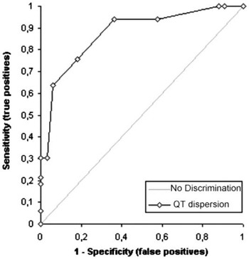
The sensitivity and specificity of the measurements of the QT dispersion in predicting acute carditis were estimated by using receiver operating characteristic curves. A QT dispersion greater than 40 milliseconds had a sensitivity of 63.6% and a specificity of 93.9% in predicting acute rheumatic carditis (Fig 1).
Receiver operating curve plot of QT dispersion in predicting rheumatic carditis.

Discussion
Since it was first described, the measurement of the QT interval dispersion has been studied as an electrocardiogram marker of ventricular repolarisation abnormality, risk of fatal arrhythmias, and therapeutic evaluation of the drugs.Reference Zabel, Klingenheben, Franz and Hohnloser15, Reference Etheridge and Shaddy16 In children, there are few published limits of normal dispersion and discordant results in clinical groups.Reference Vialle, Albalkhi, Zimmerman and Friedli17–Reference Tutar, Ocal, Imamoglu and Atalay19 The fact that we admitted sick children from all over the northeast of the country, the poorest region of the country, and many of them returned to local hospitals even during treatment, is the main reason for not assessing the QT dispersion after cure. Despite this, we used a control group, with the same baseline characteristics, to compare with the carditis group. In accordance with our results, QT dispersion showed a significant increase in children with acute rheumatic carditis when compared with normal controls. Our findings are in agreement with Polat et al,Reference Polat, Yalcin, Akdeniz, Zeybek, Erdem and Celebi10 but the QT dispersion values that we found were not as high as those. Indeed, despite the fact that the children in this study were also in their first attack of the disease, the absolute exclusion of cardiac failure, may be a reason for this difference.
Furthermore, we have not analysed the QT dispersion in rheumatic patients without carditis. As far we are aware, however, it is justified to argue that subclinical valvitis diagnosed by Doppler echocardiography should be considered as mild carditis, thus becoming a major criteria in the diagnosis of acute rheumatic fever.Reference Narula, Chandrasekhar, Shahbudin and Rahimtoola20–Reference Ozkutlu, Ayabakan and Saraçlar22 We therefore believe that rheumatic fever without cardiac involvement perhaps does not exist and decided not to include these patients in the study.
When we compared the severity of carditis, we did not find a significant difference between moderate and severe carditis and mild carditis. It is possible that in a larger study, we may find a statistical significance.
Another study by Guven et alReference Guven, Eroglu and Babaoglu23 found lower values of QT dispersion in rheumatic mitral valve prolapse when compared with patients with primary mitral prolapse. The fact that the rheumatic mitral valve prolapse is usually a late sequel, without inflammatory activity, may be a possible reason for their findings.
In our study, the assessment of QT dispersion was performed in children with rheumatic carditis during acute cardiac injury.
Although rheumatic carditis usually refers to the valve damage, myocardial impairment exists. The results seem to indicate heterogeneity of ventricular epolarisation in different regions of the myocardium, probably secondary to myocardial structural changes in the acute phase of the disease.
Finally, using a cut-off point of 40 milliseconds, we found a higher specificity in predicting acute rheumatic carditis. Such an increase in QT dispersion may be an important parameter in the diagnosis of rheumatic carditis. The assessment of regular values for QT dispersion in children provides an easier method for predicting carditis whereas it is not necessary to calculate the corrected QT dispersion in children.Reference Vialle, Albalkhi, Zimmerman and Friedli17
Conclusion
The lengthening of the QT dispersion may reflect on cardiac involvement in rheumatic fever and is a new important parameter in the diagnosis and therapeutic decision for rheumatic carditis.
Acknowledgement
We thank Dr Giordano B. Parente and Mrs Marcia Virginio for their assistance.