Introduction
Precise topographic mapping of retinal projections to central targets is a requisite of normal mammalian visual system development. The axons of neighboring retinal ganglion cells (RGCs) project to adjacent regions within the dorsal lateral geniculate nucleus (dLGN) and superior colliculus (SC) (reviewed in Reese, Reference Reese2010). Topographic specificity is guided predominantly by reciprocal molecular gradients (Sperry, Reference Sperry1963) and refined in an activity-dependent fashion (Torborg & Feller, Reference Torborg and Feller2005). The Eph family receptor tyrosine kinases and their ligands, the ephrins, are the most thoroughly characterized molecular mediators of topographic specification (McLaughlin & O’Leary, Reference McLaughlin and O’Leary2005). EphAs and ephrin-As direct the mapping of RGC axons along the anterior–posterior axis of the SC according to the position of their cell bodies on the nasotemporal axis of the retina, whereas EphBs and ephrin-Bs direct mapping along the mediolateral axis of the SC according to their retinal dorsoventral position (Hindges et al., Reference Hindges, McLaughlin, Genoud, Henkemeyer and O’Leary2002; Luo & Flanagan, Reference Luo and Flanagan2007). Ephrin signaling has also been shown necessary for topographic specificity in the dLGN (Pfeiffenberger et al., Reference Pfeiffenberger, Yamada and Feldheim2006), but the mechanisms of this patterning are less well understood, partly due to the three-dimensional structure of the nucleus.
Phr1 is an evolutionary conserved multidomain protein that plays a central role in neural development. Disruption of Phr1 orthologs is associated with defects ranging from excess synaptic growth in Drosophila (Wan et al., Reference Wan, DiAntonio, Fetter, Bergstrom, Strauss and Goodman2000; DiAntonio et al., Reference DiAntonio, Haghighi, Portman, Lee, Amaranto and Goodman2001), synaptogenesis, and axon termination defects in Caenorhabditis elegans (Schaefer et al., Reference Schaefer, Hadwiger and Nonet2000; Zhen et al., Reference Zhen, Huang, Bamber and Jin2000); abnormal retinal projections in zebrafish (D’Souza et al., Reference D’Souza, Hendricks, Le Guyader, Subburaju, Grunewald, Scholich and Jesuthasan2005); and axon guidance defects in vertebrates (Burgess et al., Reference Burgess, Peterson, Johnson, Roix, Welsh and O’Brien2004; Bloom et al., Reference Bloom, Miller, Sanes and DiAntonio2007). Complete knockout of Phr1 is neonatally lethal in the mouse (Burgess et al., Reference Burgess, Peterson, Johnson, Roix, Welsh and O’Brien2004; Bloom et al., Reference Bloom, Miller, Sanes and DiAntonio2007; Lewcock et al., Reference Lewcock, Genoud, Lettieri and Pfaff2007), and conditional knockout of Phr1 in RGCs results in an abnormal localization of the ipsilateral retinogeniculate projection that is independent of ephrin-A responsiveness and spontaneous retinal activity (Culican et al., Reference Culican, Bloom, Weiner and DiAntionio2009). Here, we report a quadrant-specific disruption of the retinotopic map in the SC, with more subtle defects in the dLGN, indicating that Phr1 is required to establish regional as well as global topography.
Materials and methods
Mice
Phr1 retinal mutant mice were described previously (Culican et al., Reference Culican, Bloom, Weiner and DiAntionio2009). Studies here were done in mice backcrossed into C57BL/6J background. Genotyping was done by PCR. Briefly, Phr1 conditional knockout mice containing a floxed allele of Phr1 were mated to mice that express Cre recombinase in RGCs under the control of the Math5 promoter (Yang et al., Reference Yang, Ding, Pan, Deng and Gan2003).
RGC labeling
RGC labeling was adapted from Simon and O’Leary (Reference Simon and O’Leary1991). A 10% solution of the fluorescent lipophilic dye, 1, 1′-dioctadecyl-3,3,3′,3′-tetramethylindocarbocyanine perchlorate (DiI; Invitrogen, Carlsbad, CA) in dimethylformamide (Sigma, St. Louis, MO), was pressure injected through a pulled glass micropipette using a Picospritzer II (General Valve Corp., Fairfield, NJ). Postnatal day 14 (P14) or older mice were anesthetized and injected with 0.1–1 μl of DiI in one eye through a small hole made in the sclera at the limbus with a 27 gauge needle. Injections were performed masked to genotype. Mice were sacrificed 6 days later (older then P20) and intercardially perfused with PBS then 4% paraformaldehyde in 0.1 M PBS. Brains were postfixed and whole-mount SCs imaged with a RoleraXR camera mounted on an Olympus BX51WI microscope (Olympus, Center Valley, PA). Brains were then embedded in 2–3% agarose (Sigma) and 100-μm coronal sections sliced on a Vibratome, mounted, and imaged. Image acquisition was performed masked to genotype.
Retrograde labeling
P14 mice were anesthetized and intercardially perfused with PBS then 4% paraformaldehyde in PBS. Brains were postfixed, and the cortex was removed. 0.1–0.5 μl of DiI was pressure injected as described above into the posterial–lateral region of the SC. After 14 days, whole-mount SC images were obtained. Brains were embedded in 2–3% agarose and 120-μm sagittal sections sliced and imaged.
Quantification and representation of data
Whole-mount SC image analysis was adapted from Pfeiffenberger et al. (Reference Pfeiffenberger, Yamada and Feldheim2006). Images of whole-mount SC were opened in Image J (v.1.41; NIH Bethesda, MD) and background fluorescence levels, determined from measurement of a nonlabeled area of the SC, were digitally subtracted. An intensity plot of the contralateral SC was obtained covering a rectangular region one-third the lateral–medial (L-M) width of the SC and equal in length to the longest anterior-posterior (A-P) dimension. All intensity plots were normalized to an A-P length of 600 pixels.
Summary of ectopic termination zones in nasal–dorsal quadrant
Termination zones from four controls and four Phr1 mutants were plotted along the anterior–posterior axis with 0 and 100 representing the posterior and anterior borders of the SC, respectively. Each point on the horizontal axis represents a single distinct termination zone along the anteroposterior extent of the nucleus, and each vertical set of points represents one animal after receiving a single retinal injection (adapted from Feldheim et al., Reference Feldheim, Kim, Bergemann, Frisen, Barbacid and Flanagan2000).
Quantification of area (in percentage) of terminal zones in SC and dLGN
The dLGN and SC were outlined manually based on gross anatomical morphology. Background fluorescence levels in the dLGN and SC were determined from measurement of a nonlabeled area of the dLGN and SC, respectively, and digitally subtracted. A threshold was set at 20% of the maximum signal intensity, and the area (in pixels) measured above that threshold was divided by the total area and reported as a percentage. For dLGN specifically, the total percentage for each animal was obtained by the averaging values from four slices (see Culican et al., Reference Culican, Bloom, Weiner and DiAntionio2009). Average area for each regional injection site was obtained by averaging the values from four animals in each condition. Errors are standard error mean. Composite panels were generated to qualitatively illustrate the extent of labeling in the dorsal–ventral and medial–lateral axis for each condition (Supplementary Fig. S1).
Results
Disrupted mapping along the nasal–dorsal axis of the Phr1 mutant retinocollicular projection
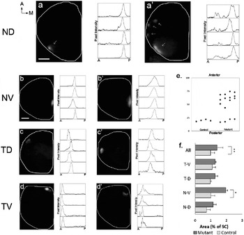
To evaluate retinocollicular connectivity in Phr1 retinal mutants, we delivered focal DiI injections into the four retinal quadrants [nasal dorsal (ND), nasal ventral (NV), temporal dorsal (TD), and temporal ventral (TV)] and assessed axon termination in the SC. In control mice, a single injection resulted in a single compact terminal zone (TZ) on the outer edge of the SC corresponding to the peripheral injection site of each retinal quadrant (Fig. 1a–1d). ND and NV retinal injections produced compact TZs located in the posterior–lateral and posterior–medial regions of the SC, respectively (Fig. 1a and 1b). The location and continuity of the ipsilateral patch is disrupted in the dLGN of Phr1 retinal mutant mice (Culican et al., Reference Culican, Bloom, Weiner and DiAntionio2009). Because RGC axons that project to the ipsilateral patch originate in the TV quadrant of the retina, we expected to also observe mislocalization of TZ in the SC from retinal axons of TV quadrant origin. But on the contrary, the most dramatic disruption was of DiI labeling from ND quadrant mutant RGCs. Labeling this quadrant lead to multiple ectopic TZs shifted anteromedially in the nucleus (Fig. 1a′ and 1e). Additionally, we observed a global disruption of local refinement of TZs in the Phr1 retinal mutant; TZs derived from the ND, as well as from the other three quadrants, were more diffuse and occupied larger areas of the SC. The total area of the SC labeled after a single injection in retina mutants was significantly larger (1.403 ± 0.351%, n = 16) than in littermate controls (0.976 ± 0.244%, n = 16) (P < 0.01) (Fig. 1f). These results thus demonstrate that loss of Phr1 in RGCs results in both a broad disruption of topographic connections along the ND retinal axis and a disruption of local TZ refinement along all retinal axes.
Phr1 retina mutants have multiple ectopic terminal zones (TZs) originating from the nasal–dorsal (ND) retina and aberrant diffuse disruption other quadrants. Retinal projections to the superior colliculus (SC) were compared between control (a–d) and Phr1 mutant (a′–d′). Multiple ectopic TZs are observed in the SC of the retinal mutant (a′) compared to a single compact TZ in control (a) with DiI injection in the ND quadrant of the retina. A summary of the topographic mapping defects from cells originating in the ND quadrant (e). Nasal–ventral (NV) (b, b′), temporal–dorsal (TD) (c, c′), and temporal–ventral (TV) (d, d′) RGCs from the mutant display more diffuse TZs compared to littermate controls. Intensity spikes indicate TZs in each intensity plot to the right. Diffuse TZs occupy a larger area (% of SC) in all quadrants (f). Mutant RGCs originating in the NV retina occupy a significantly larger area of the SC compared to control (P < 0.05). Differences in the other quadrants are not significant (TD, P ~ 0.06; ND, P ~ 0.10; TV, P ~ 0.20). Error bars represent standard error mean (s.e.m.). Total n = 32, n = 4 for each genotype and each quadrant. Scale bar = 400 μm; a, anterior; p, posterior; m, medial; d, dorsal; n, nasal; v, ventral; t, temporal.

Ectopic termination zones do not result from interstitial branching
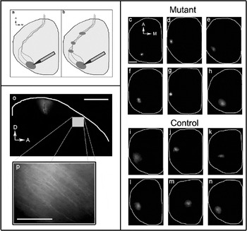
The cellular basis of ectopic TZs that arise from RGCs in the ND quadrant is unclear. One possibility is that nearest-neighbor relationships are not preserved in the Phr1 retinal mutant and project to different locations within the SC. Another possibility is that nearest-neighbor relationships are preserved but that interstitial branching of RGC axonal arbors results in representations of the same retinotopic map in multiple locations. To assess whether the ectopic patches derived from the ND projection result from multiple representations of a properly organized retinotopic map, we employed retrograde DiI labeling. Focal DiI injections were made into the posterial–lateral region of the SC, the area where ND RGCs terminate. If the disrupted mapping of ND RGCs is due to multiple representations of the normal map caused by misdirected interstitial branching, then additional ectopic TZs anterior to the injection site should be observed (Fig. 2a–2b). In both control and mutant mice, injections of various sizes and locations within the posterial–lateral region do not yield additional termination zones (Fig. 2c–2n). Furthermore, observation of labeled primary axons traveling through the most anterior edges of the SC in these animals indicate that sufficient time was allowed for dye to travel (Fig. 2o–2p). These results suggest that the disrupted mapping along the ND axis of the Phr1 mutant retinal projection is not caused by erroneous interstitial branching.
Absence of ectopic terminal zones (TZs) in controls and Phr1 mutants following DiI retrograde labeling in the posterial–lateral region in the superior colliculus (SC). Simplified diagram showing the DiI retrograde labeling (a) where anteriorly shifted TZs might result from interstitial branching (b). None of the controls or the mutant animals exhibit additional termination zone (c–n). A sagittal section of the SC shows that the DiI injection spans the entire depth of the nucleus (o). 20× magnification of the most anterior edge demonstrates that sufficient time was allowed for DiI to travel the entired span of the SC from the posterior injection site (p). Total n = 12, n = 6 for control and n = 6 for mutant. Scale bar = 400 μm; a, anterior; p, posterior; m, medial; d, dorsal; v, ventral.

Diffuse mapping along all axes in the Phr1 mutant retinogeniculate projection
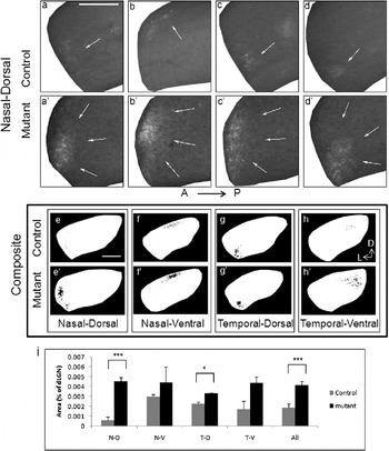
Abnormal localization of the ipsilateral retinogeniculate projection was previously demonstrated in Phr1 retinal mutants despite normal spontaneous retinal activity and eye-specific input segregation in these animals (Culican et al., Reference Culican, Bloom, Weiner and DiAntionio2009). However, it remained undetermined whether the erroneous projection resulted from a global shifting of the broad retinogeniculate map or rather as an indirect consequence of disrupted nearest-neighbor topographic connections within the ipsilateral projection. The location of RGCs along the dorsal–ventral retinal axis dictate the location of their TZs along the ventromedial–dorsolateral axis in the dLGN, and the location of ganglion cell bodies in the nasal–temporal retinal axis determine their projections along the ventrolateral–dorsomedial axis (Fig. 3a–3h; Feldheim et al., Reference Feldheim, Vanderhaeghen, Hansen, Frisen, Lu, Barbacid and Flanagan1998; Grubb et al., Reference Grubb, Rossi, Changeux and Thompson2003). Phr1 retinal mutants retain a topographically appropriate map with only minor morphological aberrations compared to controls. Mutant ND RGC TZs were confined to the area lining the most lateral aspect of the nucleus, as in controls, although they were extended diffusely along the dorsal–ventral axis (Fig. 3a′–3d′). In contrast, injections in the TV retinal quadrant, which include both contralaterally and ipsilaterally projecting RGCs, lead to normally localized TZs in the medial aspect of the nucleus. Surprisingly, no ipsilaterally projecting axons were observed to be grossly mislocated in any of our focal dye labeling experiments despite that the ipsilateral projection, which originates in the TV quadrant, was previously shown to be mislocalized in the dLGN by bulk labeling of RGCs (Culican et al., Reference Culican, Bloom, Weiner and DiAntionio2009). We did not detect any contralaterally projecting axons after TV injections. This may reflect the fact that all our injections were applied at the corneal limbus, in the most peripheral aspect of the retina, where ipsilaterally projecting RGCs are more abundant. Although Phr1 retinal mutants had a roughly normal topographical map in the dLGN, as evidenced by fairly appropriate location of TZs, we did observe more diffuse terminations in labeling from all quadrants (Fig. 3e′–3h′). Despite the similarity of this finding in both the dLGN and the SC, the disruptions found in the retinogeniculate map are more subtle than those found in the retinocollicular map, particularly regarding RGCs of the ND quadrant.
Diffuse terminal zones (TZs) in dorsal lateral geniculate nucleus (dLGN) of the Phr1 mutant. TZs are identified in coronal sections of the dLGN from anterior to posterior following DiI injection in the nasal–dorsal (ND) quadrant of the retina in control (a–d) and Phr1 mutant (a′–d′) mice. Enlarged TZs are observed in each anterior to posterior coronal section of the dLGN with injections in the ND retinal quadrant (a′–d′). Because a TZ spans multiple anterior to posterior sections, a composite image was created by collapsing each of four individual sections (a–d), using a threshold algorithm (see Materials and methods), onto a single representative section (e). This was done to normalize comparisons between mutants and controls for TZs from the ND (e, e′), nasal–ventral (f, f′), and temporal–dorsal (TD) (g, g′) quadrants. This analysis of the contralateral projection demonstrates that TZs in the mutant are appropriately located but more diffuse compared to control TZs (e′–g′). Similarly, ipsilaterally projecting temporal–ventral TZs are also more diffuse in mutant (h) than control (h′). The diffuse TZs in the mutants occupied a significantly larger area of the dLGN than in controls, particularly from the ND (P < 0.0005) and TD (P < 0.05) quadrants (i). Error bars represent standard error mean (s.e.m.). Total n = 16, n = 2 animals for each genotype and each quadrant injected. Scale bar = 200 μm; a, anterior; p, posterior; l, lateral; d, dorsal; n, nasal; v, ventral; t, temporal.

Discussion
The proper formation of topographic maps in the visual system requires spatial relationships between RGC cell bodies preserved by their projections to central targets. During development, both molecular guidance and activity-dependent mechanisms contribute to the establishment of these retinotopic maps, and abnormalities in either mechanism lead to aberrant retinotopy such as the mislocalization of ipsilaterally projecting RGCs seen in the dLGN of Phr1 retinal mutants. Since disturbed targeting of the ipsilateral retinogeniculate projection in this mouse is independent of spontaneous retinal activity and monocular segregation, we sought to determine if the defect represents a more general disruption of retinofugal mapping. Indeed, our results indicate that in addition to the ipsilaterally projecting RGCs, there is disruption of input localization from the contralateral eye to both the dLGN and the SC. Furthermore, we wondered whether the mislocalized ipsilateral projection demonstrates a reorientation of the overall topographic map or also the effect of disrupted local nearest-neighbor connectivity. Our results support both mechanisms: anterograde focal injections into the ND quadrant of the retina lead to multiple separate TZs in the SC, indicative of the latter, while the occurrence of diffuse TZs from all quadrants supports the former. Given the perturbed localization of the ipsilateral projection to the dLGN, the subtle finding of a modestly more diffuse TV quadrant projection, in contrast to the severely disturbed ND connectivity, was unexpected. This finding raises the possibility that Phr1 mediates RGC axon path finding to various central targets via multiple mechanisms.
The mechanism by which ectopic patches arise from RGCs originating in the ND quadrant is not known. One possible mechanism is that ectopic patches result from misplaced interstitial branching. In such a case, the primary axons of ND RGCs might terminate in the appropriate zone, preserving nearest-neighbor connectivity, but additional interstitial branches would be misguided and form multiple anteriorly shifted ectopic TZs. A similar mechanism has been demonstrated in the EphB2/EphB3 double mutant where RGC axons extend interstitial branches beyond the primary TZ location, in addition to a TZ in the correct location (Hindges et al., Reference Hindges, McLaughlin, Genoud, Henkemeyer and O’Leary2002). However, the failure to demonstrate multiple TZs in mutant and control SCs retrogradely labeled in the posterial–lateral region suggests an alternative mechanism for ectopic TZs in Phr1 retinal mutants. Thus, the Phr1 mutant ND phenotype may result from a topographic mapping error that disrupts nearest-neighbor connectivity. Further clarification of these possibilities will require techniques for labeling single RGC axonal arbors to assess termination zones for an individual cell in both control and Phr1 retinal mutants.
Mutant RGCs from the ND quadrant of the retina projecting to the posterolateral SC form multiple anteriorly shifted ectopic TZs. Mapping along the anterior–posterior axis of the SC is mediated by EphA/ephrin-A signaling. Gradients of EphA and ephrin-A expression run counter to each other across the mouse retina: EphAs are highly expressed in the ventral retina, while ephrin-As are enriched in the dorsal retina (Flanagan, Reference Flanagan2006). Previous studies (Culican et al., Reference Culican, Bloom, Weiner and DiAntionio2009) of the Phr1 retinal mutant focused on abnormal localization of the ipsilateral patch in the dLGN, which reflects projections from cells originating in the TV retina, a region of high EphA expression. Phr1 mutant retinal axons were found to respond to ephrin-A in vitro (Culican et al., Reference Culican, Bloom, Weiner and DiAntionio2009), indicating that EphA-mediated receptor signaling is intact. Given that ephrin-As are cell surface ligands anchored to the cell membrane via a Glycosylphosphatidylinositol (GPI) linkage and EphAs are receptor tyrosine kinases, this finding appeared to exclude a disruption of EphA/ephrin-A signaling in the Phr1 retinal mutant. However, recent findings indicate that GPI-linked ephrin-As are capable of reverse signaling (Lim et al., Reference Lim, McLaughlin, Sung, Santiago, Lee and O’Leary2008). Moreover, nasal RGCs, which express a high level of ephrin-A, erroneously map anterior to their correct termination zone in the SC when ephrin-A reverse signaling is perturbed (Hornberger et al., Reference Hornberger, Dütting, Ciossek, Yamada, Handwerker, Lang, Weth, Huf, Webel, Logan, Tanaka and Drescher1999; Lim et al., Reference Lim, McLaughlin, Sung, Santiago, Lee and O’Leary2008). A similar anteriorly shifted phenotype is seen in the Phr1 retinal mutant, specifically in the ND quadrant, leading us to hypothesize a possible role of ephrin-A reverse signaling independent of EphA forward signaling in the Phr1 mutant.
The observation of enlarged and diffuse TZs in retinofugal projections of the Phr1 mutant is curious. This phenotype is typically seen in animals with disrupted retinal wave activity. Mice lacking the β2 subunit of the Nicotinic Acetylcholine Receptor (β2−/−) display enlarged, dispersed, and patchy TZs in the dLGN and SC resulting from lack of axonal refinement (Grubb et al., Reference Grubb, Rossi, Changeux and Thompson2003; Chandrasekaran et al., Reference Chandrasekaran, Plas, Gonzalez and Crair2005). There is evidence to suggest that in addition to activity, activity-independent molecular cues regulate this process. EphBs and ephrin-Bs, for example, play an important role in axon terminal refinement through synaptogenesis, directional branch extension, and arborization (Hindges et al., Reference Hindges, McLaughlin, Genoud, Henkemeyer and O’Leary2002; McLaughlin et al., Reference McLaughlin, Hindges, Yates and O’leary2003). It is unlikely that Phr1 acts via an EphB/ephrin-B–mediated process because their role in topographic mapping has been demonstrated to mediate medial–lateral positioning and we saw no significant disruption in that anatomic axis. However, because the Phr1 retinal mutant demonstrates normal spontaneous retinal wave activity (Culican et al., Reference Culican, Bloom, Weiner and DiAntionio2009), we propose that Phr1, like the EphBs/ephrin-Bs, is an activity-independent regulator of TZ refinement. The mechanism by which Phr1 acts independently of retinal activity to direct proper retinotopic placement as well as the refinement of RCG axons within their targets is undefined. Additional studies of the molecular pathways that determine Phr1 function will help elucidate its role in controlling these cellular processes.
Acknowledgments
We are very grateful to David Simon for experimental advice and assistance. We also recognize Joseph Mertz and Oloruntoyin Falola for technical support. This work was supported by the Horncrest Foundation, the NEI (K12 EY016336), and by a Research to Prevent Blindness, Inc. Unrestricted grant, and NIH Vision Core grant (P30 EY 02687) awards to the Department of Ophthalmology and Visual Sciences at Washington University.