





Introduction
Idiopathic pulmonary fibrosis (IPF) is commonly characterized by chronic and progressive fibrosis of unknown cause (Raghu et al. Reference Raghu, Collard and Egan2011) and demonstrates a radiological pattern known as usual interstitial pneumonia (UIP) on high-resolution computed tomography (HRCT) scans. IPF shows a particularly rapid progression and poor prognosis, with an average time from diagnosis to death of ≤5 years (Cottin et al. Reference Cottin, Hirani and Hotchkin2018). In Japan, the number of IPF patients is estimated to be approximately 34,000 based on the number of recipients of designated intractable diseases for each primary disease, disease incidence surveys, and publications (Kondoh et al. Reference Kondoh, Suda and Hongo2022). On the other hand, progressive pulmonary fibrosis (PPF) is characterized by decreased respiratory function, worsening symptoms, and lung fibrosis on chest HRCT. The term “PPF” is used to refer to a group of diseases in a patient with an interstitial lung disease (ILD) other than IPF with poor prognoses that lead to death through disease exacerbation despite therapy (Raghu et al. Reference Raghu, Collard and Egan2011, Reference Raghu, Remy-jardin and Richeldi2022; Lederer and Martinez Reference Lederer and Martinez2018). PPF includes various ILDs of unknown causes other than IPF, the so-called idiopathic interstitial pneumonias (IIPs), ILD associated with connective tissue (CTD-ILD), fibrotic hypersensitivity pneumonitis, and various ILDs such as sarcoidosis, organizing pneumonia, and eosinophilic pneumonia (Cottin et al. Reference Cottin, Hirani and Hotchkin2018; Wong et al. Reference Wong, Ryerson and Guler2020; Raghu et al. Reference Raghu, Remy-jardin and Richeldi2022).
Patients with IPF or PPF frequently show respiratory symptoms such as cough, sputum, and unbearable dyspnea with disease progression (Birring et al. Reference Birring, Kavanagh and Irwin2018), which decreases their activities of daily living (ADL) and quality of life (QOL) (Swigris et al. Reference Swigris, Kuschner and Jacobs2005). In addition, patients in the end-of-life (EOL) phase experience psychological, social, and existential distress as the disease progresses and show reduced ADL, resulting in a decline in QOL (De Vries et al. Reference De Vries, Kessels and Drent2001). In these patients, pulmonary function tests may reveal reduced forced vital capacity (FVC), total lung capacity (TLC), and diffusing capacity of the lungs for carbon monoxide (DLCO) (Lederer and Martinez Reference Lederer and Martinez2018).
Management of IPF and PPF requires a combination of pharmacological and nonpharmacological treatments (Diamantopoulos et al. Reference Diamantopoulos, Wright and Vlahopoulou2018). Antifibrotic drugs inhibit the progression of the decline in lung function and prevent acute exacerbations (Swigris et al. Reference Swigris, Brown and Abdulqawi2018; Nasser et al. Reference Nasser, Larrieu and Si-mohamed2021). Acute exacerbations may occur with specific triggers, such as infection, surgical invasion, drug toxicity, and aspiration, or without specific triggers and are characterized by sudden and usually severe respiratory deterioration with new bilateral opacities on HRCT scans (Collard et al. Reference Collard, Ryerson and Corte2016; Wong et al. Reference Wong, Ryerson and Guler2020). Patients showing acute exacerbations are primarily treated with systemic steroids; however, their prognosis is poor. Pulmonary rehabilitation can improve functional exercise capacity and QOL and relieve distressing symptoms such as dyspnea in the short term, potentially offering benefits in patients with IPF (Kreuter et al. Reference Kreuter, Bendstrup and Russell2017; Dowman et al. Reference Dowman, Hill and May2021). Oxygen therapy is essential for patients with advanced disease and respiratory failure (Egan Reference Egan2011). The comprehensive care of patients with IPF should include disease treatment, symptom management, patient education, and self-management based on a strong relationship of trust between healthcare providers and patients (Lindell et al. Reference Lindell, Nouraie and Klesen2018).
Nevertheless, treating patients with unexplained chronic cough in the actual clinical setting is often difficult because evidence supporting symptom management is limited (Gibson et al. Reference Gibson, Wang and McGarvey2016). Moreover, in comparison with other lung diseases, such as lung cancer, no effective treatments for the progression of fibrosis and palliative care for ILD have been established (Ahmadi et al. Reference Ahmadi, Wysham and Lundström2016). Although a phase 2 trial has reported that cough in IPF patients can be reduced with low-dose opioids (Wu et al. Reference Wu, Spencer and Banya2024), few clinical studies on patients with IPF have clarified effective treatments and palliative care regimens (Igai Reference Igai2018). A previous study also reported inadequate EOL care in patients with ILD compared to lung cancer, since early intervention is often difficult because of a lack of symptoms, disease awareness, and variability in the clinical course (Koyauchi et al. Reference Koyauchi, Suzuki and Sato2021). Despite the known importance of changes requiring early intervention, previous studies have not demonstrated the point in the disease course at which the symptoms are likely to appear or change. No study has compared the clinical course of IPF with that of other ILDs (PPF), focusing not only on respiratory symptoms but also on psychological symptoms. Rapid determination of the presence or absence of any symptom changes within a short period in the disease trajectory may facilitate the provision of effective management and highly satisfactory palliative care at an appropriate time or at an early stage. In our clinical experience to date, IPF has a poorer prognosis and often causes more severe symptoms than PPF. Therefore, we designed a prospective study to clarify the degree of physical and psychological distress in patients with IPF and PPF and to evaluate changes in symptoms in patients with IPF and PPF over a 1-year period.
Methods
Study participants
This study included patients with interstitial pneumonia who visited or were admitted to our hospital or affiliated hospitals, including Koto Hospital, Juntendo University Hospital, and Juntendo University Urayasu Hospital. The patients had been diagnosed with interstitial pneumonia and met the definition of IPF or PPF. IPF was defined as a specific form of chronic, progressive, fibrosing interstitial pneumonia of unknown cause and was associated with the histopathological and/or radiologic patterns of UIP (Raghu et al. Reference Raghu, Collard and Egan2011). PPF was defined as at least 2 of the following 3 criteria in a patient with an interstitial pneumonia other than IPF within 12 months prior to enrollment for this examination: (1) progression of respiratory symptoms, (2) an absolute decrease in FVC of 5% or more or decrease in DLCO of 10% or more, and (3) progression of fibrosis on chest HRCT imaging (Raghu et al. Reference Raghu, Remy-jardin and Richeldi2022). We compared the findings for cases showing IPF, which has a poor prognosis and often causes severe symptoms in clinical settings, with PPF, which is also considered progressive. This was a prospective observational study based on a feasibility-driven convenience sample of consecutively enrolled patients; therefore, a formal a priori sample size calculation was not performed. We clinically needed to interpret changes in EQ-5D in the context of published minimum important difference estimates in fibrotic ILD cohorts (Tsai et al. Reference Tsai, Hur and Wong2021; Cheng et al. Reference Cheng, Chen and Cheng2024).
Exclusion criteria: Clinical diagnosis of severe mental or cognitive disorders, inability to fully understand the contents of the study protocol for the interview and examination, or inability to participate in the study as judged by the attending physician.
Criteria for discontinuation: The study was discontinued when the patient declined to participate in the study or withdrew consent, when the patient died, or when the attending physician determined that the patient was not suitable for participation in the study for other reasons.
Written consent was obtained from each eligible patient after a thorough explanation of the purpose and methods of the study. When the participants could not fully understand the contents of the study protocol, the patients’ oral consent and their surrogates’ written consent were obtained. All participants were orally explained the details of IPF or PPF by their primary physicians.
Procedures
This is a prospective study, in which patients diagnosed with IPF or PPF were consecutively recruited for 1 year from June 2021 to 2022 and followed for 1 year after enrollment. Research registration was performed within 3 weeks of consent. Each patient was observed at registration and 26 and 52 weeks after registration. “T0” was defined as the point of registration, “T1” as the point 26 weeks after registration, and “T2” as the point 52 weeks after registration. A margin of error of 4 weeks before and after treatment was allowed for T1 and T2. Respiratory symptoms, ADL, QOL, depressive conditions, and other possible explanatory factors were evaluated using validated measurements at each point. The research team performed the assessments described below at each point. The research team also noted the reasons for dropouts during the study period. The physician set the patient identification code at the time of registration, linked it to the patient chart number, and ensured that leakage of personal information was avoided. All documents and data related to this study were carefully stored and protected at each medical institution until the end of the study.
Measurements
We evaluated the changes in the symptoms and other factors of the participants. Physical evaluations were as follows: respiratory symptoms of dyspnea were evaluated by the modified British Medical Research Council (mMRC) scale (Mahler and Wells Reference Mahler and Wells1988) and rated on a 5-point scale from 0 (the mildest degree of dyspnea) to 4 (the most severe). Symptoms of cough and sputum were evaluated by visual analogue scale (VAS) (Freyd Reference Freyd1923; Nguyen et al. Reference Nguyen, Bacci and Vernon2021), which records patients’ assessment of symptom severity on a 100 mm linear scale ranging from “no cough or sputum” (0 mm) to “worst cough or sputum” (100 mm). ADL was evaluated using the Barthel Index (Mahoney and Barthel Reference Mahoney and Barthel1965). In the Barthel Index, each of the 10 items of exertion, which included eating, dressing, bathing, walking, climbing stairs, changing clothes, and toileting, was rated on a 100-point scale. In this study, the Barthel Index was categorized as 60 points or less (2), 61–90 points (1), and 91 points or more (0), and categories 1 or 2 were considered to indicate disability, while 0 indicated the absence of disability. Blood tests included measurement of the white blood cell count and the levels of C-reactive protein, total protein, albumin, liver function, renal function, creatine phosphokinase, markers of interstitial pneumonia (Krebs von den Lungen-6 [KL-6] and surfactant protein-D [SP-D]), collagen disease-related antibodies, and arterial blood gas analysis. Respiratory function tests included measurement of FVC, forced expiratory volume in 1 s, DLCO, TLC, and residual volume. The imaging tests included chest radiography and HRCT. QOL was evaluated using the EuroQol 5-dimensions 3-levels (EQ-5D-3L). The EQ-5D-3L is a self-reported survey that measures QOL across 5 domains: mobility, self-care, usual activities, pain/discomfort, and anxiety/depression. Each domain was scored on a 3-level severity ranking ranging from 1 (no problems) to 3 (extreme problems) (EuroQol Group 1990). The psychological domain of depressive symptoms was assessed using the Patient Health Questionnaire-9 (PHQ-9) for depressive symptoms (Kroenke et al. Reference Kroenke, Spitzer and Williams2001). The validity of the Japanese version of the questionnaire has been verified (Muramatsu et al. Reference Muramatsu, Miyaoka and Kamijima2018). This measure consisted of 9 items with the same 4 response options for each item: 0 (not at all) to 3 (nearly every day). Total PHQ-9 scores were stratified from lowest to highest, 0 (no depression) to 3 (severe depression), to evaluate even mild depression in detail. Medical and demographic data were obtained from the patients and medical records. Participants were also checked for vital signs, body height, and weight, in addition to completing a self-administered questionnaire. Blood examinations (including arterial blood gas analysis), 6-minute walk tests, respiratory function tests, and imaging tests were performed at T0, T1, and T2, which were performed after considering their general condition.
Statistical analysis
Univariate analysis was performed to explore potential explanatory factors associated with IPF and PPF at baseline. Intergroup differences in the categorical, nonparametric, and continuous variables were tested by the chi-square test (or Fisher’s exact test), Wilcoxon’s rank-sum test, and unpaired t-test, respectively. Factors with p < 0.10 were included in the multiple logistic regression analysis with forced entry. A significance level of p < 0.05 was adopted for all statistical analyses, and P values reported were 2-tailed. Repeated-measures analysis of variance was also performed to evaluate disease progression in IPF and PPF patients at each point during the observation period. All analyses were conducted using IBM SPSS Statistics for Windows (version 29.0).
Missing data
The primary analysis used a complete-case approach. Reasons for missingness at follow-up included clinical deterioration, transfer of doctors, and death.
Results
Demographic and clinical characteristics
Seventy potential participants were included in this study. Of these, 61 were patients who visited or were admitted to our hospital, and 9 were patients who visited or were admitted to Koto Hospital. There were no registered patients from both university hospitals. Table 1 shows the demographic and clinical characteristics of the patients. The patients were classified into IPF and PPF groups. The mean (±SD) and median age of the participants in the IPF group were 78.5 (±6.9) and 79 years, respectively. Some patients receiving home oxygen therapy were unable to perform the respiratory function tests adequately. Note that in Table 1, ADL indicates the number of patients with even a slight decline.
Demographic and clinical characteristics of the patients (n = 70)

Table 1 Long description
Patient characteristics are compared between IPF (32 people) and PPF (38 people), including demographics, symptoms, comorbidities, vital signs, blood tests, lung function, and treatments, with p values for group differences. Statistically notable differences were smoking history (88% in IPF vs 63% in PPF) and hypertension (81% in IPF vs 55% in PPF), both higher in IPF. Symptom scores showed higher cough values in IPF (about 35 vs 23), while sputum and dyspnea did not differ clearly. Age was similar (about 79 years in both groups), as were BMI (about 23), heart rate and respiratory rate, and arterial blood gases (PaO2 about 80 to 82 and PaCO2 about 39). Blood markers (white blood cells, CRP, LDH, KL-6, SP-D) and pulmonary function (FVC and percent predicted FVC, and DLCO percent predicted) were broadly comparable between groups. Treatment patterns were similar overall, with non-significant trends toward more antifibrotic drug use and home oxygen therapy in IPF. For PPF only, underlying ILD types were mostly idiopathic interstitial pneumonia (82%), with smaller proportions of CTD-ILD (13%) and rare hypersensitivity or eosinophilic pneumonia (2% each). Some rows include additional mean lines that appear inconsistent with the main summary values, so those entries should be interpreted cautiously.
BMI = Body mass index; ILD = interstitial lung disease; PPF = progressive pulmonary fibrosis; CTD-ILD = ILD associated with connective tissue; COPD = chronic obstructive pulmonary disease; ADL = activities of daily living; QOL = quality of life; PaCO2 = partial pressure of carbon dioxide; PaO2 = partial pressure of oxygen; CRP = C-reactive protein; LDH = lactate dehydrogenase; KL-6 = Krebs von den Lungen-6; SP-D = surfactant protein-D; FVC = forced vital capacity; DLCO = diffusing capacity of the lungs for carbon monoxide.
Changes observed in both groups over the course of a year
Multiple patients dropped out of the study during the 1-year follow-up period. Figure 1 shows the flow chart of this study. Patients who were eligible to continue the study were evaluated for changes in the following factors during the study period: cough, sputum, dyspnea, ADL, depression, QOL, lactate dehydrogenase, KL-6, and SP-D levels.
Flowchart of this study.

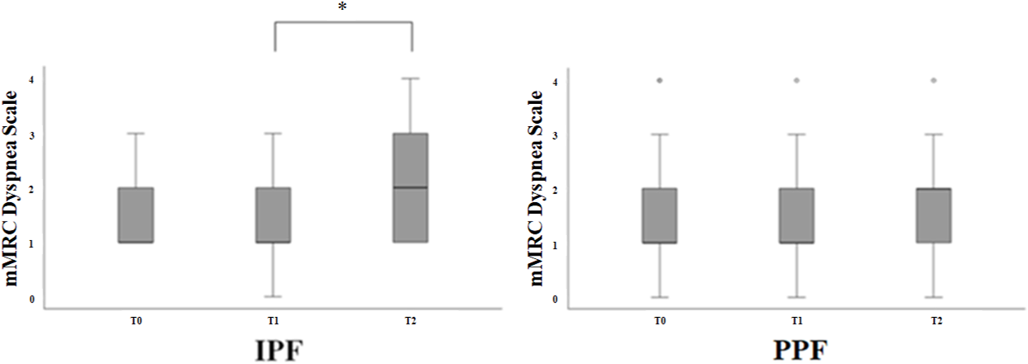
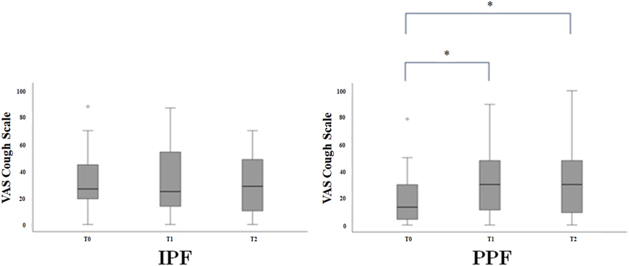
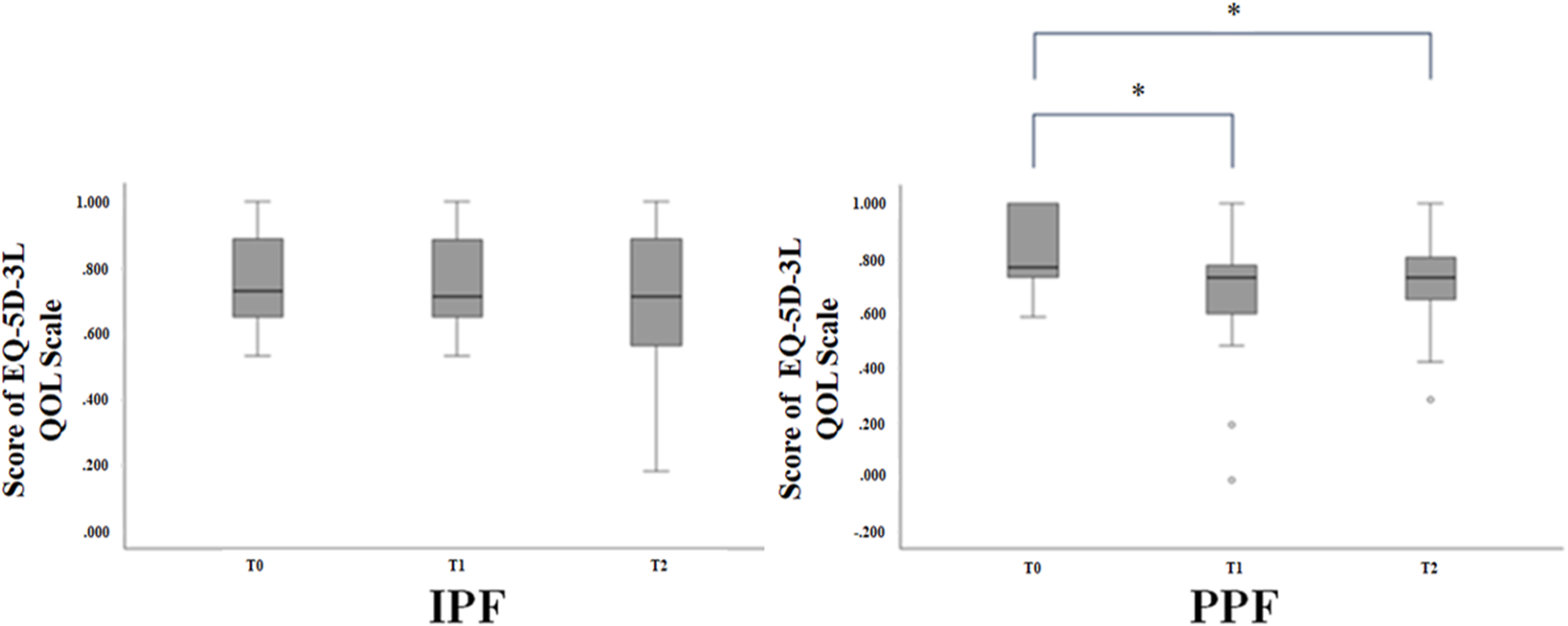
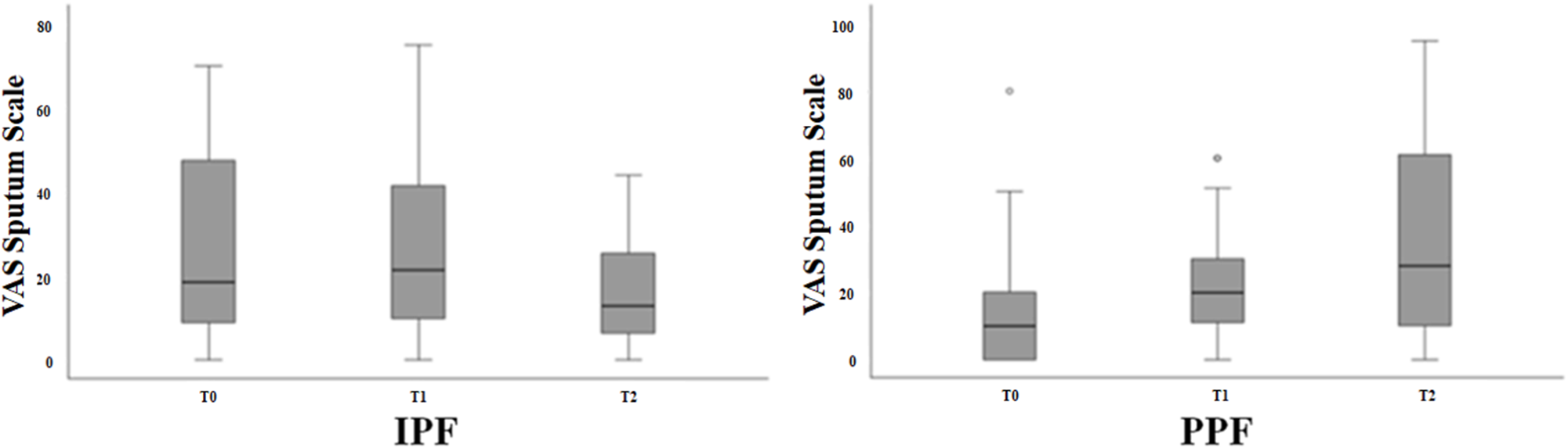
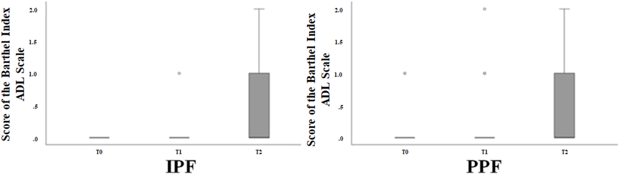
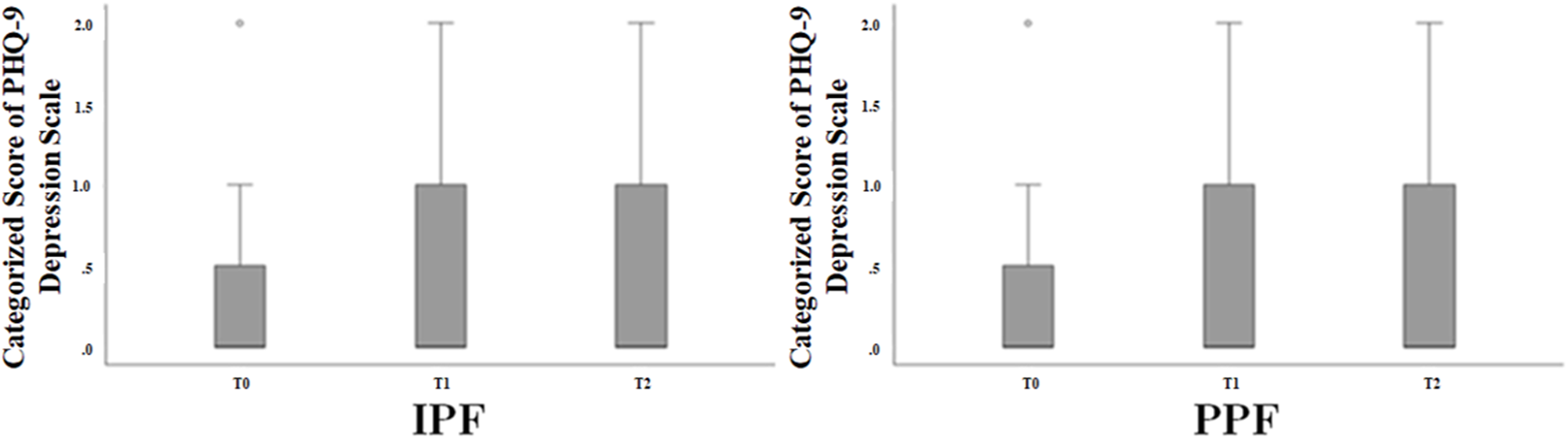
Figure 2 demonstrates that dyspnea assessed by the mMRC scale in patients with IPF worsened from T1 to T2 (p = 0.02) but not from T0 to T1, while the patients in the PPF group showed no worsening of dyspnea from T0 to T1 to T2. Patients with IPF did not show worsening of cough, whereas PPF patients showed worsening of cough over time from T0 to T1 (p = 0.01) and T2 (p = 0.02) (Figure 3). In addition, patients with IPF did not show any changes in QOL over time; however, PPF patients showed a decline in QOL from T0 to T1 (p = 0.03), and this decline was also observed from T1 to T2 (Figure 4). In contrast, both IPF and PPF groups showed no changes in sputum production, ADL, and depression over time (Figures 5–7).
Box and whisker plot analysis of changes in dyspnea in idiopathic pulmonary fibrosis (IPF) (left) and progressive pulmonary fibrosis (PPF) (right) groups. Dyspnea at T0, T1, and T2 was assessed by the modified British Medical Research Council (mMRC) scale. * indicates p values less than 0.01.

Box and whisker plot analysis of changes in cough in idiopathic pulmonary fibrosis (IPF) (left) and progressive pulmonary fibrosis (PPF) (right) groups. Cough at T0, T1, and T2 was assessed by visual analogue scale (VAS). * indicates p values less than 0.01.

Box and whisker plot analysis of changes in quality of life (QOL) in idiopathic pulmonary fibrosis (IPF) (left) and progressive pulmonary fibrosis (PPF) (right) groups. QOL at T0, T1, and T2 was assessed by the EuroQol 5-dimensions 3-levels (EQ-5D-3L). * indicates p values less than 0.01.

Box and whisker plot analysis of changes in sputum in idiopathic pulmonary fibrosis (IPF) (left) and progressive pulmonary fibrosis (PPF) (right) groups. Sputum at T0, T1, and T2 was assessed by visual analogue scale (VAS).

Box and whisker plot analysis of changes in activities of daily living (ADL) in idiopathic pulmonary fibrosis (IPF) (left) and progressive pulmonary fibrosis (PPF) (right) groups. ADL at T0, T1, and T2 were assessed by the Barthel Index.

Box and whisker plot analysis of changes in depression in idiopathic pulmonary fibrosis (IPF) (left) and progressive pulmonary fibrosis (PPF) (right) groups. Depression at T0, T1, and T2 was assessed by the Patient Health Questionnaire-9 (PHQ-9).

Discussion
To clarify the degree of distress in patients with progressive ILD, we examined each factor at the beginning of the study and evaluated the changes in symptoms over the course of 1 year in the IPF and PPF groups. Since IPF has a poor prognosis, tends to show variable respiratory symptoms or distress, and often results in acute exacerbations in daily clinical practice, we decided to evaluate the symptom changes separately in IPF and PPF groups in the present study.
Our study demonstrated that while patients with IPF showed only worsening of dyspnea, those with PPF showed worsening of cough and reduced QOL during the 1-year follow-up period. IPF is the most widely studied and most common ILD with a poor prognosis (Cottin et al. Reference Cottin, Hirani and Hotchkin2018). Although several outcomes reached statistical significance, clinical interpretability requires consideration of MCID. For dyspnea VAS (0–100 mm), the observed change exceeded reported MCID thresholds (≈5 mm in fibrotic ILD; ≈9–10 mm in broader chronic breathlessness cohorts) (Bevanda et al. Reference Bevanda, Mok and Lin2024). For the mMRC scale, MCID in IPF/PPF is not well established; therefore, we reported the proportion with a ≥1-grade change as an additional clinically interpretable summary. This limitation should be considered when interpreting the clinical impact of small mean shifts in this ordinal measure (Aronson et al. Reference Aronson, Danoff and Russell2021). PPF is characterized by respiratory symptoms and disease progression, which result in a decline in ADL and QOL (Swigris et al. Reference Swigris, Kuschner and Jacobs2005). Our study showed worsening of dyspnea in patients with IPF from 6 months to 1 year after recruitment. No notable changes in the administration of steroid-containing medications for respiratory symptoms were noted during the research period; however, half of the patients with IPF had been receiving home oxygen therapy since the start of this study. Despite the lack of clarity regarding the therapeutic usefulness of treatment including steroids, opioids, respiratory rehabilitation, background factors, and individual characteristics, the finding showing worsening of dyspnea over 6 months may be an indicator for follow-up period and symptom management.
In contrast to the findings for the IPF group, PPF patients showed worsening cough and a temporary decline in QOL in our study. Cough is a common symptom of chronic pulmonary diseases, and neither IPF nor PPF is an exception. Chronic coughing increases the disease burden in patients. A previous study found that cough predicts disease progression in patients with IPF (Kondoh et al. Reference Kondoh, Suda and Hongo2022). Although the cause of cough in PPF patients has not yet been understood, underlying inflammation or fibrosis and comorbidities may be involved. Another study showed that the mechanism underlying cough in IPF may involve mechanical distortion associated with lung parenchymal fibrosis (Jones et al. Reference Jones, Hilldrup and Hope-gill2011). The role of some forms of airway inflammation or the physical condition and surrounding environmental factors at the time of the assessment was also undeniable. Although one report on cough in patients with IPF indicated the important roles of increased cough reflex sensitivity, gastroesophageal reflux, and airway inflammation (Jones et al. Reference Jones, Hilldrup and Hope-gill2011), the specific management of cough in patients with PPF has not been reported in previous studies. To the best of our knowledge, general antitussive therapy does not improve the chronic cough caused by PPF. Although a persistent cough is an extremely important factor worth consideration in the palliative care phase, evidence regarding the effectiveness of therapeutic agents, including opioids, is insufficient. It has been demonstrated that opioids are effective for cough in IPF (Wu et al. Reference Wu, Spencer and Banya2024), but their efficacy for PPF has not been demonstrated. Therefore, a comprehensive multidisciplinary approach consisting of treatment and care is required.
QOL in patients with PPF declined 6 months after the start of the study and showed no trend of improvement within the study period. In contrast, no reduction in QOL was observed in the patients with IPF in our study. Reductions in QOL can be attributed to several reasons, but cough may have had an impact in our study. Nevertheless, prolonged coughing eventually leads to a loss of strength and secondary sarcopenia, which may occur in both IPF and PPF patients. Half of the patients with pulmonary sarcoidosis, which is included in PPF, had chronic cough, which is associated with a poor QOL (Sinha et al. Reference Sinha, Lee and Rafferty2016). Other studies have shown that PPF patients with cough had higher odds of lower cough-specific QOL than patients with IPF (Lee et al. Reference Lee, White and Freiheit2022). Our study suggests that this trend may be applicable to PPF patients over a relatively short period.
The results indicated that the trajectory of PPF may be like that of cancer, which deteriorates in a relatively short period of time during the final stages of the disease (Lynn Reference Lynn2001). Although a previous study investigated QOL in patients with PPF (Swigris et al. Reference Swigris, Kuschner and Jacobs2005), none of the existing studies have reported significant reductions. The decline in QOL for each disease that constituted PPF could not be examined individually in the present study; however, some of the PPF patients who entered the study were diagnosed with nonspecific interstitial pneumonia or CTD-ILD. Thus, while the medical condition of the primary disease may have had an impact, other factors could not be identified in our study.
Symptoms of sputum, ADL, and depression did not differ over time in IPF and PPF patients. One reason for this is that patients with IPF as well as the PPF patients, such as those with IIPs and sarcoidosis, experienced a persistent dry cough with low sputum volume (Vigeland et al. Reference Vigeland, Hughes and Horton2017; Raghu et al. Reference Raghu, Remy-jardin and Richeldi2022), making it difficult to be aware of changes in sputum symptoms over time. Another reason is that patients with IPF tended to have lower ADL at the time of registration, whereas the PPF patients often had a background of cancer. IPF is often associated with lung cancer, and no prospective studies have reported significantly more lung cancer complications in patients with PPF than in those with IPF. Although PPF may also be strongly associated with smoking and exposure to carcinogens, like in IPF (Naccache et al. Reference Naccache, Gibiot and Monnet2018), we could not determine why the PPF group had more cancers.
Although some patients were receiving home respiratory rehabilitation as part of their nonpharmacological treatment, we could not exclude the possibility that the addition of respiratory care may have already influenced the findings at the beginning of this study or the absence of symptom changes over the course of 1 year.
This study has several limitations. First, the sample size was smaller than the preliminary estimate. The number of registered patients did not increase, contrary to our expectations, owing to a decrease in the number of outpatients and difficulties in coming to the hospital because of the explosive spread of COVID-19. A large number of patients were unable to undergo pulmonary function testing at T1 and T2 in this study, making sufficient data analysis difficult: 24 patients (including 5 deaths, 4 withdrawals of consent due to COVID-19, 13 with severe physical conditions, and 2 transitioning to home-visit medical care) were at T1, and 17 patients (including 5 deaths, 3 withdrawals of consent due to COVID-19, 6 with severe physical conditions, and 3 transitioning to home-visit medical care) were at T2. Since pulmonary functional decline is one of the defining criteria for PPF and an objective outcome measure, the absence of these data was considered a significant limitation. The modest sample size and substantial attrition by T2 (including deterioration and death) limited statistical power for between-group comparisons and may have introduced selection bias, as complete-case analyses rely on strong assumptions about the missing-data mechanism. Therefore, nonsignificant between-group differences may reflect limited power (type II error), and estimates should be interpreted with emphasis on effect sizes and confidence intervals. Second, patients with missing values who dropped out of the study were also excluded. Since the hospital with the largest number of enrolled cases was a medical institution specializing in the elderly, comorbidities such as cognitive decline or household-related factors may have affected the data collection. Furthermore, since the study included patients with IPF and PPF, some participants dropped out during the 1-year follow-up because they died or were unable to visit the hospital due to disease progression. In some cases, even though the disease had progressed and an antifibrotic drug was indicated, patients did not use expensive antifibrotic diseases due to complications or financial reasons. Third, to ensure that the findings were more aligned with real-world conditions, we did not set strict inclusion criteria. Although almost all outpatients were enrolled, some may have had advanced diseases and may have already undergone home oxygen therapy or rehabilitation, potentially causing a selection bias. In addition, PPF patients with more background cancer complications compared to IPF patients may have also caused selection bias. Therefore, the changes in symptoms during the follow-up period may not have been reflected in the data. Finally, we assessed dyspnea and QOL using the mMRC and EQ-5D, respectively. Considering the researchers’ workload, we used an evaluation scale that is commonly used in daily clinical practice. However, the EQ-5D-3L used to assess QOL in this study may have had some inherent limitations. It has been suggested that the EQ-5D-5L – which was not used in this study – not only improves sensitivity in detecting deviations from perfect health compared to the EQ-5D-3L due to its more detailed descriptive framework but also enhances specificity in accurately characterizing health states that are close to perfect health but slightly impaired (Thompson and Turner Reference Thompson and Turner2020). On the other hand, when using the EQ-5D-3L to evaluate severe diseases where a floor effect is a concern, clinical changes might have been captured in greater detail by using disease-specific QOL measures in conjunction with it – for example, the St. George’s Respiratory Questionnaire for respiratory diseases. Furthermore, the use of another health-related QOL measure capable of covering a lower score range, such as the Short Form 6 Dimensions, should have been considered.
Conclusions
IPF and PPF are characterized by progressive respiratory symptoms and are highly distressing to patients; no sufficiently effective therapeutic strategies have been established to date. Respiratory symptoms are directly related to the patient’s daily life and lead to reduced QOL. Although a variety of treatments and care are being provided in clinical settings, evidence supporting the suitability of these modalities for individual patients may be unavailable. This study, in which several symptoms were assessed over a short period, highlighted the significance of identifying and assessing the patient’s distress as early as possible. Additional research is required to understand how IPF and PPF affect the lives of patients over time.
Acknowledgments
The authors gratefully acknowledge the support of the study participants, their families, medical staff, and physicians. We also would like to thank Editage (https://www.editage.jp) for English language editing.
Author contributions
Conceived and designed the experiments: T.U., K.S., K.T. Performed the experiments: T.U., K.S., K.M., S.H., M.F., H.I. Analyzed the data: T.U., K.S., K.M., S.H., M.F., H.I., K.T. Wrote the paper: T.U., K.S., K.M., S.H., M.F., H.I., K.T.
Funding
The authors declare no funding.
Competing interests
The authors declare that they have no competing interests.
Ethical approval
This study was approved by the Ethics Committee of the Juntendo Tokyo Koto Geriatric Medical Center, Tokyo, Japan, and was conducted in accordance with the Declaration of Helsinki.



 Open access

