Introduction
The family Rhabditidae Örley (Reference Örley1880) represents one of the most diverse group of free-living nematodes, found in a wide range of habitats including decaying organic matter rich in bacteria, soil, and freshwater, as well as in association with live creatures (particularly insects) underscoring their ecological significance (Andrássy Reference Andrássy1983). The systematics, phylogeny, ecology, and biology of Rhabditidae genera have been reviewed by several authors (e.g., Sudhaus Reference Sudhaus2023; Sudhaus and Fitch Reference Sudhaus and Fitch2001; Sudhaus and Schulte Reference Sudhaus and Schulte1989; Scholze and Sudhaus Reference Scholze and Sudhaus2011). Scholze and Sudhaus (Reference Scholze and Sudhaus2011) listed 38 genera for the family. Later, two members were also added (Sudhaus Reference Sudhaus2023).
Oscheius was established by Andrássy (Reference Andrássy1976) with the type species O. insectivorus (Körner, Reference Körner1954) Andrássy, Reference Andrássy1976, as the monotypic genus. It currently includes 39 species (Sudhaus Reference Sudhaus2023). The species of the genus are morphologically divided into two intragenus groups: the Dolichura-group, comprising 16 species, and the Insectivorus-group, comprising 23 species (Abolafia and Peña-Santiago Reference Abolafia and Peña-Santiago2019; Sudhaus Reference Sudhaus2023). The Dolichura-group is characterized by a peloderan bursa and thin tubular spicules tips, while the Insectivorus-group is distinguished by a leptoderan or pseudopeloderan bursa and crochet needle-shaped spicules. Some Oscheius spp. have been regarded as facultative entomopathogens of different insects (Dillman et al. Reference Dillman, Chaston, Adams, Ciche, Goodrich-Blair, Stock and Sternberg2012; Torres-Barragan et al. Reference Torres-Barragan, Suazo, Buhler and Cardoza2011; Zhang et al. Reference Zhang, Liu, Tan, Wang, Qiao, Yedid, Dai, Qiu, Yan, Tan, Su, Lai and Gao2012), a biological feature mainly related to their association with Serratia spp. (Nguyen and Hunt Reference Nguyen and Hunt2007; Torres-Barragan et al. Reference Torres-Barragan, Suazo, Buhler and Cardoza2011; Zhang et al. 2009). In recent years, the pathogenicity of several Oscheius species has been investigated on different insects including O. carolinensis Ye, Torres-Barragan & Cardoza, Reference Ye, Torres-Barragan and Cardoza2010; O. cyrus Kuhestani, Karimi, Shokoohi & Makhdoumi 2023; O. myriophilus Poinar, 1986; O. niazii Tabassum & Shahina, Reference Tabassum and Shahina2010; and O. siddiqii Tabassum & Shahina, Reference Tabassum and Shahina2010 from the Insectivorus-group, and O. onirici Torrini, Mazza, Carletti, Benvenuti, Roversi, Fanelli, De Luca, Troccoli & Tarasco Reference Torrini, Mazza, Carletti, Benvenuti, Roversi, Fanelli, De Luca, Troccoli and Tarasco2015, from the Dolichura-group (Castro-Ortega et al. Reference del Rocio Castro-Ortega, Caspeta-Mandujano, Suárez-Rodríguez, Peña-Chora, Ramírez-Trujillo, Cruz-Pérez, Sosa and Hernández-Velázquez2020; Karimi et al. Reference Karimi, Yoshiga and Habibi2011; Tabassum and Shahina Reference Tabassum and Shahina2010; Alghanimi et al., Reference Alghanimi, Talebi, Gholami Ghavamabad and Majid2025; Torrini et al. Reference Torrini, Mazza, Carletti, Benvenuti, Roversi, Fanelli, De Luca, Troccoli and Tarasco2015; Ye et al. Reference Ye, Torres-Barragan and Cardoza2010; Zhou et al. Reference Zhou, Yang, Wang, Bao, Wang, Hou, Lin, Yedid and Zhang2017).
Morphological and morphometric data are currently available only for the type population of Oscheius insectivorus.
In Iran, research on the identification, ecology, and biology of Oscheius species has been limited. Formerly O. tipulae Lam & Webster, Reference Lam and Webster1971 was reported from Iran (Hassani-Kakhki et al. Reference Hassani-Kakhki, Karimi and Hosseini2013). O. rugaoensis (Zhang et al. Reference Zhang, Liu, Tan, Wang, Qiao, Yedid, Dai, Qiu, Yan, Tan, Su, Lai and Gao2012) Darsouie, Karimi & Shokoohi, Reference Darsouei, Karimi and Shokoohi2014 was isolated and reported from white grub larvae (Polyphylla adspersa Motschulsky, 1853) (Coleoptera: Scarabaeidae) (Darsouei et al. Reference Darsouei, Karimi and Shokoohi2014). Later, three species, O. necromenus Sudhaus & Schulte, Reference Sudhaus and Schulte1989; O. onirici; and O. tipulae Lam & Webster Reference Lam and Webster1971, were isolated and reported in association with bark beetle galleries in city of Tehran (Valizadeh et al. Reference Valizadeh, Goldasteh, Rafiei-Karahroodi and Pedram2017). Characterization of the Iranian isolates of O. myriophilus and O. rugaoensis, recovered from northern Iran, and their pathogenicity as biocontrol agents against the larvae of two invasive forest pests under laboratory conditions, was recently published (Gholami Ghavamabad et al. Reference Gholami Ghavamabad, Talebi, Mehrabadi, Farashiani and Pedram2021, Reference Gholami Ghavamabad, Talebi, Pedram, Farashiani and Mehrabadi2024). Oscheius cyrus (belonging to the Insectivorus-group) is the last species described from Iran.
The poplar leaf beetle, Chrysomela populi L. (Col.: Chrysomelidae), is a significant pest of poplar trees, impacting nurseries and plantations by attacking the leaves (Urban Reference Urban2006). It dominates Palaearctic regions and causes severe economic losses in European and Asian countries including Turkey (Aslan and Özbeck Reference Aslan and Özbek1999), Iran (Damanabi et al. Reference Damanabi, Kazemi and Akrami1977), and India (Thakur Reference Thakur1999). Adult beetles overwinter in various shelters such as leaf litter (Zareh et al. Reference Zareh, Ahmadi and Alishah1984), grass tufts (Loi Reference Loi1970), or soil (Damanabi et al. Reference Damanabi, Kazemi and Akrami1977). Upon reaching their host, they begin feeding immediately, causing severe damage to tree buds at the late unfolding of leaves. The high population of the pest prevents the development of planted cuttings. Females deposit their eggs on the underside of apical leaves. First instar larvae are gregarious and feed on leaves, while second- and third instar larvae feed individually. The insects most often pupate on the underside of the leaves. Under favorable conditions, two or sometimes three overlapping generations can develop annually (Urban Reference Urban2006).
Various control strategies have been studied on this pest including employing predators (Teodorescu Reference Teodorescu1980), parasitoids (Lotfalizadeh and Ahmadi Reference Lotfalizadeh and Ahmadi1998), neogregarine pathogen (Ophryocystis anatoliensis (Apicomplexa) (Yaman Reference Yaman2017), pathogens (Yaman et al. Reference Yaman, Yener and Celik2016), chemical controls (De Tillesse et al. Reference De Tillesse, Nef, Charles, Hopkin and Augustin2007), and breeding resistant poplar varieties (Tahriri Adabi et al. Reference Tahriri Adabi, Sadeghi and Bagheri2012).
Entomopathogenic nematodes often infect coleopterans (Poinar Reference Poinar, Jolivet, Petitpierre and Hsiao1988; Toguebaye et al. Reference Toguebaye, Marchand and Bouix1988; Yaman et al. Reference Yaman, Özcan, Radek, Linde and Lipa2011), serving as environmentally friendly biocontrol agents. In recent years, entomopathogenic nematodes (EPNs) have been commercially produced (Shapiro-Ilan et al. Reference Shapiro-Ilan, Hiltpold, Lewis, Hajek and Shapiro-Ilan2017), primarily targeting soil-dwelling insects (Wright et al. Reference Wright, Peters, Schroer and Fife2005). Studies have also explored their efficacy in foliar applications (Brusselman et al. Reference Brusselman, Beck, Pollet, Temmerman, Spanoghe, Moens and Nuyttens2012; Gholami Ghavamabad et al. Reference Gholami Ghavamabad, Talebi, Pedram, Farashiani and Mehrabadi2024; Schroer and Ehlers Reference Schroer and Ehlers2005). EPNs have been used against Chrysomelidae pests on various plant hosts, such as the Colorado potato beetle, Leptinotarsa decemlineata (Say) (Trdan et al. Reference Trdan, Vidrih, Andjus and Laznik2009); the cereal leaf beetle, Oulema melanopus (L.) (Laznik et al. Reference Laznik, Tóth, Lakatos, Vidrih and Trdan2010); the alder leaf beetle, Agelastica alni (L.) (Bayramoglu et al. Reference Bayramoglu, Demir, Inan and Demirbag2018); and the cabbage stem flea beetle, Psylliodes chrysocephala (L.) (Godina et al. Reference Godina, Vandenbossche, Schmidt, Sender, Tambe, Touceda-González and Ehlers2023).
Some species of EPNs are widely distributed over the world; however, using native EPNs is often preferred for their higher efficacy and lower environmental risk (Campos-Herrera Reference Campos-Herrera2015; Matuska-Łyżwa et al. Reference Matuska-Łyżwa, Duda, Nowak and Kaca2024; Shapiro-Ilan and Dolinski Reference Shapiro-Ilan, Dolinski and Campos-Herrera2015; Sun et al. Reference Sun, Zhang, Song, Zheng, Wei, Gu, Cui, Hu, Yoshiga, Abd-Elgawad and Ruan2021).
In the study of Dillman et al. (Reference Dillman, Chaston, Adams, Ciche, Goodrich-Blair, Stock and Sternberg2012), some criteria were proposed so that an isolate of nematode could be regarded as an EPN, and two Oscheius spp. were approved as EPNs.
During our samplings in the forests of northern Iran, a population of the genus Oscheius was recovered from inside the body of a live scarab beetle collected in natural forests of Golestan province, which was identified as O. insectivorus. This study aims to characterize the Iranian isolate of this species using morphological, morphometric, and molecular criteria, and to assess its potential as a biocontrol agent against the larvae of two pests, Chrysomela populi and Galleria mellonella L., under laboratory conditions.
Materials and methods
Nematode isolation, culture, and morphological observation
Several specimens of Oscheius insectivorus (females, males, juveniles) were recovered from the inside body of a live scarab beetle collected from natural forests of Jafarabad village, Golestan province, northern Iran. The insect was rinsed with autoclaved distilled water and dissected, and the body pieces were placed on water agar in a 6-cm Petri dish plate. After four days, a number of 10 female and juvenile specimens of the appeared population on cadaver were transferred onto a new water agar plate, including a live larva of Galleria mellonella. In another test, a mature female was separately transferred onto another water agar plate, and all plates (the original plate including the cadaver, the plate including 10 females and juveniles and the plate including one female specimen) were stored at 27–28°C.
For morphological observations, the nematode specimens were heat-killed by adding boiling 4% formalin solution, transferred to anhydrous glycerin (De Grisse Reference De Grisse1969), mounted on permanent slides, and examined using a Nikon Eclipse E600 light microscope. Photographs were taken using an Olympus DP72 digital camera attached to an Olympus BX51 microscope powered with differential interference contrast (DIC).
Infective juveniles isolation for pathogenicity tests
Nematodes were cultured on G. mellonella larvae on water agar plates. Two to five days after inoculation, the appeared IJs were recovered by rinsing the plate using distilled water.
Pathogenicity of Oscheius insectivorus against Galleria mellonella and Chrysomela populi
A colony of Galleria mellonella (wax moth) was obtained from the Conservation and Protection Research Laboratory, Research Institute of Forests and Rangelands of Iran, and cultured on a diet consisting of wheat flour, yeast powder, beeswax, glycerine, and honey in a growth chamber (28°C, 60% R.H., 16:8 h L:D) as described by Woodring and Kaya (Reference Woodring and Kaya1988).
Different developmental stages of Chrysomala populi were collected from a poplar plantation at Alborz Research Station of the Research Institute of Forests and Rangelands (RIFR) in Iran, on Populus nigra in May–June 2024. Larvae, pupae, and adults were kept in plastic containers (diameter 20 cm, height 10 cm) in growth chambers (22°C, 70% R.H., 16:8 h L:D). Fresh leaves of P. nigra were provided daily, and old leaves were replaced with fresh ones.
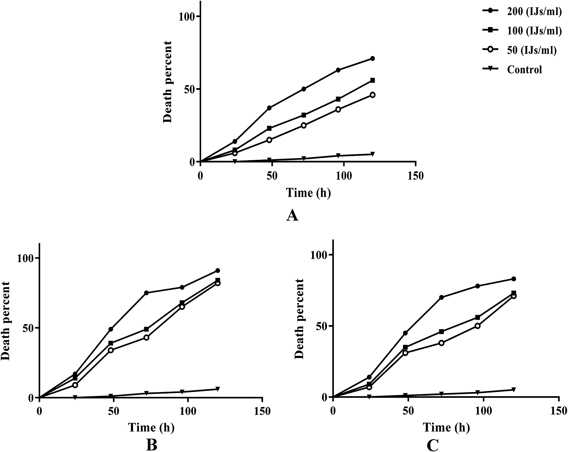
The susceptibility of fifth instar larvae of G. mellonella, and third instar larvae and adults of C. populi to Oscheius insectivorus, was assessed following the method of Kaya and Stock (Reference Kaya and Stock1997). For this, the bottom of each 6-cm Petri dish plate was covered with wet filter paper, and a single larva or adult insect (larvae for G. mellonella, larvae and adults for C. populi) was placed inside the plate. The larvae and adult insects were exposed to O. insectivorus at concentrations of 50, 100, and 200 infective juveniles (IJs) per milliliter of distilled water (IJs/ml) (the concentrations were selected after preliminary tests; data not shown), applied to the filter paper. Control groups received 1 mL of distilled water. The experiment was conducted five times. In each replicate, 20 insects were infected with infective juveniles, with each insect placed in a separate Petri dish. This resulted in a total of 100 insects/Petri dishes. The pooled data of 100 insects/Petri dishes were used to calculate the corresponding graphs. Plates were placed in plastic containers in a dark growth chamber at 25 ± 2°C, 60% R.H., and 16:8 h L:D.
Insect mortality and infection signs, such as changes in larval body color, were recorded daily for four consecutive days. Dead individuals were transferred to white traps to confirm nematode-induced mortality and to see nematodes appearing on the cadaver. Mortality curves (logrank Mantel–Cox test) from laboratory experiments were analyzed using the Kaplan–Meier method with GraphPad Prism (version 6.07, GraphPad Software, Inc.). LC50 and LC90 values of EPNs were estimated by probit analysis using SPSS Statistics v22.
Molecular methods and phylogenetic analyses
A single nematode specimen of the propagated population on either of the aforementioned plates (on moth cadaver or pure culture of a single female) was picked out and transferred to a small drop of TE buffer (10 mM Tris-Cl, 0.5 mM EDTA; pH 9.0, Qiagen) on a clean slide, studied under light microscope, and squashed using a clean coverslip. The suspension was collected by adding 50 μl TE buffer. DNA samples were stored at −20°C until used as PCR templates. Primers for partial amplification of SSU rDNA were forward primer 988F (5´–CTCAAAGATTAAGCCATGC–3´) and reverse primer 1912R (5´–TTTACGGTCAGAACTAGGG–3´), forward primer 1813F (5´–CTGCGCGAGAGGTGAAAT–3´) and reverse primer 646R (5´–ACCTACTGATACCTTGTTACGACTTTT–3´) (Holterman et al. Reference Holterman, Van Der Wurff, Den Elsen, Van Megen, Bongers, Holovachov, Bakker and Helder2006). The LSU rDNA D2-D3 expansion segment was amplified using the forward primer D2Ab (5´–ACAAGTACCGTGAGGGAAAGTTG–3´) and reverse primer D3B (5´–TCGGAAGGAACCAGCTACTA–3´) (De Ley et al. Reference De Ley, Félix, Frisse, Nadler, Sternberg and Thomas1999). The PCR products were sequenced in both directions using the same primers used in PCR with an ABI3730XL sequencer. The newly obtained sequences were deposited into the GenBank database under the accession numbers PQ061487 for SSU; and PQ061488, PQ061489, and PQ061490 for LSU rDNA D2-D3 sequences.
The newly obtained sequences were compared with those of other nematode species available in GenBank database using the BLAST homology search program. For reconstruction of the phylogenetic relationships, two independent SSU and LSU datasets were prepared. The selected DNA sequences were aligned using Clustal X2 (Larkin et al. Reference Larkin, Blackshields, Brown, Chenna, Mcgettigan, McWilliam, Valentin, Wallace, Wilm, Lopez, Thompson, Gibson and Higgins2007) for both SSU and LSU fragments. The poorly aligned positions and divergent regions of SSU and LSU datasets were eliminated manually using MEGA 7.0 (Kumar et al. Reference Kumar, Stecher and Tamura2016) software. The model of base substitution was selected using MrModeltest.2 (Nylander Reference Nylander2004). The Bayesian analyses were performed using MrBayes v3.1.2 (Ronquist and Huelsenbeck Reference Ronquist and Huelsenbeck2003) running the chains for 5,000,000 generations for SSU and 4,000,000 for LSU datasets. After discarding burn-in samples, the remaining samples were retained for further analyses. The Markov chain Monte Carlo (MCMC) method within a Bayesian framework was used to estimate the posterior probabilities of the phylogenetic trees using the 50% consensus majority rule (Larget and Simon Reference Larget and Simon1999). Adequacy of the posterior sample size was evaluated using autocorrelation statistics as implemented in Tracer v1.6 (Rambaut and Drummond Reference Rambaut and Drummond2009). The SSU sequence of the species Myolaimus byersi Giblin-Davis, Kanzaki, De Ley, Williams, Schierenberg, Ragsdale, Zeng & Center, Reference Giblin-Davis, Kanzaki, De Ley, Williams, Schierenberg, Ragsdale, Zeng and Center2010 was used as outgroup in SSU, and sequences of species Poikilolaimus oxycerca De Man, Reference De Man1895 and Buetschlinema nidrosiensis (Allgén Reference Allgén1933) Sudhaus, Reference Sudhaus2011 were used as outgroups in LSU phylogenies. The output files of the phylogenetic program were visualized using Dendroscope V3.2.8 (Huson and Scornavacca Reference Huson and Scornavacca2012) and were digitally drawn in the CorelDRAW® software version 20.
Results
Culturing of the recovered population
The three plates including the culture of nematodes on body cadaver of the dissected insect, the plate including one specimen on water agar, and the plate including the inoculated G. mellonella larvae with ten nematodes were studied separately. The plate including the cadaver of the scarabid beetle host yielded a high population of nematodes (females, males, juveniles) after four days. The inoculated G. mellonella larvae was killed after three days, and the IJs appeared four days after larvae death. Females and males also appeared concomitantly. A mixed population of different stages of nematode appeared in the plate, including the single specimen of female nematode on water agar after 20 days.
Description
Iranian isolate of Oscheius insectivorus
(Figure 1)
Photomicrographs of Iranian isolate of Oscheius insectivorus (Körner, Reference Körner1954) Andrássy, Reference Andrássy1976; A: Female anterior body region; B: Bacteria in anterior part of intestine; C: Female tail; D: Infective juveniles anterior body end; E: Infective juveniles tail; F: Ventral view of male bursa and genital papillae; G: Lateral view of posterior end of male; H: Spicules in lateral view.

Measurements, Table 1
Morphometrics of Iranian isolate of Oscheius insectivorus (Körner, Reference Körner1954) Andrássy, Reference Andrássy1976 recovered from Golestan province, north Iran, and comparison with data of the type population

Female
Medium to large size nematodes. Body fusiform, slightly ventrally curved after fixation. Cuticle appears smooth under light microscope, bearing fine transverse striae visible at the entire body, about 1 μm thick at almost entire body. Lateral field with six lines. Deirids in secretory-excretory (S-E) pore level. S-E pore in 83.7 to 95.1% of neck length from the anterior end. Phasmids at about the middle of tail. Labial region continuous with body contour, with a slight depression, separating it from adjacent body. Six separate lips with sensilla in labial plate. Stoma rhabditoid, tubular, not highly sclerotized, 1.0–1.2 times the lip region width long, or 2.8 to 4.2 times longer than broad. Pharynx rhabditoid, procorpus and metacorpus (=corpus) merged, subcylindrical, 2.7 to 3.5 times longer than isthmus, muscular, slightly swollen posteriorly, isthmus narrower than metacorpus, basal bulb rounded with developed valve. Cardia distinct, cordiform to rounded. Nerve ring at about first part of the isthmus or 73.3 to 81.0 % of neck length from the anterior end. S-E pore slightly posterior to nerve ring. Reproductive system didelphic-amphidelphic, each gonad reflexed at tip, including mature eggs, vulva slightly swollen, at 46.9–57.4% of body. In old females, hatched juveniles fill inside the body. Intestine simple, rectum and anus functional, rectum 1.1 to 2.0 times of anal body width. Tail elongate conical with sharp tip.
Male
Generally similar to females, slightly shorter. Reproductive system rhabditoid, monodelphic, spicules long, testis reflexed ventrally at tip. Tail with conoid anterior part that is flanked by the bursa, and short and thin posterior filiform section that is located outside of the bursa. Bursa leptoderan, nine pairs of genital papillae (1 + 1 + 1/3 + 3 + ph): three pairs pre-cloacal (GP1–GP3) and six pairs post-cloacal, with three pairs at anterior part of tail length (GP4–GP6) and three pairs (GP7–GP9) at the terminal length; GP1, GP2, and GP3 are placed apart; space between GP1 and GP2 greater than the space between GP2 and GP3. Spicules paired, symmetrical, separate, slightly curved ventrally and showing hook-like tip. Gubernaculum sclerotized, slightly curved. Tail conoid, ending to a spike-like differentiation.
Infective juveniles
Straight when heat-killed, fusiform. The cuticular sheath conspicuous. Lip region rounded. Stoma tubular, about 6.8 to 8.3 times longer than wide (excluding the sheath at the apex). Reproductive system lacking. Tail elongate conical, ending with a sharp tip.
Remark
The Iranian isolate of O. insectivorus was recovered from inside body of an unidentified scarabid beetle. It has a shorter body (1103–1942 vs. 1979–3205 μm), lower body width (65–102 vs. 125–169 μm), and lower b value (5.0–8.3 vs. 8.8–13.5) in females, and shorter males (890–1145 vs. 1587–3252 μm) with less body width (43.5–56.8 vs. 76–159 μm), lower b value (4.5–5.7 vs. 7.3–13.4), shorter spicules (60.0–74.5 vs. 85–118 μm), and shorter gubernaculum (27.3–32.3 vs. 38–54 μm) compared to the type population. The IJs of Iranian isolate were, however, longer (790–985 vs. 666–725 μm) and had greater body width (35.7–48.9 vs. 30–35 μm) compared to the type population (Körner Reference Körner1954).
Molecular phylogenetic analyses
Partial sequencings of SSU and LSU rDNA D2-D3 fragments of Iranian isolate yielded one 1205 nucleotide long SSU, and three 484, 564, and 565 nucleotide long LSU D2-D3 sequences. The BLAST search using the newly generated SSU sequence (PQ061487) revealed it has a 100% identity with the SSU sequence of Oscheius insectivorus deposited into the GenBank database under the accession number AF083019. The blast searching using the longest fragment of newly generated sequence LSU D2-D3 (PQ061488) revealed it has 100% identity with two sequences of the same locus of Oscheius insectivorus deposited into the Genbank database under the accession numbers PP087421 and EU195968.
Figure 2 shows the SSU phylogenetic tree reconstructed using 48 ingroup and outgroup sequences. In this tree, two intragenus groups of this genus (i.e., the Insectivorus- and Dolichura-groups) each formed independent clades, in sister relation to each other with a high (0.95) Bayesian posterior probability (BPP). The newly generated SSU sequence of Iranian isolate formed a clade with the sequence AF083019 assigned to an American isolate of O. insectivorus.
Bayesian 50% majority rule consensus tree of Iranian isolate of Oscheius insectivorus (Körner, Reference Körner1954) Andrássy, Reference Andrássy1976 Based on SSU sequences under GTR + I + G model. Bayesian posterior probability values more than 0.50 are given for appropriate clades. The new sequence is indicated in bold.

Figure 3 represents the Bayesian phylogenetic tree inferred using 45 ingroup and outgroup LSU sequences. In this tree, the included sequences formed two separate Insectivorus- and Dolichura clades, in maximally supported relation to each other. The three sequences of the Iranian isolate and the sequence EU195968 assigned to an American isolate of O. insectivorus occupied the same clade inside Insectivorus clade.
Bayesian 50% majority rule consensus tree of Iranian isolate of Oscheius insectivorus (Körner, Reference Körner1954) Andrássy, Reference Andrássy1976 based on LSU rDNA D2-D3 sequences under GTR + I + G model. Bayesian posterior probability values more than 0.50 are given for appropriate clades. The new sequences are indicated in bold.

Pathogenicity of Iranian isolate of O. insectivorus on Galleria mellonella larvae; and adults and larvae of Chrysomela populi
Application of Oscheius insectivorus against G. mellonella and C. populi under laboratory conditions resulted in the significant mortality of these pests compared to the control (Figure 4). The mortality rates increased by increasing the exposure time and IJs concentration. In the bioassay against fifth instar larvae of G. mellonella, O. insectivorus caused 50% mortality at the dosage of 200 IJs/ml within 72 h post-exposure. At lower nematode concentrations of 100 IJs/ml and 50 IJs/ml, mortality rates were 32% and 25%, respectively, at the same time point (Log-rank (Mantel–Cox) test, χ2=95.78, df=3, P<0.0001). Third instar larvae of Chrysomela populi exhibited a mortality rate of 75%, 49%, and 43% at concentrations of 200 IJs/ml, 100 IJs/ml, and 50 IJs/ml, respectively, 72 hours post-exposure (Log-rank (Mantel–Cox) test, χ2=177.9, df=3, P<0.0001). Mortality rates for adults of C. populi were 70%, 46%, and 38% at the dosage of 200 IJs/ml, 100 IJs/ml, and 50 IJs/ml, respectively, over the same time points (Log-rank (Mantel–Cox) test, χ2=141.9, df=3, P<0.0001). Table 2 shows the LC50 and LC90 values at 72 hours post-exposure with the nematode. The lowest LC50 value (77.71) was observed for the third instar larvae of C. populi, whereas the highest value (217.04) was for fifth instar larvae of G. mellonella.
Mortality percent of Galleria mellonella L. larvae and Chrysomela populi L. larvae and adults after exposure to different doses of Oscheius insectivorus in 24–120 h post-exposure in laboratory bioassays. (A) Fifth instar larvae of Galleria mellonella exposed to nematode; (B) Third instar larvae of Chrysomela populi exposed to nematode; (C) Adults of Chrysomela populi exposed to the nematode. Log-rank test (Mantel–Cox) on GraphPad Prism software was performed to determine statistical significance for the death curves, All P values less than 0.0001 are summarized with four asterisks.

LC50 and LC90 values of Iranian isolate of Oscheius insectivorus (Körner, Reference Körner1954) Andrássy, Reference Andrássy1976 on the Galleria mellonella and Chrysomela populi at 72 h post-treatment

Discussion
During the present study, a population of O. insectivorus was recovered from inside body of a scarab beetle. The population was reared on the cadaver of the host insect on water agar plate, water agar plate only, and Galleria mellonella. In previous studies, similar cultures have established on wax moth as a suitable host (Gholami Ghavamabad et al. Reference Gholami Ghavamabad, Talebi, Mehrabadi, Farashiani and Pedram2021; Karimi et al. Reference Karimi, Rezaei and Shokoohi2018; Kuhestani et al. Reference Kuhestani, Karimi, Shokoohi and Makhdoumi2022; Lephoto and Gray Reference Lephoto and Gray2019).
The body length of females of the recovered populations from three cultures ranged from 1.1 to 2.6 mm (two female specimens with body length 2.2 and 2.6 mm were observed in one of the cultures. These specimens were used for DNA extraction). The short- and long-body specimens were separately sequenced for their LSU D-D3, resulting in identical sequences, approving the cultures were pure. As already presented (see Remarks), Iranian isolates of O. insectivorus (cultured using three different methods) have smaller body size in comparison with the type population. Currently, no data are available for other populations of the species (i.e., those populations that have sequenced for their ribosomal markers and are available at GenBank). The variation of size in this group of nematodes was already recorded for other species of the genus (e.g., in O. myriophilus (Demir et al. Reference Demir, Demirbağ and Erbaş2017; Gholami Ghavamabad et al. Reference Gholami Ghavamabad, Talebi, Mehrabadi, Farashiani and Pedram2021) and O. tipulae (Karimi et al. Reference Karimi, Rezaei and Shokoohi2018; Sultana et al. Reference Sultana, Waghmare and Pankaj2022)). In line with previous phylogenies (Abolafia and Peña-Santiago Reference Abolafia and Peña-Santiago2019; Gholami Ghavamabad et al. Reference Gholami Ghavamabad, Talebi, Mehrabadi, Farashiani and Pedram2021), the presently resolved SSU and LSU trees confirmed the genus is monophyletic and two groups/clades of genus, the Insectivorus, and Dolichura-group, form two clades of the genus.
The Iranian isolate of O. insectivorus caused mortality on both Galleria mellonella larvae and Chrysomela populi larvae and adults under laboratory conditions. Previous studies have also highlighted the biocontrolling potential of Oscheius species like O. rugaoensis and O. onirici on G. mellonella. Oscheius rugaoensis caused a larval mortality rate of 95% at a concentration of 284 IJs/ml (Zhang et al. Reference Zhang, Liu, Tan, Wang, Qiao, Yedid, Dai, Qiu, Yan, Tan, Su, Lai and Gao2012; Zhou et al. Reference Zhou, Yang, Wang, Bao, Wang, Hou, Lin, Yedid and Zhang2017), while O. onirici killed 46% of G. mellonella larvae within a week (Torrini et al. Reference Torrini, Mazza, Carletti, Benvenuti, Roversi, Fanelli, De Luca, Troccoli and Tarasco2015). In contrast, a 50% mortality rate of fifth instar larvae of G. mellonella was observed at 200 IJs/ml concentration after 72 hours in the present study. Additionally, it was revealed that O. insectivorus has a high virulence against C. populi larvae compared to its adults and G. mellonella larvae. Specifically, the 200 IJs/ml dosage resulted in 17% to 91% mortality rates on third-instar larvae of C. populi within 24–120 hours post-exposure. The isolation of O. insectivorus from scarabeid beetle and its remarkable effectiveness against C. populi may indicate its potential suitability for targeting soil-dwelling coleopteran insects.
New data on pathogenicity and effectiveness of EPNs against Chrysomela populi were presented in this study. Formerly, the efficacy of biocontrolling fungi and bacteria has been investigated for controlling this pest. Beauveria bassiana has previously been studied for its effectiveness against this pest, and significant mortality in adult beetles was achieved when applied in the field or in the laboratory (Gindin et al. Reference Gindin, Levski, Glazer and Soroker2006). Metarhizium anisopliae, another fungus, reduced the population of the pest (Zimmermann Reference Zimmermann2007). Bacillus thuringiensis (Bt), is another biological agent that showed efficacy against this pest (Marques et al. Reference Marques, Santos, Castro, Moscardini, Rosseto, Silva and Babcock2019).
Oscheius myriophilus was previously recovered in association with the third instar larvae of Popillia japonica Newman (Coleoptera Scarabaeidae) in north Italy, and the cultured population showed a maximum mortality rate of 54% in Galleria mellonella larvae. (Goda et al. Reference Goda, Mirzaei and Brunetti2020). Based on available data, Oscheius spp. have the potential of biocontrolling insect pests and are regarded as either facultative, potential, or approved entomopathogenic nematodes (Dillman et al. Reference Dillman, Chaston, Adams, Ciche, Goodrich-Blair, Stock and Sternberg2012; Torres-Barragan et al. Reference Torres-Barragan, Suazo, Buhler and Cardoza2011; Zhang et al. Reference Zhang, Liu, Tan, Wang, Qiao, Yedid, Dai, Qiu, Yan, Tan, Su, Lai and Gao2012). In the most recent study, the effectiveness of Oscheius myriophilus in controlling of Bactrocera zonata (Saunders, Reference Saunders1842) (Diptera: Tephritidae) is approved (Alghanimi et al., Reference Alghanimi, Talebi, Gholami Ghavamabad and Majid2025).
The free-living nature of Oscheius spp. (i.e., having no obligatory host-dependent phase), especially those species proven by their entomopathogenic ability, allows their persistence in soil and could propose them as promising biocontrol agents.
Financial support
Tarbiat Modares University.
Competing interest
The authors have no competing interests.
Ethical standard
The authors assert that all procedures contributing to this work comply with the ethical standards of the relevant national and institutional guides on the care and use of laboratory animals.