





Significant outcomes
-
• Convergent neuroimaging, biochemical, and metabolomic evidence indicates that bipolar disorder involves systemic bioenergetic dysregulation, linking mitochondrial dysfunction, oxidative stress, and purinergic imbalance to core pathophysiological mechanisms.
-
• Integration of peripheral mitochondrial markers with cerebral metabolic imaging supports a multidimensional model of BD that may enable biologically informed patient stratification and mechanism-based therapeutic targeting.
-
• Emerging bioenergetic and immunometabolic interventions, including mitochondrial cofactors and purinergic modulators, represent a translational avenue towards precision psychiatry grounded in restoration of energetic homeostasis.
Limitations
-
• Substantial heterogeneity in study populations, illness phase, pharmacotherapy exposure, and metabolic comorbidities limits cross-study comparability and may confound interpretation of bioenergetic biomarkers.
-
• Many findings derive from relatively small, single-centre studies lacking standardised protocols for sample collection, imaging acquisition, and biomarker quantification, restricting reproducibility and generalizability.
-
• The predominance of cross-sectional designs constrains causal inference regarding whether bioenergetic alterations represent trait markers, state-dependent phenomena, or consequences of chronic illness progression.
Introduction
Bipolar disorder (BD) affects approximately 1–2% of the global population and is associated with a marked reduction in quality of life, elevated suicide risk, and numerous somatic comorbidities (Wang et al., Reference Wang, Li, Su and Bai2015; van der Wijk et al., Reference van der Wijk, Bélteczki and Döme2024). The aetiology of BD is multifactorial, encompassing genetic, epigenetic, neurochemical, neuroinflammatory, and metabolic components. Mitochondrial dysfunction has emerged as a potentially important mechanism involved in BD pathophysiology (Andreazza, Reference Andreazza2019; Ahmed et al., Reference Ahmed, MahmoudianDehkordi, Bhattacharyya, Arnold, Liu, Neavin, Moseley, Thompson, Williams, Louie, Skime, Wang, Riva-Posse, McDonald, Bobo, Craighead, Krishnan, Weinshilboum, Dunlop, Millington, Rush, Frye and Kaddurah-Daouk2020).
The brain is an energetically costly organ, consuming 20–25% of all biochemical energy produced in the body, despite comprising only 2–3% of total body mass (Glausier, Reference Glausier2025). It relies on the balance between three key bioenergetic systems: the phosphocreatine (PCr) shuttle, glycolysis, and mitochondrial oxidative phosphorylation (OXPHOS) (Young et al., Reference Young, Kim, Andreazza and Kim H.K.2018). Disruption of any of these systems leads to neuronal energy deficits, oxidative stress, and redox imbalance, processes that may underlie the mood instability characteristic of BD (Andreazza, Reference Andreazza2019; Ahmed et al., Reference Ahmed, MahmoudianDehkordi, Bhattacharyya, Arnold, Liu, Neavin, Moseley, Thompson, Williams, Louie, Skime, Wang, Riva-Posse, McDonald, Bobo, Craighead, Krishnan, Weinshilboum, Dunlop, Millington, Rush, Frye and Kaddurah-Daouk2020). The energetic imbalance not only impairs synaptic signalling but may also contribute to neuronal vulnerability, neuroprogression, and cognitive decline observed in chronic stages of the illness.
Mitochondria, often described as the ‘powerhouses of the cell’, play a fundamental role not only in ATP generation but also in maintaining redox homeostasis, regulating the production of reactive oxygen species (ROS), initiating apoptotic signalling, and synthesising neurotransmitters and signalling metabolites (Wang et al., Reference Wang, Li, Su and Bai2015; Adzic et al., Reference Adzic, Brkic, Bulajic, Mitic and Radojcic2016; Raitiere, Reference Raitiere2022; van der Wijk et al., Reference van der Wijk, Bélteczki and Döme2024). Mitochondrial dysfunction includes decreased activity in the electron transport chain, increased oxidative damage, decreased antioxidant defense, decreased ATP levels, and decreased mitochondrial potential (Kolar et al., Reference Kolar, Kleteckova, Brozka and Vales2021). It promotes pro-inflammatory cascades and disturbs synaptic plasticity, mechanisms increasingly recognised as central to the pathophysiology of BD (Andreazza, Reference Andreazza2019; Ahmed et al., Reference Ahmed, MahmoudianDehkordi, Bhattacharyya, Arnold, Liu, Neavin, Moseley, Thompson, Williams, Louie, Skime, Wang, Riva-Posse, McDonald, Bobo, Craighead, Krishnan, Weinshilboum, Dunlop, Millington, Rush, Frye and Kaddurah-Daouk2020; Anderson et al., Reference Anderson, Almulla, Reiter and Maes2023).
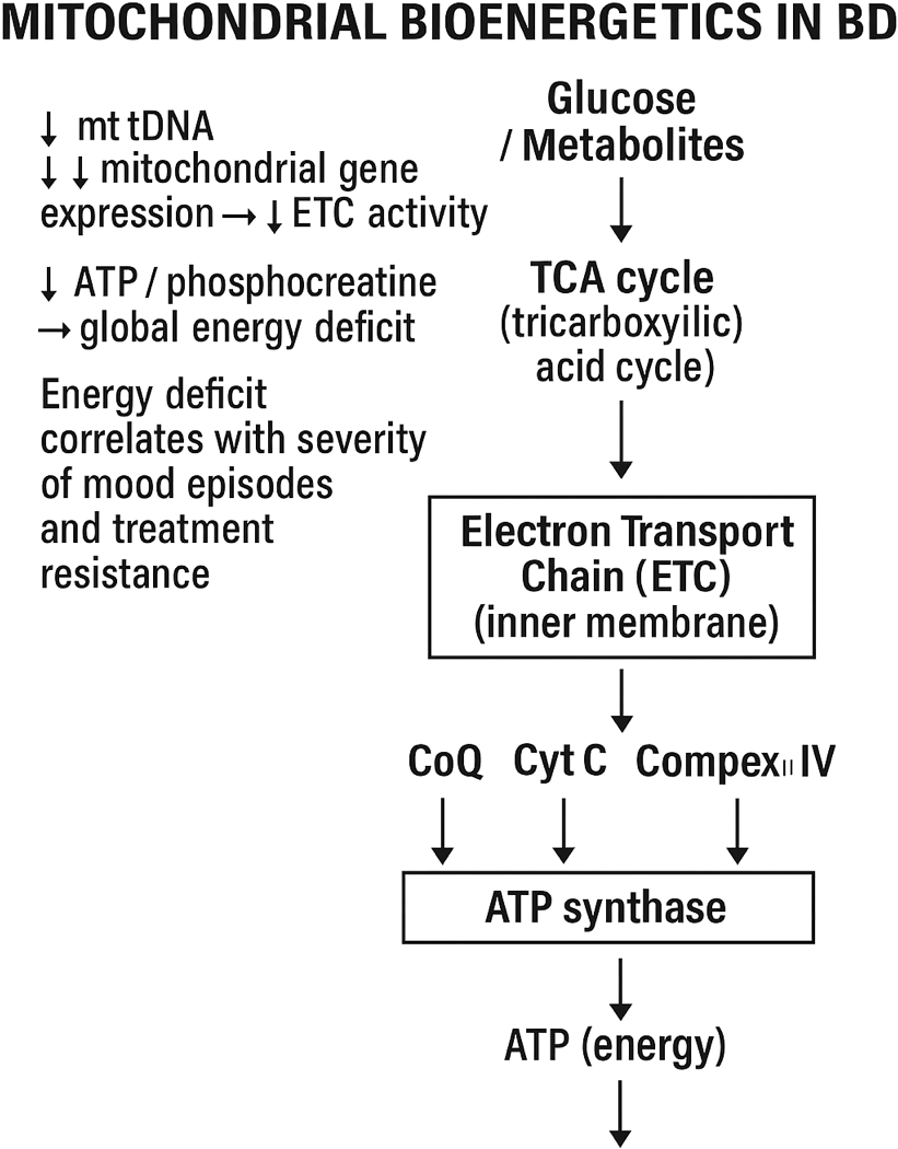
Consistent evidence supports altered mitochondrial bioenergetics in BD. Both postmortem and peripheral blood analyses have demonstrated reduced mitochondrial DNA (mtDNA) copy number, decreased expression of mitochondrial genes, and impaired electron transport chain (ETC) activity (Yoshimi et al., Reference Yoshimi, Futamura, Bergen, Iwayama, Ishima, Sellgren, Ekman, Jakobsson, Pålsson, Kakumoto, Ohgi, Yoshikawa, Landén and Hashimoto2016; Yamaki et al., Reference Yamaki, Otsuka, Numata, Yanagi, Mouri, Okazaki, Boku, Horai, Ohmori, Shirakawa, Sora and Hishimoto2018). Moreover, neuroimaging and magnetic resonance spectroscopy (MRS) studies reveal decreased PCr and ATP levels, reflecting a global energy deficit that may correspond with mood episode severity and treatment resistance (Benevenuto et al., Reference Benevenuto, Scaini, Barichello, Fries, Kennon, Andrews, Nix, Zunta-Soares, Valvassori, Soares and Quevedo2019) (Figure 1).
The figure shows the structure of the electron transport chain (ETC) in the internal. mitochondrial membrane, which results in ATP production. TCA cycle, tricarboxylic acid cycle; CoQ, coenzyme Q; Cyt C, cytochrome C.

Despite advances in pharmacotherapy, current treatments for BD remain only partially effective, and therapeutic selection is largely guided by clinical observation rather than biological criteria. As a result, many patients experience prolonged periods of inadequate treatment and heightened risk of relapse or treatment resistance (Anglin, Reference Anglin2016). The urgent need for objective biomarkers to support diagnosis, monitor disease progression, and predict therapeutic response has therefore become a critical focus in contemporary BD research (Liang et al., Reference Liang, Chen, Xiao, Wang and Wang2022).
Mitochondrial and metabolic markers represent particularly promising candidates, as they reflect core energetic deficits, oxidative stress, and redox imbalance inherent to BD pathology (Benevenuto et al., Reference Benevenuto, Scaini, Barichello, Fries, Kennon, Andrews, Nix, Zunta-Soares, Valvassori, Soares and Quevedo2019).These include alterations in mitochondrial respiration, ATP turnover, lactate levels, antioxidant enzyme activity, and circulating metabolites involved in energy metabolism. Integrating metabolic, genomic, transcriptomic, and neuroimaging data offers unprecedented potential to move towards precision psychiatry. Such an approach enables the identification of biologically defined subtypes (‘biotypes’) of BD and facilitates the development of personalised therapeutic strategies, encompassing pharmacological, nutritional, and lifestyle interventions aimed at restoring optimal neuronal metabolism and mitochondrial function (Licinio and Wong, Reference Licinio and Wong2023).
Ultimately, a deeper understanding of the energetic underpinnings of BD could bridge the gap between mechanistic insights and clinical translation, providing the foundation for precision therapeutics that target metabolic and mitochondrial dysfunction as core disease mechanisms rather than secondary phenomena. This review aims to summarize current evidence linking energetic metabolism to BD pathophysiology and discuss emerging biomarkers and therapeutic strategies targeting mitochondrial dysfunction.
Literature search strategy
This narrative review was informed by a structured literature search conducted according to selected PRISMA 2020 principles (Page et al., Reference Page, McKenzie, Bossuyt, Boutron, Hoffmann, Mulrow, Shamseer, Tetzlaff, Akl, Brennan, Chou, Glanville, Grimshaw, Hróbjartsson, Lalu, Li, Loder, Mayo-Wilson, McDonald, McGuinness, Stewart, Thomas, Tricco, Welch, Whiting and Moher2021).
In the first stage, we identified online repositories of biomedical literature, including Scopus, EMBASE, PubMed, and MEDLINE. Articles were retrieved from these databases using the following search strategy: [(bipolar disorder) AND ((energetic metabolism) OR (mitochondria))]. Duplicate records were automatically removed using the ZOTERO reference manager (Page et al., Reference Page, McKenzie, Bossuyt, Boutron, Hoffmann, Mulrow, Shamseer, Tetzlaff, Akl, Brennan, Chou, Glanville, Grimshaw, Hróbjartsson, Lalu, Li, Loder, Mayo-Wilson, McDonald, McGuinness, Stewart, Thomas, Tricco, Welch, Whiting and Moher2021).
Further screening of the retrieved records was performed according to predefined inclusion and exclusion criteria.
Inclusion criteria:
-
(a) full-text articles;
-
(b) published in English;
-
(c) published in peer-reviewed journals;
-
(d) human studies;
-
(e) original research articles;
-
(f) meta-analyses.
Exclusion criteria:
-
(a) diagnoses other than bipolar disorder;
-
(b) non-human studies;
-
(c) lack of full-text availability;
-
(d) case reports;
-
(e) studies involving children or adolescents;
-
(f) studies with comorbidities;
-
(g) studies on neurodegeneration;
-
(h) other non-relevant topics;
-
(i) non-English articles;
-
(j) duplicate records.
Additionally, a manual search was conducted to identify specific biomarkers mentioned in the text, yielding 28 additional publications.
Publications meeting all the above criteria underwent a full-text review. Figure 2 presents a schematic representation of the publication selection process.
Flow diagram of a selection of energetic metabolism in bipolar disorder publications according to. PRISMA guidelines. *Record excluded using automated tools built-in database and bibliography manager. **Record excluded by a researcher.

Integrated mechanisms of energetic dysfunction in BD
Redox imbalance, inflammatory pathways
Oxidative stress represents one of the central pathophysiological mechanisms in bipolar disorder (BD), linking mitochondrial dysfunction with widespread neuronal, immune, and metabolic abnormalities This persistent oxidative burden contributes to progressive molecular damage and functional deterioration across both central and peripheral systems in BD (Lu et al., Reference Lu, Yang, Zhao, Zhang, Sun, Liao, Kang, Feng, Sun and Yue2024).
Oxidative stress not only causes molecular damage but also directly interferes with mitochondrial energy metabolism. Excess ROS oxidise components of the electron transport chain (ETC), particularly complexes I and III, leading to reduced ATP synthesis and increased electron leakage, which further amplifies ROS production (Karabatsiakis et al., Reference Karabatsiakis, Böck, Salinas-Manrique, Kolassa, Calzia, Dietrich and Kolassa2014). ROS-mediated oxidation of mitochondrial lipids and DNA disrupts mitochondrial biogenesis and dynamics, contributing to the decreased efficiency of oxidative phosphorylation consistently observed in BD (Gimenez-Palomo et al., 2024). Dysregulation of antioxidant defense systems, including key enzymes such as superoxide dismutase, catalase (CAT), and glutathione peroxidase, results in the accumulation of oxidative damage in lipids, proteins, and nucleic acids, promoting mitochondrial dysfunction, impaired cellular signalling, and apoptosis (Jiménez-Fernández et al., Reference Jiménez-Fernández, Gurpegui, Garrote-Rojas, Gutiérrez-Rojas, Carretero and Correll2021; Valvassori et al., Reference Valvassori, Bavaresco, Feier, Cechinel-Recco, Steckert, Varela, Borges, Carvalho-Silva, Gomes, Streck and Quevedo2018; Kim et al., Reference Kim, Vadodaria, Lenkei, Kato, Gage, Marchetto and Santos2019).
From a neurobiological perspective, chronic oxidative stress disrupts mitochondrial function by reducing ATP production, altering mitochondrial membrane potential, and increasing mitochondrial permeability transition pore opening. These changes impair synaptic plasticity, enhance neuronal apoptosis, and dysregulate glutamatergic and dopaminergic neurotransmission- pathways strongly implicated in mood regulation and cognitive performance in BD (Kim et al., Reference Kim, Vadodaria, Lenkei, Kato, Gage, Marchetto and Santos2019; Kahyacı Kılıç et al., Reference Kahyacı Kılıç, Çinar, Görgülü and Sönmez2020; Romano et al., Reference Romano, Serviddio, Calcagnini, Villani, Giudetti, Cassano and Gaetani2017; van der Wijk et al., Reference van der Wijk, Bélteczki and Döme2024). The cumulative effect is neuroprogression, characterised by increasing structural and functional brain alterations over the course of the illness (Berk et al., Reference Berk, Kapczinski, Andreazza, Dean, Giorlando, Maes, Yücel, Gama, Dodd, Dean, Magalhães, Amminger, McGorry and Malhi2011; Cyrino et al., Reference Cyrino, Delwing-de Lima, Ullmann and Maia2021; Föcking et al., Reference Föcking, Dicker, Lopez, Hryniewiecka, Wynne, English, Cagney and Cotter2016).
At the systemic level, oxidative stress acts as a key activator of pro-inflammatory signalling cascades, including the nuclear factor kappa-light-chain-enhancer of activated B cells (NF-κB) pathway, which promotes the expression of inflammatory cytokines such as TNF-α, IL-1β, and IL-6 (Jones et al., Reference Young and Juruena2021; Kahyacı Kılıç et al., Reference Kahyacı Kılıç, Çinar, Görgülü and Sönmez2020; van der Wijk et al., Reference van der Wijk, Bélteczki and Döme2024). This establishes a self-reinforcing feedback loop between mitochondrial dysfunction, redox imbalance, and inflammation. Mitochondria also influence redox-sensitive signalling networks that regulate melatonin and kynurenine pathways, both of which modulate neuroinflammatory responses and neurotoxicity (Anderson et al., Reference Anderson, Jacob, Bellivier and Alexis Geoffroy2016). The interplay of these systems provides a mechanistic explanation for the overlap between metabolic, circadian, and affective disturbances frequently observed in BD (Andreazza, Reference Andreazza2019; Ahmed et al., Reference Ahmed, MahmoudianDehkordi, Bhattacharyya, Arnold, Liu, Neavin, Moseley, Thompson, Williams, Louie, Skime, Wang, Riva-Posse, McDonald, Bobo, Craighead, Krishnan, Weinshilboum, Dunlop, Millington, Rush, Frye and Kaddurah-Daouk2020; Jiménez-Fernández et al., Reference Jiménez-Fernández, Gurpegui, Garrote-Rojas, Gutiérrez-Rojas, Carretero and Correll2021; Ogawa et al., Reference Ogawa, Hattori, Ota, Hidese, Miyakawa, Matsumura, Yokota, Ishida, Matsuo, Yoshida, Yamazaki, Goodenowe and Kunugi2020).
Glucose metabolism and insulin resistance
Energy dysregulation in BD is not limited to mitochondrial dysfunction in neurons and peripheral cells but also involves substantial disturbances in glucose metabolism and the development of insulin resistance. These alterations consequently increase the risk of comorbid obesity, type 2 diabetes mellitus, and metabolic syndrome (Ahmed et al., Reference Ahmed, MahmoudianDehkordi, Bhattacharyya, Arnold, Liu, Neavin, Moseley, Thompson, Williams, Louie, Skime, Wang, Riva-Posse, McDonald, Bobo, Craighead, Krishnan, Weinshilboum, Dunlop, Millington, Rush, Frye and Kaddurah-Daouk2020; Miola et al., Reference Miola, Alvarez-Villalobos, Ruiz-Hernandez, De Filippis, Veldic, Prieto, Singh, Sanchez Ruiz, Nunez, Resendez, Romo-Nava, McElroy, Ozerdem, Biernacka, Frye and Cuellar-Barboza2023; Yu et al., Reference Yu, Oz and Sethi2023). Patients with BD frequently exhibit elevated fasting glucose and insulin levels, impaired glucose tolerance, and abnormal insulin responses, indicating deficits in energy homeostasis regulation in both peripheral tissues and the central nervous system (Campbell and Campbell, Reference Campbell and Campbell2019, Reference Campbell and Campbell2020).
Dysregulation of glucose metabolism in BD may directly affect brain function by limiting energy availability in key regions such as the prefrontal cortex and hippocampus, which are critically involved in mood regulation, cognitive processing, and impulse control. Glucose deficiency in neuronal tissue may exacerbate mood instability, contribute to cognitive deficits, and increase vulnerability to manic and depressive episodes. Simultaneously, these metabolic disturbances may enhance oxidative stress, activate pro-inflammatory pathways, and impair mitochondrial function, forming pathological feedback loops between energy metabolism and CNS functioning (Cyrino et al., Reference Cyrino, Delwing-de Lima, Ullmann and Maia2021).
At the systemic level, insulin resistance promotes chronic inflammation by increasing the production of pro-inflammatory cytokines and chemokines, including TNF-α, IL-6, and MCP-1, which can cross the blood–brain barrier, modulate microglial activity, and disrupt neurotransmission (Bortolasci et al., Reference Bortolasci, Voigt, Turner, Mohebbi, Gray, Dodd, Walder, Berk, Cotton, Malhi, Ng, Dowling, Sarris and Dean2020; Kageyama et al., Reference Kageyama, Deguchi, Kasahara, Tani, Kuroda, Inoue and Kato2022, Reference Kageyama, Okura, Sukigara, Matsunaga, Maekubo, Oue, Ishihara, Deguchi and Inoue2025).This process further destabilises cerebral energy balance, increasing the risk of disease relapse and cognitive decline (Papageorgiou and Filiou, Reference Papageorgiou and Filiou2024). Moreover, these metabolic abnormalities often correlate with the metabolic phenotype of BD patients, including hyperphagia, central obesity, and dyslipidemia, emphasizing the need to consider metabolic parameters in the comprehensive assessment and treatment of BD (Miola et al., Reference Miola, Alvarez-Villalobos, Ruiz-Hernandez, De Filippis, Veldic, Prieto, Singh, Sanchez Ruiz, Nunez, Resendez, Romo-Nava, McElroy, Ozerdem, Biernacka, Frye and Cuellar-Barboza2023; Yu et al., Reference Yu, Oz and Sethi2023).
Understanding the interplay among glycemic dysregulation, insulin resistance, mitochondrial dysfunction, and oxidative stress is crucial for developing targeted therapeutic interventions. Such approaches should integrate pharmacological modulators of metabolism with dietary and lifestyle strategies to restore energy balance and stabilize mood in individuals with BD.
purinergic pathways
Disturbances in purinergic signalling represent a mechanism contributing to the neurobiological alterations observed in BD. These abnormalities are closely integrated with mitochondrial impairment, oxidative stress, and dysregulated inflammatory activity. The purinergic system encompasses two primary classes of receptors: P1 receptors responsive to adenosine, and P2 receptors responsive to ATP. The P2 family is further categorised into ionotropic P2X channels and metabotropic P2Y receptors (Ortiz et al., Reference Ortiz, Ulrich, Zarate and Machado-Vieira2015). In addition to receptor signalling, altered metabolism of purine derivatives such as adenosine and uric acid (UA), along with dysfunction of enzymes including xanthine dehydrogenase (XDH) and adenosine deaminase (ADA), has been implicated in the pathophysiology of BD (El Sabbagh et al., Reference El Sabbagh, Machado, Pappis, Beroncal, Ji, Nader, Chander, Choi, Duong, Jeong, Panizzutti, Bortolasci, Szatmari, Carlen, Hahn, Attisano, Berk, Walder and Andreazza2025; Malewska et al., Reference Malewska, Rybakowski and Rybakowski2024). Clinical and molecular studies reveal that individuals with BD often show elevated plasma levels of adenosine and ATP, accompanied by increased expression of P2X7 receptors in microglial and immune cells. This receptor overactivation promotes the release of pro-inflammatory cytokines, particularly IL-1β, IL-6, and TNF-α – driving a sustained inflammatory response within both the peripheral and central nervous systems (Ahmed et al., Reference Ahmed, MahmoudianDehkordi, Bhattacharyya, Arnold, Liu, Neavin, Moseley, Thompson, Williams, Louie, Skime, Wang, Riva-Posse, McDonald, Bobo, Craighead, Krishnan, Weinshilboum, Dunlop, Millington, Rush, Frye and Kaddurah-Daouk2020; Skokou et al., Reference Skokou, Oikonomakis, Andreopoulou, Kypreos, Gourzis and Halaris2023; Malewska-Kasprzak et al., Reference Malewska-Kasprzak, Sikorski, Dmitrzak-Weglarz and Rybakowski2025; Young et al., Reference Young, Kim, Andreazza and Kim H.K.2018). The inflammatory model of BD proposes that chronic immune activation contributes to illness progression, as reflected in consistently elevated serum IL-6 levels among affected patients (Ozkaya et al., Reference Ozkaya, Gürbüzer, Tozoğlu, Akyildirim and Mercantepe2025). Inflammasomes, cytosolic multiprotein complexes that detect cellular stress or pathogen-related signals, have also been recognised as crucial mediators linking immunity and neuronal dysfunction in BD (Świerczyński and Fichna, Reference Świerczyński and Fichna2021), (Malewska-Kasprzak et al., Reference Malewska-Kasprzak, Jowik and Tyszkiewicz-Nwafor2023). In particular, hyperactivation of the NLRP3 inflammasome within microglia induces chronic neuroinflammation that exacerbates mood instability and cognitive decline. Sustained activation of this pathway disrupts neuronal signalling and plasticity, leading to alterations in dopaminergic, serotonergic, and glutamatergic neurotransmission (Kim, Reference Kim2022; Han et al., Reference Han, Li, Chen, Wang, Bao, Huang, Liu and Chen2023).
Excessive stimulation of P2X7 receptors further amplifies inflammatory cascades and contributes to oxidative stress–related neuronal damage. Together with mitochondrial dysfunction, these processes facilitate apoptosis and synaptic loss (Glausier, Reference Glausier2025; Malewska et al., Reference Malewska, Rybakowski and Rybakowski2024; Malewska-Kasprzak et al., Reference Malewska-Kasprzak, Permoda and Rybakowski2022). Such neuroinflammatory and metabolic alterations correlate strongly with symptom severity and cognitive impairment in BD.
Additionally, purinergic dysregulation influences cellular energy dynamics. Overactivation of ATP/P2X7 signalling can elevate metabolic demand and accelerate ATP depletion within neurons and peripheral cells (Bertocci et al., Reference Bertocci, Yushmanov, Moon, Abdul-Waalee, Versace, Phillips, Birmaher, Hetherington and Pan2021; Malewska-Kasprzak et al., Reference Malewska-Kasprzak, Permoda-Osip and Rybakowski2019; Scott and McClung, Reference Scott and McClung2023). The purinergic system also interacts with redox-sensitive pathways, including those dependent on the NAD+/NADH ratio, thereby modulating oxidative phosphorylation efficiency. Persistent disturbances in these interconnected mechanisms – when combined with chronic inflammation – create self-reinforcing loops between immune activation, oxidative imbalance, and impaired energy metabolism (Lindberg et al., Reference Lindberg, Shan, Ayers-Ringler, Oliveros, Benitez, Prieto, McCullumsmith and Choi2015; Ahmed et al., Reference Ahmed, MahmoudianDehkordi, Bhattacharyya, Arnold, Liu, Neavin, Moseley, Thompson, Williams, Louie, Skime, Wang, Riva-Posse, McDonald, Bobo, Craighead, Krishnan, Weinshilboum, Dunlop, Millington, Rush, Frye and Kaddurah-Daouk2020; Permoda-Pachuta et al., Reference Permoda-Pachuta, Malewska-Kasprzak, Skibińska, Rzepski and Dmitrzak-Węglarz2023) (Figure 3).
Inflammatory and purinergic pathways in BD: Interac9ons between uric acid, P2X7,. adenosine, xanthine oxidase, and adenosine deaminase. P2X7, P2X7 receptor; ADO, adenosine; ADA, deaminase adenosine; XDH, xanthine oxidase; UA, uric acid; IL-1, interleukin.

Figure 3. Long description
A diagram of the inflammatory and purinergic pathways in bipolar disorder. Microglia contain the NRLP inflammasome, which includes NRLP3, ASC, and Nacht, leading to the activation of pro-caspase and pro IL-1, resulting in IL-1 beta. P2X7 activation is shown near the microglia. Astrocytes release ATP, which is involved in energetic stress. Uric acid (UA) and adenosine (ADO) interact through xanthine oxidase (XDH) and adenosine deaminase (ADA), contributing to neuroinflammation and exacerbation of bipolar disorder.
Neuroplasticity and glutamatergic–Astrocytic function in bipolar disorder
Energy dysregulation represents a pathological feature of BD, profoundly influencing synaptic plasticity, glutamatergic signalling, and astrocytic function. Impairments in mitochondrial bioenergetics within both neurons and astrocytes disrupt neuron–astrocyte metabolic coupling, an essential mechanism for maintaining glutamate synthesis, recycling, and uptake. Consequently, limited ATP availability compromises the activity of astrocytic glutamate transporters, leading to excessive extracellular glutamate accumulation and excitotoxic stress (Anderson et al., Reference Anderson, Almulla, Reiter and Maes2023).
Astrocytic dysfunction is further characterised by downregulation of excitatory amino acid transporters EAAT1 and EAAT2, reducing the efficiency of synaptic glutamate clearance. The resulting glutamate excess promotes overactivation of NMDA receptors, particularly at extrasynaptic sites, which induces sustained calcium influx, oxidative stress, and activation of pro-apoptotic signalling cascades. In contrast, synaptic NMDA receptor activation supports neuronal survival and adaptive synaptic remodelling, highlighting the location-dependent duality of NMDA receptor function. Under physiological conditions (Rybakowski et al., Reference Rybakowski, Permoda-Osip and Bartkowska-Sniatkowska2017).
Energy deficits in BD also constrain the synthesis and release of neurotrophic factors, particularly brain-derived neurotrophic factor (BDNF), a key regulator of synaptic growth, dendritic spine formation, and neuronal resilience. Reduced BDNF availability has been consistently associated with cognitive dysfunction, emotional dysregulation, and the severity of both manic and depressive episodes. Collectively, these disturbances underscore the interdependence between cellular energy metabolism, glutamatergic homeostasis, and astrocytic support in maintaining neuroplastic integrity (Permoda-Pachuta et al., Reference Permoda-Pachuta, Malewska-Kasprzak, Skibińska, Rzepski and Dmitrzak-Węglarz2023; Rybakowski et al., Reference Rybakowski, Permoda-Osip and Bartkowska-Sniatkowska2017).
Taken together, the mechanisms described above support several of the leading contemporary hypotheses of bipolar disorder. The mitochondrial hypothesis is reinforced by evidence of impaired oxidative phosphorylation, reduced ATP availability, altered redox balance, and disrupted neuron, astrocyte metabolic coupling, all of which may contribute to neuronal vulnerability and mood instability. Simultaneously, the immunological hypothesis is supported by persistent activation of inflammatory pathways, including NF-κB signalling, increased pro-inflammatory cytokine production, and NLRP3 inflammasome activation, which interact bidirectionally with mitochondrial dysfunction and oxidative stress. In parallel, the purinergic hypothesis is substantiated by abnormalities in ATP-mediated signalling, altered adenosine and UA metabolism, and enhanced P2X7 receptor activity, linking cellular energy deficits with neuroinflammatory processes (Malewska et al., Reference Malewska, Permoda-Osip, Kasprzak, Niemiec and Rybakowski2017; Malewska-Kasprzak et al., Reference Malewska-Kasprzak, Dmitrzak-Weglarz and Rybakowski2026; Malewska-Kasprzak et al., Reference Malewska-Kasprzak, Dmitrzak-Weglarz and Rybakowski2026). Rather than representing independent models, these hypotheses appear highly interconnected, converging on a common framework in which bioenergetic dysfunction, immune activation, and purinergic dysregulation mutually reinforce one another and contribute to the development and progression of BD. This integrated perspective may provide a useful conceptual basis for biomarker development and the design of mechanism-based therapeutic interventions.
Bioenergetic dysregulation in bipolar disorder – Neuroimaging and biochemical evidence
Neuroimaging indicators of energetic dysfunction
Neuroimaging studies employing proton (1H) and phosphorus (31P) MRS, as well as functional near-infrared spectroscopy (fNIRS), have provided convergent evidence for impaired mitochondrial oxidative metabolism in individuals with BD (Bertocci and Diler, Reference Bertocci and Diler2025; Colasanti et al., Reference Colasanti, Russo, Örzsik, Imrak, Kartar, Caddye, Pizzamiglio, Pitceathly, Asllani and Ronen2025; Dudley et al., Reference Dudley, Lee, Durling, Strakowski and Eliassen2015; Föcking et al., Reference Föcking, Dicker, Lopez, Hryniewiecka, Wynne, English, Cagney and Cotter2016).
Across multiple studies, significant reductions in NAD+, citrate, and other intermediates of the tricarboxylic acid (TCA) cycle have been reported, indicating compromised neuronal capacity for efficient energy generation via oxidative phosphorylation (Yoshimi et al., Reference Yoshimi, Futamura, Bergen, Iwayama, Ishima, Sellgren, Ekman, Jakobsson, Pålsson, Kakumoto, Ohgi, Yoshikawa, Landén and Hashimoto2016; Du et al., Reference Du, Yuksel, Chouinard, Huynh, Ryan, Cohen and Öngür2018; Campbell and Campbell, Reference Campbell and Campbell2019; Ahmed et al., Reference Ahmed, MahmoudianDehkordi, Bhattacharyya, Arnold, Liu, Neavin, Moseley, Thompson, Williams, Louie, Skime, Wang, Riva-Posse, McDonald, Bobo, Craighead, Krishnan, Weinshilboum, Dunlop, Millington, Rush, Frye and Kaddurah-Daouk2020). In parallel, decreased concentrations of PCr and adenosine triphosphate (ATP) within the prefrontal cortex, along with slower creatine kinase forward reaction rate constants (CK kf), reflect a limited energetic reserve and a diminished adaptive capacity of neurons to meet increased metabolic demands (Stork and Renshaw, Reference Stork and Renshaw2005; Bertocci et al., Reference Bertocci, Yushmanov, Moon, Abdul-Waalee, Versace, Phillips, Birmaher, Hetherington and Pan2021).These findings collectively suggest a global impairment in cerebral energy transfer systems that may underlie affective instability and cognitive dysfunction in BD.
Importantly, such energetic abnormalities are observed not only during manic or depressive episodes but also persist during euthymic phases, suggesting that mitochondrial dysfunction represents a trait-like feature rather than a mere state-dependent phenomenon (Bertocci et al., Reference Bertocci, Yushmanov, Moon, Abdul-Waalee, Versace, Phillips, Birmaher, Hetherington and Pan2021). This persistence supports the hypothesis that impaired bioenergetics may serve as an early predictor of relapse risk and a stable pathophysiological marker throughout the course of the disorder.
The advent of ultra–high-field magnetic resonance imaging (7T MRI) has further advanced our ability to map metabolic alterations with greater spatial and spectral resolution. These high-resolution studies reveal abnormal cortical accumulation of lactate, consistent with impaired oxidative phosphorylation and a metabolic shift towards anaerobic glycolysis (Yoshimi et al., Reference Yoshimi, Futamura, Bergen, Iwayama, Ishima, Sellgren, Ekman, Jakobsson, Pålsson, Kakumoto, Ohgi, Yoshikawa, Landén and Hashimoto2016; Ahmed et al., Reference Ahmed, MahmoudianDehkordi, Bhattacharyya, Arnold, Liu, Neavin, Moseley, Thompson, Williams, Louie, Skime, Wang, Riva-Posse, McDonald, Bobo, Craighead, Krishnan, Weinshilboum, Dunlop, Millington, Rush, Frye and Kaddurah-Daouk2020; Bertocci et al., Reference Bertocci, Yushmanov, Moon, Abdul-Waalee, Versace, Phillips, Birmaher, Hetherington and Pan2021). Lactate accumulation is interpreted as a compensatory response to reduced mitochondrial ATP production, reflecting an adaptive yet inefficient attempt to maintain energy supply under mitochondrial stress. Such findings are particularly significant as they link functional neuroimaging markers to the biochemical signatures of mitochondrial distress, bridging molecular and systems-level evidence.
Converging biochemical data reinforce these neuroimaging observations. Elevated oxidative stress, reduced activity of mitochondrial electron transport chain complexes, and disturbances in purinergic homeostasis have been consistently reported in BD, underscoring the multifaceted nature of energetic dysfunction in this disorder (Yoshimi et al., Reference Yoshimi, Futamura, Bergen, Iwayama, Ishima, Sellgren, Ekman, Jakobsson, Pålsson, Kakumoto, Ohgi, Yoshikawa, Landén and Hashimoto2016; Ahmed et al., Reference Ahmed, MahmoudianDehkordi, Bhattacharyya, Arnold, Liu, Neavin, Moseley, Thompson, Williams, Louie, Skime, Wang, Riva-Posse, McDonald, Bobo, Craighead, Krishnan, Weinshilboum, Dunlop, Millington, Rush, Frye and Kaddurah-Daouk2020; Bertocci et al., Reference Bertocci, Yushmanov, Moon, Abdul-Waalee, Versace, Phillips, Birmaher, Hetherington and Pan2021). Moreover, abnormalities in brain bioenergetics have been associated with alterations in neurovascular coupling and impaired cortical oxygen utilisation, providing a broader physiological context for understanding cognitive and affective symptoms in BD (Dudley et al., Reference Dudley, Lee, Durling, Strakowski and Eliassen2015)
Integration of neuroimaging findings with mitochondrial functional assays and oxidative stress markers offers a multidimensional framework for characterising BD pathophysiology. The identification of neuroenergetic markers as quantifiable biomarkers enables early recognition of patients at elevated relapse risk, objective monitoring of treatment response to metabolic and pharmacological interventions, and the design of precision therapeutic strategies aimed at restoring normal cerebral bioenergetics (Du et al., Reference Du, Yuksel, Chouinard, Huynh, Ryan, Cohen and Öngür2018; Ahmed et al., Reference Ahmed, MahmoudianDehkordi, Bhattacharyya, Arnold, Liu, Neavin, Moseley, Thompson, Williams, Louie, Skime, Wang, Riva-Posse, McDonald, Bobo, Craighead, Krishnan, Weinshilboum, Dunlop, Millington, Rush, Frye and Kaddurah-Daouk2020; Bertocci et al., Reference Bertocci, Yushmanov, Moon, Abdul-Waalee, Versace, Phillips, Birmaher, Hetherington and Pan2021).
In this context, neuroimaging-derived measures of mitochondrial metabolism may serve as a critical interface between molecular, metabolic, and clinical dimensions of BD research, facilitating the development of a coherent and mechanistically informed model of disease progression (Giménez-Palomo et al., Reference Giménez-Palomo, Guitart-Mampel, Roqué, Sánchez, Borràs, Meseguer, García-García, Tobías, Valls-Roca, Anmella, Valentí, Olivier, de Juan, Ochandiano, Andreu, Radua, Verdolini, Berk, Vieta, Garrabou, Roca, Alsina-Restoy and Pacchiarotti2024).
Peripheral and cellular markers
Mitochondrial dysfunction in BD extends beyond the central nervous system, encompassing peripheral cells and tissues. This systemic nature of bioenergetic disruption offers a unique opportunity to monitor mitochondrial processes through accessible peripheral biomarkers, thereby facilitating translational research and clinical application. Studies of peripheral blood mononuclear cells (PBMCs) and leukocytes have consistently demonstrated significant impairments in the activity of mitochondrial respiratory chain complexes I and IV, leading to decreased ATP synthesis and increased generation of reactive oxygen species (ROS) (Bertocci et al., Reference Bertocci, Yushmanov, Moon, Abdul-Waalee, Versace, Phillips, Birmaher, Hetherington and Pan2021; Gimenez-Palomo et al., 2024; Young et al., Reference Young, Kim, Andreazza and Kim H.K.2018). This imbalance promotes chronic oxidative stress and subclinical inflammatory states, which may interact with neural circuits to influence disease progression, episode recurrence, and treatment resistance.
An important index of cellular redox homeostasis – the NAD+/NADH ratio – provides valuable insights into the energetic state of peripheral cells. Decreased NAD+/NADH ratios observed in BD patients indicate a limited capacity for oxidative phosphorylation and impaired regeneration of cellular energy pools. These alterations also suggest deficits in antioxidant defense mechanisms, which are essential for maintaining metabolic stability and protecting against oxidative injury (Bertocci et al., Reference Bertocci, Yushmanov, Moon, Abdul-Waalee, Versace, Phillips, Birmaher, Hetherington and Pan2021; Gimenez-Palomo et al., 2024; Young et al., Reference Young, Kim, Andreazza and Kim H.K.2018). In parallel, reduced levels of oxidised cytochrome c oxidase (oxCOX) in peripheral tissues reflect diminished efficiency of oxidative phosphorylation and reduced mitochondrial competence, supporting the notion of a systemic impairment in electron transport across multiple biological compartments.
Recent metabolomic analyses have further identified alterations in peripheral lipid metabolism, particularly in ethanolamine plasmalogens and phosphatidylethanolamines, key phospholipids involved in membrane structure, redox signalling, and mitochondrial dynamics. Changes in these compounds observed in blood plasma of BD patients suggest disrupted lipid remodelling and oxidative vulnerability of cellular membranes, providing an additional dimension to the systemic bioenergetic dysregulation associated with the disorder.
Markers of oxidative stress, such as malondialdehyde (MDA) and 8-hydroxy-2’-deoxyguanosine (8-OHdG), are elevated both in serum and in peripheral cells of BD patients, reflecting persistent activation of oxidative and proinflammatory pathways (Andreazza, Reference Andreazza2019; Ahmed et al., Reference Ahmed, MahmoudianDehkordi, Bhattacharyya, Arnold, Liu, Neavin, Moseley, Thompson, Williams, Louie, Skime, Wang, Riva-Posse, McDonald, Bobo, Craighead, Krishnan, Weinshilboum, Dunlop, Millington, Rush, Frye and Kaddurah-Daouk2020; Gimenez-Palomo et al., 2024). Elevated MDA levels indicate increased lipid peroxidation, whereas 8-OHdG serves as a marker of oxidative DNA damage – both contributing to mitochondrial genome instability and progressive energetic failure. The persistence of these markers even during euthymic phases underscores the systemic and trait-like nature of mitochondrial and oxidative dysregulation in BD, extending beyond localised neuronal effects to encompass the entire organism’s metabolic response to chronic stress (Andreazza, Reference Andreazza2019; Ahmed et al., Reference Ahmed, MahmoudianDehkordi, Bhattacharyya, Arnold, Liu, Neavin, Moseley, Thompson, Williams, Louie, Skime, Wang, Riva-Posse, McDonald, Bobo, Craighead, Krishnan, Weinshilboum, Dunlop, Millington, Rush, Frye and Kaddurah-Daouk2020). Elevated levels of 8-OHdG and MDA have been also associated with greater severity of manic and depressive episodes, longer illness duration, and higher relapse frequency. In contrast, normalisation of antioxidant enzyme activity during remission suggests that dynamic redox balance may reflect illness phase and treatment response. These associations highlight the translational potential of oxidative biomarkers as objective indicators of disease activity and therapeutic efficacy (Andreazza, Reference Andreazza2019; Ahmed et al., Reference Ahmed, MahmoudianDehkordi, Bhattacharyya, Arnold, Liu, Neavin, Moseley, Thompson, Williams, Louie, Skime, Wang, Riva-Posse, McDonald, Bobo, Craighead, Krishnan, Weinshilboum, Dunlop, Millington, Rush, Frye and Kaddurah-Daouk2020; Jiménez-Fernández et al., Reference Jiménez-Fernández, Gurpegui, Garrote-Rojas, Gutiérrez-Rojas, Carretero and Correll2021; Ogawa et al., Reference Ogawa, Hattori, Ota, Hidese, Miyakawa, Matsumura, Yokota, Ishida, Matsuo, Yoshida, Yamazaki, Goodenowe and Kunugi2020).
Circulating mitochondrial DNA (mtDNA) has also emerged as a promising biomarker of systemic inflammation and cellular stress. Elevated plasma mtDNA levels in BD may reflect enhanced mitochondrial turnover, impaired mitophagy, or leakage of mitochondrial components into the extracellular space, which can activate innate immune responses through toll-like receptor signaling (Cordeiro et al., Reference Cordeiro, Lima, Fries, Zunta-Soares, Soares, Quevedo and Scaini2023; Shao et al., Reference Shao, Zou, Kennedy, Dimick, Andreazza, Young, Goncalves, MacIntosh and Goldstein2024; Teng et al., Reference Teng, Xu, Chen, Qiu, Li, Chen, Tang, Xiang, Wang, Tan and Wu2024). These findings strengthen the concept of a bidirectional relationship between mitochondrial dysfunction and inflammation, positioning mtDNA as a potential bridge between metabolic dysregulation and immune activation in BD pathophysiology.
Recent studies confirm that peripheral mitochondrial biomarkers also reflect metabolic vulnerability in BD. Elevated plasma lactate has been associated with higher fasting glucose, triglycerides, and blood pressure, indicating a link between mitochondrial energy imbalance and metabolic syndrome. In contrast, circulating cell-free mitochondrial DNA showed no such association, suggesting that lactate may serve as a more sensitive marker of systemic energetic stress. Mitochondrial-targeted metabolomics further revealed distinct metabolic patterns in patients with concurrent lactate elevation and metabolic syndrome, highlighting changes in TCA cycle intermediates and redox balance (Zachos et al., Reference Zachos, Choi, Godin, Chernega, Kwak, Jung, Aouizerate, Aubin, Bellivier, Belzeaux-R, Courtet, Dubertret, Etain, Haffen, Lefrere, Llorca, Olié, Polosan, Samalin, Schwan, Roux, Barau, Richard, Tamouza, Leboyer and Andreazza2024).
In recent studies, bipolar patients exhibited significant alterations in key purinergic enzymes, including ADA, XDH, and the P2X7 receptor. Concentrations of XDH, adenosine, and ADA were significantly lower in both manic and depressive episodes, as well as during remission, compared to healthy controls, suggesting trait-dependent dysfunction of purinergic signaling. Conversely, UA levels were elevated, particularly in female patients, consistent with enhanced purine catabolism and oxidative burden. Importantly, P2X7 and NACHT levels were markedly increased after treatment in both manic and depressive states, indicating persistent activation of immune and inflammasome pathways despite symptomatic improvement. These findings support the hypothesis that purinergic dysregulation contributes to the chronic inflammatory milieu observed in BD and may modulate mitochondrial and energetic homeostasis. Moreover, XDH and ADA have been proposed as potential biomarkers for distinguishing mood episode polarity and monitoring treatment response, while persistent P2X7 activation could represent a biological substrate of relapse vulnerability. Gender differences – such as higher baseline ADA, NACHT, and P2X7 in women – further emphasise the modulatory role of hormonal and metabolic factors in purinergic signalling within BD pathology (Malewska et al., Reference Malewska, Rybakowski and Rybakowski2024; Malewska-Kasprzak et al., Reference Malewska-Kasprzak, Sikorski, Dmitrzak-Weglarz and Rybakowski2025; Malewska-Kasprzak et al., Reference Malewska-Kasprzak, Permoda and Rybakowski2022).
Additionally, neurotrophic and inflammatory markers provide complementary insights into BD pathophysiology. Decreased levels of BDNF in serum and plasma of BD patients have been associated with impaired neuronal plasticity, cognitive deficits, and mood episode severity. Elevated proinflammatory cytokines, including interleukin-6 (IL-6), interleukin-1β (IL-1β), and tumour necrosis factor-alpha (TNF-α), are frequently observed in both manic and depressive episodes, reflecting chronic systemic inflammation that may exacerbate mitochondrial dysfunction (Machado-Vieira et al., Reference Machado-Vieira, Courtes, Zarate, Henter and Manji2023).
From a translational perspective, the assessment of mitochondrial and oxidative biomarkers in peripheral cells offers a minimally invasive, reproducible, and clinically feasible approach for precision psychiatry. Monitoring redox ratios (NAD+/NADH), mitochondrial respiratory activity (oxCOX), and oxidative stress markers (MDA, 8-OHdG), metabolic indicators such as plasma lactate and purinergic mediators including ADA, XDH, and P2X7 receptor levels could enable early detection of relapse risk, real-time evaluation of pharmacological and metabolic interventions, and refined patient stratification in clinical trials (Cikankova et al., Reference Cikankova, Sigitova, Zverova, Fisar, Raboch and Hroudova2017; Shao et al., Reference Shao, Zou, Kennedy, Dimick, Andreazza, Young, Goncalves, MacIntosh and Goldstein2024).
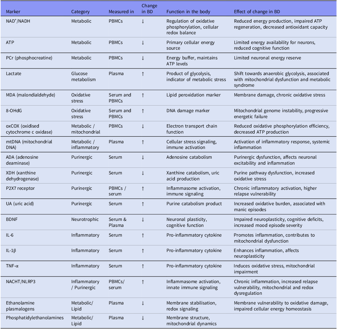
When integrated with neuroimaging findings and detailed clinical phenotyping, peripheral mitochondrial markers contribute to a unified systems-level model of BD pathophysiology – one that connects central and peripheral energetic dysfunction. This model not only enhances mechanistic understanding but also provides a scientific basis for developing precision therapeutic strategies aimed at restoring mitochondrial function, reducing oxidative stress, and promoting long-term mood stability (Ahmed et al., Reference Ahmed, MahmoudianDehkordi, Bhattacharyya, Arnold, Liu, Neavin, Moseley, Thompson, Williams, Louie, Skime, Wang, Riva-Posse, McDonald, Bobo, Craighead, Krishnan, Weinshilboum, Dunlop, Millington, Rush, Frye and Kaddurah-Daouk2020; Bertocci et al., Reference Bertocci, Yushmanov, Moon, Abdul-Waalee, Versace, Phillips, Birmaher, Hetherington and Pan2021; Gimenez-Palomo et al., 2024) (Table 1).
Peripheral and cellular markers in bipolar disorder (BD)

Table 1. Long description
A table with 18 rows and 5 columns. The columns are labeled Marker, Category, Measured in, Change in BD, Function in the body, and Effect of change in BD. The table lists different markers such as NAD+/NADH, ATP, PCr, Lactate, MDA, 8-OHdG, oxCOX, mtDNA, ADA, XDH, P2X7 receptor, UA, BDNF, IL-6, IL-1β, TNF-α, NACHT/NLRP3, Ethanolamine plasmalogens, and Phosphatidylethanolamines. Each row provides details on the category, where the marker is measured, the change observed in bipolar disorder, its function in the body, and the effect of this change in bipolar disorder.
Therapeutic implications and experimental interventions
In recent years, there has been increasing interest in therapies aimed at improving mitochondrial function and overall energy homeostasis in patients with BD (Lopresti and Jacka, Reference Lopresti and Jacka2015).
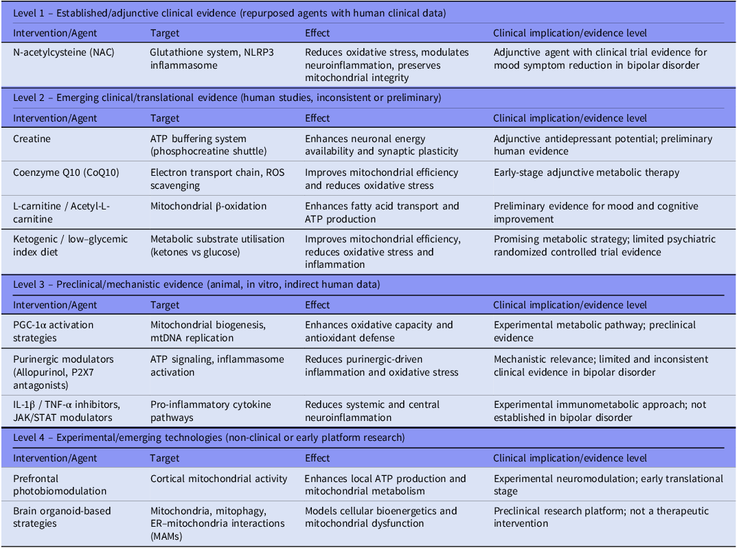
For clarity, the interventions discussed in this section are stratified according to their level of translational and clinical evidence. Level 1 includes established adjunctive interventions supported by clinical trial data in bipolar disorder. Level 2 comprises emerging clinical or translational approaches with preliminary or inconsistent human evidence. Level 3 includes preclinical or mechanistic interventions supported mainly by animal or in vitro studies or indirect clinical data. Level 4 refers to early experimental or platform-based approaches that are not yet applicable to clinical practice. This classification is intended to distinguish between differing stages of evidence and does not imply equivalence in clinical efficacy or readiness for implementation.
The therapeutic implications of mitochondrial dysfunction in BD should be interpreted within the context of established pharmacological treatment. Mood stabilisers, antipsychotics, and antidepressants remain the cornerstone of BD management, whereas mitochondrial, metabolic, and purinergic interventions currently have an adjunctive role. Notably, several standard treatments appear to influence pathways discussed in this review. Lithium, for example, has been associated with improved mitochondrial bioenergetics, reduced oxidative stress, modulation of mitochondrial dynamics, and normalisation of multiple mitochondrial abnormalities reported in BD (Balaç et al., Reference Balaç, Dal Yöntem, Karacicek, Aksahin, Pisanu, Hun Senol, Manchia, Meloni, Damri, Agam, Özerdem, Genc, Squassina and Ceylan2026). These findings suggest that mitochondrial dysfunction may represent a biologically relevant component of BD pathophysiology and a mechanism through which established treatments exert part of their therapeutic effects. Consequently, emerging bioenergetic interventions should be viewed as complementary to, rather than replacements for, current pharmacological approaches.
Among natural and semi-synthetic mitochondrial compounds, creatine plays a role as a phosphate buffer, enabling rapid ATP regeneration via the creatine kinase system, particularly in brain regions with high energy demands such as the prefrontal cortex and hippocampus (Young et al., Reference Young, Kim, Andreazza and Kim H.K.2018). Studies indicate that creatine supplementation not only enhances neuronal energy reserves but also supports synaptic plasticity, stabilises glutamatergic neurotransmission, and strengthens neuroprotective pathways, leading to improved cognitive function and reduction of depressive symptoms in BD patients (Berk et al., Reference Berk, Turner, Malhi, Ng, Cotton, Dodd, Samuni, Tanious, McAulay, Dowling, Sarris, Owen, Waterdrinker, Smith and Dean2019; Ahmed et al., Reference Ahmed, MahmoudianDehkordi, Bhattacharyya, Arnold, Liu, Neavin, Moseley, Thompson, Williams, Louie, Skime, Wang, Riva-Posse, McDonald, Bobo, Craighead, Krishnan, Weinshilboum, Dunlop, Millington, Rush, Frye and Kaddurah-Daouk2020; Juneja et al., Reference Juneja, Bhuchakra, Sadhukhan, Mehta, Niharika, Thareja, Nimmakayala and Sahu2024).
Coenzyme Q10 (CoQ10), an essential component of the electron transport chain, functions both as a cofactor in ATP synthesis and as a lipophilic antioxidant, reducing mitochondrial ROS production (Pradhan et al., Reference Pradhan, Singh and Singh2021; Anderson et al., Reference Anderson, Almulla, Reiter and Maes2023; Yu et al., Reference Yu, Oz and Sethi2023). CoQ10 supplementation may attenuate oxidative stress, enhance mitochondrial membrane potential, and limit neuroinflammatory processes, making it a promising bioenergetic therapy for BD (Yoshimi et al., Reference Yoshimi, Futamura, Bergen, Iwayama, Ishima, Sellgren, Ekman, Jakobsson, Pålsson, Kakumoto, Ohgi, Yoshikawa, Landén and Hashimoto2016; Bertocci et al., Reference Bertocci, Yushmanov, Moon, Abdul-Waalee, Versace, Phillips, Birmaher, Hetherington and Pan2021).
Carnitine, in the forms of L-carnitine and acetyl-L-carnitine, plays a role in the transport of long-chain fatty acids into mitochondria for β-oxidation, thereby directly supporting ATP production and lipid homeostasis (Brennan et al., Reference Brennan, Jensen, Hudson, Coit, Beaulieu, Pope, Renshaw and Cohen2013; Adzic et al., Reference Adzic, Brkic, Bulajic, Mitic and Radojcic2016; Anderson et al., Reference Anderson, Jacob, Bellivier and Alexis Geoffroy2016; Bernard et al., Reference Bernard, Tamouza, Godin, Berk, Andreazza and Leboyer2025). Impaired carnitine metabolism is associated with reduced energetic efficiency and the accumulation of toxic lipid intermediates, contributing to neuronal stress and mood instability (Büttiker et al., Reference Büttiker, Weissenberger, Esch, Anders, Raboch, Ptacek, Kream and Stefano2023; Gabriel et al., Reference Gabriel, Oliveira, Bruna De Martella, Berk, Brietzke, Jacka and Lafer2023). Preclinical evidence suggests that carnitine enhances mitochondrial respiration, decreases neurotoxic lipid accumulation, and promotes synaptic plasticity, potentially translating into cognitive and mood stabilisation benefits in BD (Andreazza, Reference Andreazza2019; Gabriel et al., Reference Gabriel, Oliveira, Bruna De Martella, Berk, Brietzke, Jacka and Lafer2023; Wu et al., Reference Wu, Chang, Lin, Liu and Chen2025).
N-acetylcysteine (NAC) serves as a precursor of glutathione, the principal intracellular antioxidant and exhibits potent anti-inflammatory properties (Brennan et al., Reference Brennan, Jensen, Hudson, Coit, Beaulieu, Pope, Renshaw and Cohen2013; Berk et al., Reference Berk, Turner, Malhi, Ng, Cotton, Dodd, Samuni, Tanious, McAulay, Dowling, Sarris, Owen, Waterdrinker, Smith and Dean2019). NAC reduces oxidative stress by modulating neuroinflammatory pathways, including inhibition of the NLRP3 inflammasome (Kim, Reference Kim2022), preserves mitochondrial integrity, and restores redox signalling in neurons and glial cells. Clinical studies have demonstrated that NAC may alleviate both depressive and manic symptoms, particularly in patients with elevated oxidative and inflammatory markers, supporting its role as an adjunctive therapy in BD management (Ahmed et al., Reference Ahmed, MahmoudianDehkordi, Bhattacharyya, Arnold, Liu, Neavin, Moseley, Thompson, Williams, Louie, Skime, Wang, Riva-Posse, McDonald, Bobo, Craighead, Krishnan, Weinshilboum, Dunlop, Millington, Rush, Frye and Kaddurah-Daouk2020; Scaini, Reference Scaini2023; van der Wijk et al., Reference van der Wijk, Bélteczki and Döme2024).
In parallel, metabolic interventions that modulate energy and inflammatory homeostasis are gaining attention. Ketogenic diets and low–glycemic index nutritional strategies shift cellular metabolism from glucose dependence towards fatty acid and ketone body utilisation – particularly β-hydroxybutyrate, an efficient mitochondrial substrate for ATP synthesis that also reduces ROS generation and neuroinflammatory signalling (Bortolasci et al., Reference Bortolasci, Voigt, Turner, Mohebbi, Gray, Dodd, Walder, Berk, Cotton, Malhi, Ng, Dowling, Sarris and Dean2020; Bertocci et al., Reference Bertocci, Yushmanov, Moon, Abdul-Waalee, Versace, Phillips, Birmaher, Hetherington and Pan2021; Longhitano et al., Reference Longhitano, Finlay, Peachey, Swift, Fayet-Moore, Bartle, Vos, Rudd, Shareef, Gordon, Azghadi, Campbell, Sethi, Palmer and Sarnyai2024; Sethi et al., Reference Sethi, Wakeham, Ketter, Hooshmand, Bjornstad, Richards, Westman, Krauss and Saslow2024). Such interventions show potential for improving mood stability, cognitive function, and neuronal resilience, especially when combined with pharmacotherapy (Valvassori et al., Reference Valvassori, Bavaresco, Feier, Cechinel-Recco, Steckert, Varela, Borges, Carvalho-Silva, Gomes, Streck and Quevedo2018; Campbell and Campbell, Reference Campbell and Campbell2020).
At the molecular level, activation of PGC-1α (peroxisome proliferator-activated receptor gamma coactivator 1-alpha) represents a promising strategy to enhance mitochondrial biogenesis and oxidative capacity by promoting mtDNA replication, electron transport chain efficiency, and antioxidant mechanisms (Anderson et al., Reference Anderson, Jacob, Bellivier and Alexis Geoffroy2016; Calarco et al., Reference Calarco, Keppetipola, Kumar, Shipper and Lobo2023, Reference Calarco, Keppetipola, Kumar, Shipper and Lobo2024; Zachos et al., Reference Zachos, Choi, Godin, Chernega, Kwak, Jung, Aouizerate, Aubin, Bellivier, Belzeaux-R, Courtet, Dubertret, Etain, Haffen, Lefrere, Llorca, Olié, Polosan, Samalin, Schwan, Roux, Barau, Richard, Tamouza, Leboyer and Andreazza2024). Alterations in mtDNA copy number have been observed in both medicated and unmedicated BD patients (Meloni et al., Reference Meloni, Paribello, Pinna, Contu, Ardau, Chillotti, Congiu, Gennarelli, Minelli, Buson, Severino, Pisanu, Manchia and Squassina2025; Yamaki et al., Reference Yamaki, Otsuka, Numata, Yanagi, Mouri, Okazaki, Boku, Horai, Ohmori, Shirakawa, Sora and Hishimoto2018), with evidence suggesting that mtDNA levels may correlate with treatment response and cognitive performance (Chang et al., Reference Chang, Chen, Lin, Chen, Liu, Lin and Chang2022; Chung et al., Reference Chung, Ahn, Kim and Joo2022). Elevated circulating mtDNA may also serve as a biomarker of systemic inflammation (Tsui et al., Reference Tsui, Gabreski, Hein and Pierchala2015; Das et al., Reference Das, Hjelm, Galke, Morgan, Omidsalar, Sequeira, Schatzberg, Barchas, Lee, Myers, Watson, Akil, Bunney and Vawter2022; Teng et al., Reference Teng, Xu, Chen, Qiu, Li, Chen, Tang, Xiang, Wang, Tan and Wu2024). Pharmacological, nutraceutical, and exercise-based interventions targeting PGC-1α activation have shown improvements in mitochondrial function and oxidative stress resilience in preclinical BD models (Wu et al., Reference Wu, Chang, Lin, Liu and Chen2025).
Furthermore, anti-inflammatory and immunomodulatory agents constitute a complementary therapeutic direction. Overactivation of the NLRP3 inflammasome and elevated pro-inflammatory cytokines (IL-1β, TNF-α) directly impact mitochondrial function, oxidative stress, and glutamatergic neurotoxicity (Kim et al., Reference Kim, Andreazza, Elmi, Chen and Young2016; Berk et al., Reference Berk, Turner, Malhi, Ng, Cotton, Dodd, Samuni, Tanious, McAulay, Dowling, Sarris, Owen, Waterdrinker, Smith and Dean2019; Kim et al., Reference Kim, Chen and Andreazza2015; Malewska-Kasprzak et al., Reference Malewska-Kasprzak, Sikorski, Dmitrzak-Weglarz and Rybakowski2025; Malewska-Kasprzak et al., Reference Malewska-Kasprzak, Jowik and Tyszkiewicz-Nwafor2023). Targeted agents such as IL-1β or TNF-α inhibitors, JAK/STAT pathway modulators may restore bioenergetic balance, reduce neuroinflammation (Bernard et al., Reference Bernard, Tamouza, Godin, Berk, Andreazza and Leboyer2025; Pereira et al., Reference Pereira, Chavarria, Vian, Ashton, Berk, Marx and Dean2018, Reference Chung, Ahn, Kim and Joo2022; Malewska-Kasprzak et al., Reference Malewska-Kasprzak, Permoda-Osip and Rybakowski2019; Zachos et al., Reference Zachos, Choi, Godin, Chernega, Kwak, Jung, Aouizerate, Aubin, Bellivier, Belzeaux-R, Courtet, Dubertret, Etain, Haffen, Lefrere, Llorca, Olié, Polosan, Samalin, Schwan, Roux, Barau, Richard, Tamouza, Leboyer and Andreazza2024). Purinergic pathway modulators, including inhibitor of XDH (allopurinol) and P2X7 receptor antagonists, also show potential in attenuating chronic inflammasome activation, reducing oxidative stress, and modulating pro-inflammatory signalling within neural tissue (Berk et al., Reference Berk, Turner, Malhi, Ng, Cotton, Dodd, Samuni, Tanious, McAulay, Dowling, Sarris, Owen, Waterdrinker, Smith and Dean2019; Bernard et al., Reference Bernard, Tamouza, Godin, Berk, Andreazza and Leboyer2025). Moreover, modulation of glutamate transmission and promotion of neuroplasticity, such as low-dose ketamine, compounds stimulating BDNF signalling may restore synaptic equilibrium and ameliorate cognitive and affective symptoms in BD (Malewska et al., Reference Malewska, Permoda-Osip and Bartkowska-Śniatkowska2016; Permoda-Pachuta et al., Reference Permoda-Pachuta, Malewska-Kasprzak, Skibińska, Rzepski and Dmitrzak-Węglarz2023; Rybakowski et al., Reference Rybakowski, Permoda-Osip and Bartkowska-Sniatkowska2017).
Translationally, integrating dietary, molecular, and immunomodulatory approaches provides a multidimensional framework for modulating bioenergetic dysfunction in BD. Personalised interventions based on mitochondrial, oxidative, and metabolic biomarkers may help identify patients most likely to benefit from specific therapies, forming the foundation of precision psychiatry (Anderson et al., Reference Anderson, Jacob, Bellivier and Alexis Geoffroy2016; Bertocci et al., Reference Bertocci, Yushmanov, Moon, Abdul-Waalee, Versace, Phillips, Birmaher, Hetherington and Pan2021; Yu et al., Reference Yu, Oz and Sethi2023).
Building upon the understanding of BD’s complex bioenergetic pathophysiology, innovative therapeutic strategies are emerging that combine pharmacological, bioenergetic, and experimental cellular interventions.
Non-invasive interventions, such as prefrontal photobiomodulation, aim to enhance mitochondrial metabolism and increase local ATP availability in the prefrontal cortex, potentially improving cognitive and mood regulation (Zorzo et al., Reference Zorzo, Rodríguez-Fernández, Martínez and Arias2024; Cao et al., Reference Cao, Xu, Li, Qiu, Chen, Wu and Cai2025) Experimental preclinical models, including brain organoids, are being used to investigate strategies that support mitochondrial function, modulate mitophagy, and regulate endoplasmic reticulum–mitochondria interactions (MAMs), which are crucial for oxidative stress control, calcium homeostasis, and inflammasome activation (Coronel et al., Reference Coronel, García-Moreno, Siendones, Barrero, Martínez-Delgado, Santos-Ocaña, Liste and Cascajo-Almenara2024; El Sabbagh et al., Reference El Sabbagh, Machado, Pappis, Beroncal, Ji, Nader, Chander, Choi, Duong, Jeong, Panizzutti, Bortolasci, Szatmari, Carlen, Hahn, Attisano, Berk, Walder and Andreazza2025; Pereira et al., Reference Pereira, Marques, Resende, Batista, Macedo, Pais, Melo, Madeira and Pereira2022, Reference Pereira, Marques, Resende, Serrano-Cuñarro, Batista, Macedo, Pais, Melo, Madeira, Cruz, Cavadas and Pereira2023; O’Donnell et al., Reference O’Donnell, Barrett, O’Connor and Gonzalez-Lima2023).
Collectively, these approaches aim not only to restore energy dysfunction but also to mitigate secondary neuroinflammatory and neurotoxic processes. The integration of mitochondrial, purinergic, and neuroplasticity-modulating therapies offers a pathway towards more effective, targeted, and individualised treatments, potentially improving both the disease trajectory and quality of life in patients with BD (Table 2).
Therapeutic implications in bipolar disorder stratified by evidence level and clinical maturity

Table 2. Long description
The table categorizes interventions for bipolar disorder into four levels based on evidence and clinical maturity. It has four main sections: Level 1, Level 2, Level 3, and Level 4, each with columns for Intervention/Agent, Target, Effect, and Clinical implication/evidence level. Level 1 includes N-acetylcysteine (NAC) targeting the glutathione system and NLRP3 inflammasome, reducing oxidative stress and neuroinflammation. Level 2 lists Creatine, Coenzyme Q10, L-carnitine/ Acetyl-L-carnitine, and a Ketogenic/low-glycemic index diet, targeting ATP buffering, electron transport chain, mitochondrial β-oxidation, and metabolic substrate utilization respectively, with effects enhancing neuronal energy availability, mitochondrial efficiency, and reducing oxidative stress. Level 3 includes PGC-1α activation strategies, Purinergic modulators, and IL-1β/TNF-α inhibitors targeting mitochondrial biogenesis, ATP signaling, and pro-inflammatory cytokine pathways, enhancing oxidative capacity and reducing inflammation. Level 4 features Prefrontal photobiomodulation and Brain organoid-based strategies targeting cortical mitochondrial activity and cellular bioenergetics, enhancing local ATP production and modeling mitochondrial dysfunction.
Perspectives of precision therapy
Modern psychiatry is increasingly shifting towards a precision-based approach, in which the treatment of bipolar disorder (BD) is guided not only by clinical symptomatology but primarily by the individual biological profile of the patient, incorporating genetic, molecular, immunological, and metabolic parameters (Drevets, Reference Drevets2015).
The integration of data derived from genomic, transcriptomic, proteomic, and metabolomic studies enables the identification of distinct biological subtypes of BD with heterogeneous pathophysiological mechanisms. This facilitates targeted therapy selection, enhancing treatment efficacy while minimising adverse effects (Scaini et al., Reference Scaini, Andrews, Lima, Benevenuto, Streck and Quevedo2021; Gimenez-Palomo et al., 2024, Reference Scaini, Andrews, Fries, Zunta-Soares, Soares and Quevedo2020a, Reference Scaini, Fries, Andrews, Sanches, Soares and De Quevedo2020b, Reference Scaini, Fries, Valvassori, Zeni, Zunta-Soares, Berk, Soares and Quevedo2017a, Reference Scaini, Fries, Valvassori, Zeni, Zunta-Soares, Berk, Soares and Quevedo2017b, Reference Scaini, Barichello, Fries, Kennon, Andrews, Nix, Zunta-Soares, Valvassori, Soares and Quevedo2019)
Analysis of gene variants encoding mitochondrial proteins, antioxidant enzymes, neurotransmitter transporters, and proinflammatory cytokines allows for the identification of patients particularly responsive to bioenergetic, immunomodulatory, or neuroprotective interventions, crucial for the personalisation of treatment strategies (Tachi et al., Reference Tachi, Ohi, Nishizawa, Soda, Fujikane, Hasegawa, Kuramitsu, Takai, Muto, Sugiyama, Kitaichi, Hashimoto, Ikeda and Shioiri2023). Longitudinal studies have further supported the causal link between energetic deficits and mood episodes (Tachi et al., Reference Tachi, Ohi, Nishizawa, Soda, Fujikane, Hasegawa, Kuramitsu, Takai, Muto, Sugiyama, Kitaichi, Hashimoto, Ikeda and Shioiri2023).
The rapid development of mitochondrial functional biomarkers enables real-time assessment of neuronal energetic state and therapeutic response monitoring (Holper et al., Reference Holper, Ben-Shachar and Mann2019; Tsujii et al., Reference Tsujii, Otsuka, Okazaki, Yanagi, Numata, Yamaki, Kawakubo, Shirakawa and Hishimoto2019).Such an approach requires advanced data integration algorithms that combine clinical, neuroimaging, metabolic, and immunological findings, thereby enabling personalised therapeutic frameworks based on underlying disease mechanisms rather than surface-level symptomatology (Jones and Nemeroff, Reference Jones, Vecera, Pinjari and Machado-Vieira2021).
An essential element of precision psychiatry lies in combined intervention strategies, wherein pharmacological, nutraceutical, and lifestyle modifications act synergistically. The integration of a ketogenic diet, mitochondrial-targeted supplementation, and regular physical activity can synergistically enhance neuronal metabolism, promote mitochondrial biogenesis, reduce oxidative stress, and modulate inflammatory signaling, leading to sustained stabilisation of mood and cognitive function
Personalised implementation of such strategies, guided by continuous biomarker monitoring and dynamic adjustment of interventions, may substantially improve treatment outcomes and reduce relapse risk in both manic and depressive episodes (Gimenez-Palomo et al., 2024; El Soufi El Sabbagh et al., Reference El Soufi El Sabbagh, Kolinski Machado, Pappis, Leigh Beroncal, Ji, Nader, Ravi Chander, Choi, Duong, Jeong, Panizzutti, Bortolasci, Szatmari, Carlen, Hahn, Attisano, Berk, Walder and Andreazza2025; Hroudová et al., Reference Hroudová, Fišar, Hansíková, Kališová, Kitzlerová, Zvěřová, Lambertová and Raboch2019; Kuperberg et al., Reference Kuperberg, Greenebaum and Nierenberg2021; Liang et al., Reference Liang, Chen, Xiao, Wang and Wang2022; Morris et al., Reference Morris, Walder, McGee, Dean, Tye, Maes and Berk2017, Reference Morris, Maes, Berk, Carvalho and Puri2020; Pollicino et al., Reference Pollicino, Veronese, Dominguez and Barbagallo2023).
The development of digital technologies and wearable devices enables the collection of behavioural and physiological data in real time, including activity patterns, sleep rhythms, heart rate variability, and glycemic parameters (Morris et al., Reference Morris, Walder, McGee, Dean, Tye, Maes and Berk2017). Integrating these data with biomarker profiles allows the creation of predictive models of mood episode recurrence and facilitates proactive, individualised therapeutic adjustments (Jones and Nemeroff, Reference Jones, Vecera, Pinjari and Machado-Vieira2021). This digital integration provides clinicians with tools for early relapse prediction, continuous treatment monitoring, and optimisation of therapeutic response, forming a central pillar of precision psychiatry in BD (Mansur et al., Reference Mansur, Lee, McIntyre and Brietzke2020).
Looking ahead, the application of gene therapy and epigenetic modulation holds promise for the selective regulation of gene expression associated with mitochondrial dysfunction, stress metabolism, and neuroinflammatory pathways (Ceylan et al., Reference Ceylan, Arat-Çelik and Aksahin2024; Kato, Reference Kato2017). When combined with bioenergetic and immunomodulatory therapies, such approaches may allow not only symptomatic relief but also true modification of disease progression and long-term improvement of patient quality of life (Kato, Reference Kato2017).
Ultimately, the integration of genetics, metabolomics, immunology, neuroimaging, and digital monitoring paves the way for fully personalised therapeutic trajectories, reframing BD not as a singular diagnostic entity but rather as a spectrum of complex, multidimensional bioenergetic–immunological disorders that demand flexible, mechanism-based treatment strategies (Gimenez-Palomo et al., 2024; Freyberg et al., Reference Freyberg, Andreazza, McClung and Phillips2025; Holper et al., Reference Holper, Ben-Shachar and Mann2019).
Challenges and limitations
Research on bioenergetic disturbances in bipolar disorder (BD) faces numerous challenges related to the phenotypic heterogeneity of patients. The diversity of symptoms, disease course, age at onset of episodes, and variability in treatment response significantly complicates the identification of consistent and reproducible biomarkers that could serve as universal diagnostic or prognostic indicators (Koning et al., Reference Koning, McDonald, Bambokian, Gomes, Vorstman, Berk, Fabe, McIntyre, Milev, Mansur and Brietzke2023). An additional issue is the influence of pharmacotherapy and comorbid metabolic disorders, which may modify bioenergetic and immunological parameters, making it difficult to interpret study results and limiting the ability to unambiguously attribute observed changes to the primary pathophysiology of BD (Aguglia et al., Reference Aguglia, Natale, Conio, De Michiel, Lechiara, Pastorino, Fusar-Poli, Costanza, Amerio, Amore and Serafini2023; Kuperberg et al., Reference Kuperberg, Greenebaum and Nierenberg2021). Many publications also emphasise limitations arising from small sample sizes and lack of replication in independent cohorts, which reduce generalizability of findings and hinder the development of clinical standards (Lopresti and Jacka, Reference Lopresti and Jacka2015; Lam et al., Reference Lam, Xu, Yeo, Cheah and Ling2023; Malewska-Kasprzak et al., Reference Malewska-Kasprzak, Permoda-Pachuta, Skibińska, Malinowska-Kubiak, Rybakowski and Dmitrzak-Węglarz2024; Malewska-Kasprzak et al., Reference Malewska-Kasprzak, Permoda-Osip and Rybakowski2019; Malewska-Kasprzak et al., Reference Malewska-Kasprzak, Skibińska and Dmitrzak-Węglarz2024; Malewska et al., Reference Malewska, Rybakowski and Rybakowski2024). Another challenge remains the absence of standardised methodologies for biological sample collection and biomarker analysis, which makes cross-study comparisons problematic and may lead to divergent interpretations (Codoñer-Franch et al., Reference Codoñer-Franch, Gombert, Martínez-Raga and Cenit2023; Menéndez Valle et al., Reference Menéndez Valle, Julio, Bermejo Millo, Antuña, Potes, Vega Naredo, Caballero, García-Portilla and Coto Montes2021). Consequently, despite numerous promising observations, research on bioenergetics in BD requires cautious interpretation and the development of standardised protocols to ensure consistency, reproducibility, and potential clinical implementation (Lopresti and Jacka, Reference Lopresti and Jacka2015).
Summary and conclusions
Bioenergetic dysfunction appears to be an important component of the pathophysiology of bipolar disorder, integrating mitochondrial impairment, oxidative stress, purinergic dysregulation, and broader metabolic abnormalities into a coherent systems-level framework. Converging evidence from neuroimaging, peripheral biomarkers, and molecular studies supports the presence of both central and systemic energy deficits, suggesting that these alterations are not merely secondary phenomena but may reflect core disease mechanisms (Codoñer-Franch et al., Reference Codoñer-Franch, Gombert, Martínez-Raga and Cenit2023; Jones et al., Reference Young and Juruena2021).
The integration of metabolic and mitochondrial markers, such as NAD+/NADH balance, PCr/ATP ratios, electron transport chain activity, and purinergic signaling components, offers promising avenues for biologically informed patient stratification and the development of mechanism-based interventions. These approaches may ultimately contribute to a shift towards precision psychiatry in bipolar disorder, linking biological heterogeneity with targeted therapeutic strategies (Ganesh et al., Reference Ganesh, Venkatasubramani, Raimundo, Gonzalez and Gonzalez2022; Giménez-Palomo et al., Reference Giménez-Palomo, Guitart-Mampel, Roqué, Sánchez, Borràs, Meseguer, García-García, Tobías, Valls-Roca, Anmella, Valentí, Olivier, de Juan, Ochandiano, Andreu, Radua, Verdolini, Berk, Vieta, Garrabou, Roca, Alsina-Restoy and Pacchiarotti2024; Koning et al., Reference Koning, McDonald, Bambokian, Gomes, Vorstman, Berk, Fabe, McIntyre, Milev, Mansur and Brietzke2023).
However, current evidence remains largely exploratory, with significant limitations related to heterogeneity, small sample sizes, and lack of standardisation across studies. Consequently, most bioenergetic interventions should presently be considered adjunctive or investigational, requiring validation in large-scale, longitudinal, multicenter studies before clinical implementation.
Data availability statement
Data sharing is not applicable to this article as no new datasets were generated or analysed during the current study.
Funding statement
study received no specific grant from any funding agency, commercial or not-for-profit sectors
Competing interests
The authors declare no conflicts of interest.
Protocol registration
No review protocol was prospectively registered.
AI statement
No artificial intelligence tools were used in the preparation of this manuscript.
Figures statement
All figures were prepared at high resolution (≥300 dpi) and include standardised legends describing all abbreviations and statistical parameters.



 Open access

