Aneurysm of the fetal arterial duct usually occurs in the third trimester of pregnancy. It’s prevalence was once thought to be low, but more recent studies have documented rates of greater than 8%. Reference Tseng and Jan1,Reference Jan, Hwang, Fu, Chai and Chi2 Rarely, it has been associated with devastating sequelae, including death, arterial duct rupture, and thromboembolic events such as a stroke. Reference Aly, Contreras, Honjo and Villemain3,Reference Dyamenahalli, Smallhorn and Geva4 The true incidence of these outcomes is unknown. This presents a difficult clinical conundrum, as monitoring options for sequelae are limited, given that these events occur without prior warning. Serial echocardiography can document closure of the duct and resolution of the aneurysm, but cannot assess risk to a degree of certainty useful when counselling parents. This is a retrospective echocardiographic study to assess the frequency of negative sequelae related to fetally diagnosed arterial duct aneurysm, in order to better counsel families as to the clinical importance of this finding.
Methods
With Institutional Review Board approval, a retrospective review of the Mt. Sinai echocardiogram databases (EchoLAN, Syngo) was performed, querying for fetal echocardiograms with a concern for an arterial duct aneurysm that were performed between October 2008 and December 2021. Patients were included if there was clinical concern for arterial duct aneurysm and visual confirmation of dilation of the ductal arch. Previously reported values were used to determine normal ductal dimensions. Reference Mielke and Benda5 Patients were excluded if there were insufficient fetal echocardiographic images available, or if there was severe associated CHD that would require surgical intervention. Fetal echocardiogram images were reviewed. Fetal and postnatal demographics were assessed, including indication for fetal echocardiogram, gestational age at diagnosis, maternal age, and presence of maternal diabetes. Given prior evidence that maternal type A blood was a risk factor for fetal arterial duct aneurysm, maternal blood type was also recorded. Ductal arch characteristics were recorded, including aortic end dimension, pulmonary end dimension, and maximal duct dimension, aortic arch and descending aorta dimensions, main pulmonary artery dimension, maximum arterial duct velocity, and arterial duct pulsatility index, whether the arterial duct was curved or tortuous, and whether it protruded towards the left side of the fetus. If multiple fetal echocardiograms were performed, ductal arch was assessed at the time a concern was first raised for aneurysm and at the last available fetal echocardiogram prior to delivery. Chart review of the Epic medical record system was performed to determine whether there was evidence of postnatal compromise, including demise related to the arterial duct, thromboembolic events including stroke, spontaneous rupture of the aneurysm, evidence of vocal cord injury, whether any ductal intervention was performed, and whether there was any associated genetic disorder or connective tissue disorder identified. Descriptive statistics were performed.
Results
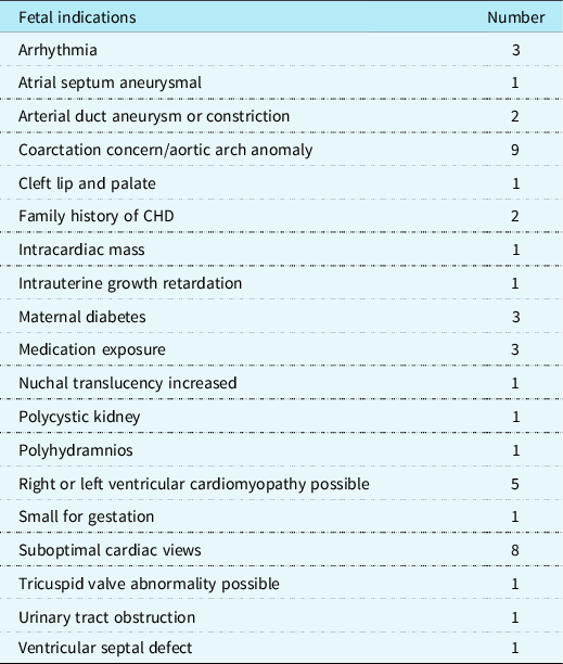
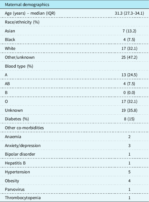
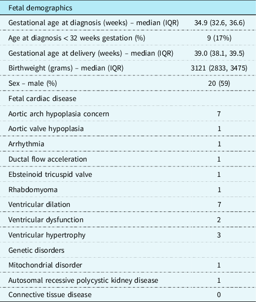
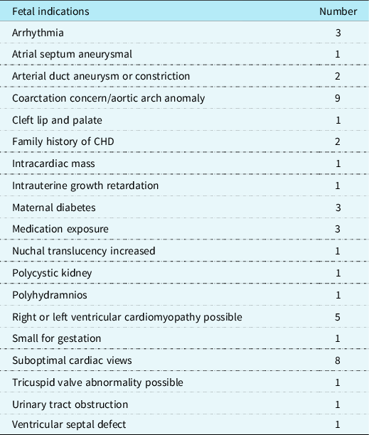
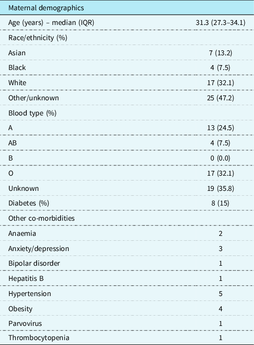
Fifty-three patients met the inclusion and exclusion criteria. Two additional patients were excluded due to severe associated CHD (hypoplastic left heart syndrome and tetralogy of Fallot). Thirty-four of these patients had postnatal data available. Seven fetuses had a repeat echocardiogram. Of these, the images of one fetal echocardiogram were too limited to make accurate measurements. The last echocardiogram occurred at a median of 5.4 weeks (IQR 3.3–7.5) after the first echocardiogram that demonstrated an arterial duct aneurysm. Table 1 lists the indications for each study. Maternal demographics are described in Table 2. Mothers were 31 years old (IQR 27.3–34.1) at diagnosis. Eight patients (15%) had diabetes, 17 (39.5%) had type O blood, and 13 (30.2%) had type A blood. Table 3 displays the fetal demographics. The diagnosis of an arterial duct aneurysm was made at a median of 34.9 weeks (IQR 32.6, 36.6) gestation, with 9 (17%) occurring before 32 weeks gestation. Twenty-five (47%) had concern for an associated cardiac findings, most of which were relatively minor. Twenty fetuses (59% of those with available data) were male.
Fetal indications
Maternal demographics
Fetal demographics
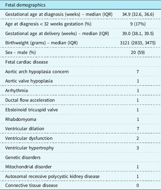
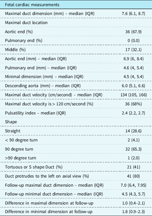
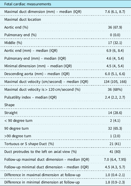
Table 4 describes the fetal echocardiographic findings. The median maximal dimension of the duct was 7.6 mm (IQR 6.1, 8.7) at the initial diagnosis. The maximal dimension was at the aortic end in 67.9%, the middle in the remainder, with no fetuses noted to have the maximal dimension at the pulmonary end. The duct protruded to the left in 80% of the cases when assessed in an axial plane at the three-vessel view. Thirty-six fetuses (68%) had ductal flow velocity greater than 120 cm/second. All of the fetuses had a maximal dimension greater than the 95% for gestational age. Five fetuses had pulsatility index of < 1.9, though only two of these had a maximal velocity greater than 120 cm/second. There was a median of 1 mm increase in the maximal dimension and 1.8 mm increase in the minimal dimension at the last echocardiogram prior to delivery. None of the pregnancies resulted in termination. The median gestational age at delivery was 39.0 weeks (IQR 38.1, 39.5). The median birthweight was 3121 g (IQR 2833, 3475). The newborns were followed for a median of 76 days of life (IQR 7.5, 368). Of 26 newborns with available data, the patent arterial duct was confirmed to be closed at a median of 6.5 days of life (IQR 2, 14). None of the fetuses were found to have associated connective tissue disease postnatally, and 2 were found to have genetic disorders not previously reported in association with fetal arterial duct aneurysm. No newborn was found to have associated symptoms or sequelae attributed to the ductal aneurysm, including no documented cases of ductal-related postnatal demise, ductal rupture, vocal cord injury, thromboembolic event, or stroke. None required ductal intervention. Three newborns died due to unrelated causes including hypertrophic cardiomyopathy, mitochondrial disease, and renal disease.
Fetal cardiac measurements
Discussion
Fetal arterial duct aneurysms present an important quandary for clinicians. While they are usually benign, self-resolving, and may even be considered a normal variant, they rarely cause devastating outcomes. Monitoring for resolution of the aneurysm postnatally may be reasonable in some circumstances, but even with monitoring the ductal size, it is not possible to predict whether and when an arterial duct aneurysm will cause a problem, such as a stroke or rupture, as these events occur suddenly. Better understanding the risk of a negative outcome will help clinicians assist patients in calibrating their level of concern.
Arterial duct aneurysm has been reported in association with Marfan Syndrome, Ehlers-Danlos Syndrome, Loeys-Dietz Syndrome, Larsen Syndrome, Smith-Lemli-Opitz Syndrome, Williams Syndrome, Trisomy 21, Trisomy 13, and MHY11 mutation. Reference Dyamenahalli, Smallhorn and Geva4,Reference Siddiqui, Polanco and DiLorenzo6–Reference van den Berg, Helbing, van Beynum and Krasemann9 The aetiology is unknown. It has been proposed that there is delayed closure of the aortic end, exposing the duct to high systemic pressure, though this does not explain cases documented prenatally. Other proposals suggest a deficiency of elastin or abnormal extracellular matrix deposition within the artery wall. Reference Aly, Contreras, Honjo and Villemain3,Reference Dyamenahalli, Smallhorn and Geva4,Reference Kirks, McCook, Serwer and Oldham10 It may also be caused by necrosis and mucoid degeneration of the medial layer of the duct. Reference Weichert, Hartge and Axt-Fliedner11
Previously reported risk factors for fetal arterial duct aneurysm include maternal diabetes, large fetus for gestational age, and maternal type A blood. Reference Jan, Hwang, Fu, Chai and Chi2,Reference Weichert, Hartge and Axt-Fliedner11 The mean gestational age for development of ductal aneurysm was 36.9 weeks gestation in one study, slightly later than the current study. Reference Tseng and Jan1 That study reported an average maximal diameter of 8 mm at initial diagnosis, similar to our findings. Like our study, no significant intracardiac findings were associated with the arterial duct aneurysm. Though, several patients in our study had concerns for aortic arch hypoplasia or ventricular dilation, and a few had concern for ventricular dysfunction or hypertrophy. Of note, a plurality of patients had Type O blood, rather than Type A, unlike data previously reported. Also, the average birthweight for our patient population was normal, and not large for gestational age as previously reported.
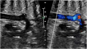
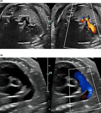
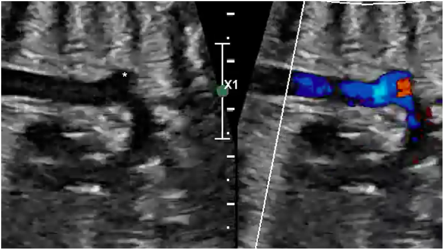
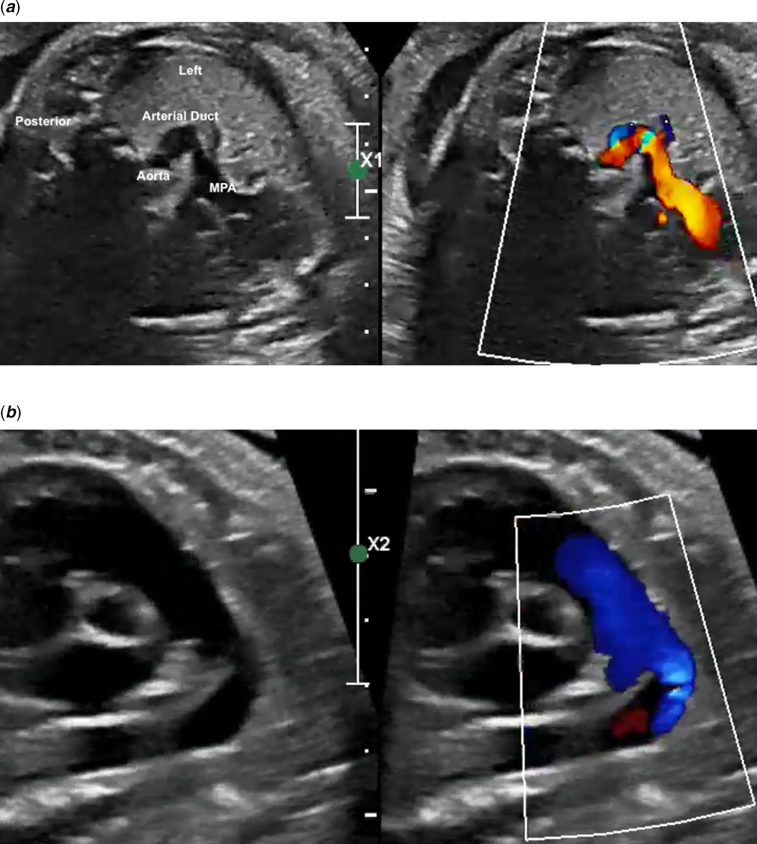
Diagnosis is often incidental, but may also be due to presenting symptoms. Sometimes a “ductal bump” can be seen on chest X-ray, which may be confused for pneumonia. Reference Jan, Hwang, Fu, Chai and Chi2,Reference Dyamenahalli, Smallhorn and Geva4,Reference Chodhary, Tomar and Radhakrishnan12 While it was previously reported that ductal aneurysm is rare in the second trimester, our study found that 17% of fetuses had concerns raised prior to 32 weeks gestation. Reference Sheridan, Michelfelder and Choe13 The duct is typically larger at its aortic aspect. It is best viewed from a sagittal view and a three-vessel view. Reference Oh and Jeung14,Reference Jackson, Sandor, Lim, Duncan and Potts15 The dilation may be fusiform or saccular, and typically protrudes leftward from the aortic arch in the three-vessel view. Reference Dyamenahalli, Smallhorn and Geva4 It is important to differentiate a ductal aneurysm with mild dilation associated with ductal constriction, though there can be overlap with fetuses demonstrating both findings. In this series, all fetuses had a maximal dimension greater than previously published normal values for gestational age. In the current study, we found that, especially with larger ducts, when viewed from a sagittal plane, the duct often extended from the proximal descending aorta posteriorly, rather than anteriorly, as expected in a normal heart (Fig 1). The duct may be tortuous, straight, or have a bend, sometimes greater than 90 degrees. Reference Dyamenahalli, Smallhorn and Geva4 By angiography, ducts were consistently found to be largest at the aortic end, and blood flow was noted to be swirling within the aneurysm by colour Doppler. In the current study, most ducts had a 90 degree turn or greater, and the aortic aspect was the largest aspect of the duct (Fig 2a and b).
Sagittal view. Note the aortic end of the arterial duct (asterisk) arising from the posterior aspect of the proximal descending aorta, as opposed to the anterior aspect expected in a normal heart.
( a ) Axial view. Note that the arterial duct bulges outward toward the fetus’s left side, the pulmonary aspect is relatively narrow, and the aortic aspect is relatively wide. It makes a 90 degree turn. ( b ) In a sagittal view, one sees the prominence of the ductal arch.
The natural history of a fetal arterial duct aneurysm is usually benign. It is thought that most self-resolve by thrombosing off and narrowing at the aortic aspect. Reference Weichert, Hartge and Axt-Fliedner11,Reference Xu, Delpey, Finel and Pennanéach16,Reference Masood, Bokowski, Kazmouz and Amin17 One study reported that 70% were closed by 35 days of life. Of the remainder, thrombosis was noted starting within 3 to 10 days of life and the aneurysm resolved by 1 month of life. Reference Jan, Hwang, Fu, Chai and Chi2 This confirmed a prior study which showed that in 15 of 20 patients with arterial duct aneurysm, the duct was closed by 20 days of life and 9 had thrombosis documented. Reference Dyamenahalli, Smallhorn and Geva4
An early study published in 1991, reported a high morbidity rate, with 31% of affected patients having complications by 2 months of age and 66% having complications between 2 months of age and 15 years of age. Reference Lund, Jensen and Hjelms18 However, this study was likely affected by selection bias. A more recent study showed that only 4 out of 24 patients developed symptoms. Reference Dyamenahalli, Smallhorn and Geva4 One patient died due to sequelae of the arterial duct aneurysm. In addition to thromboembolic events and rupture, it has been associated with recurrent laryngeal nerve compression causing vocal cord injury with stridor and dyspnoea, phrenic nerve injury with associated hemi-diaphragmatic paralysis, and left pulmonary artery obstruction from a thrombus extending from the duct into the left pulmonary artery. Reference Lund, Jensen and Hjelms18 It may also present with a unilateral wheeze due to bronchial compression. Reference Brock, Nussbaum, Shows, Nguyen and Setty19 Rare association with coarctation has been reported. Reference Hayashi, Yoshizawa and Yoshikawa20 This has also been reported to have caused fetal hydrops. Reference Aly, Contreras, Honjo and Villemain3,Reference Sheridan, Michelfelder and Choe13,Reference Fripp, Whitman, Waldhausen and Boal21 The current study, with a relatively large cohort of patients, did not find any associated sequelae.
Reported approaches to clinical follow-up and treatment are variable, and consensus has not been reached. With such a low morbidity rate, it may be reasonable to advise the family of the low but non-zero risks associated and not recommend any follow-up postnatally. This is reasonable, especially when the arterial duct is only mildly dilated. Others may choose to monitor the arterial duct until it closes or during the first 3 months of life, mainly to be able to reassure the family once the arterial duct aneurysm resolves. A large, multi-centre study will better delineate which approach is optimal. Some advise starting anticoagulation for severe, symptomatic aneurysms, Reference Aly, Contreras, Honjo and Villemain3 while others empirically ligate and divide the duct. Reference Aly, Contreras, Honjo and Villemain3,Reference Siddiqui, Polanco and DiLorenzo6 Still others recommend surgical treatment if the child presents after the newborn period, if the aneurysm is associated with connective tissue disease, if the thrombus extends from the arterial duct to an adjacent vessel, if there are signs of thromboembolism, or if there is compression of adjacent structures. Reference Dyamenahalli, Smallhorn and Geva4,Reference Xu, Delpey, Finel and Pennanéach16,Reference Manuel, Kumar and Jose22 Surgical treatment would include both ligation and resection of the duct to eliminate the risk of rupture. Reference Dyamenahalli, Smallhorn and Geva4 It is likely best to wait until after 6 weeks of life before intervening, given the data demonstrating spontaneous resolution prior to this in many affected patients. For adults presenting with sequelae of an arterial duct aneurysm, endovascular or hybrid approaches with a vascular plug may be considered. Reference Psathas, Katsargyris, Lioudaki, Moris, Doulaptsis and Klonaris23,Reference Balanescu, Linte, Dregoesc, Marc and Iancu24 Interventional occlusion has also been reported in the paediatric population. Reference Morgan, Yim, Hayes, Martin, Hamilton and Stuart25
This study was limited by its retrospective nature and by being a chart review study. While the images of each fetal echocardiogram were re-reviewed for the study, imaging windows in late gestation are limited, making accurate, reproducible measurements difficult. Also, the study could not eliminate the possibility that patients with postnatal complications presented to other institutions.
This is a large echocardiographic study that did not demonstrate any clinically significant sequelae of fetally diagnosed aneurysm of the arterial duct. It indicates that the finding is indeed usually benign, and that families can be counselled accordingly. Postnatal follow-up is likely not necessary for most patients, as there is little it would add to patient benefit. In the cases of very large aneurysms, postnatal follow-up may be indicated so that families can be reassured once the duct resolves. In either case, families should be informed of the risks and advised to see a cardiologist should the newborn develop unexplained respiratory or thromboembolic-related symptoms.
Supplementary material
To view supplementary material for this article, please visit https://doi.org/10.1017/S1047951122001925
Acknowledgements
None.
Financial support
This research received no specific grant from any funding agency, commercial, or not-for-profit sectors.
Conflict of interest
David Ezon, MD is CEO of Tapestry Insight, Inc. The company’s activities are unrelated to the content of this study.
Ethical standards
Not applicable.