Introduction
Posttraumatic stress disorder (PTSD) is a severe and debilitating mental illness that may develop after experiencing a traumatic event. The Diagnostic and Statistical Manual of Mental Disorders (DSM–5) defines a traumatic event as an experience that possesses the threat of death or serious injury to the self or others. 1 The event also induces feelings of intense fear, helplessness, or terror. Some events that frequently precede PTSD include physical assault, sexual assault, motor vehicle accidents, and combat experience.Reference Villarreal, Hamilton and Petropoulus 2 PTSD is characterized by symptoms of increased arousal, intrusive reexperiencing, avoidant behaviors, and negative changes in mood and/or cognition. 1 The National Comorbidity Survey Replication (NCS–R) estimated lifetime prevalence of PTSD among U.S. adults to be 6.8%.Reference Kessler, Berglund, Delmer, Jin, Merikangas and Walters 3 Current PTSD prevalence (within 12 months) was estimated at 3.5%.Reference Kessler, Chiu, Demler, Merikangas and Walters 4
Improving technology has caused a surge of interest in studying the neuroanatomic correlates of PTSD using magnetic resonance imaging (MRI). One structure that has received substantial research is the hippocampus. It has been shown to play a role in learning, memory storage, and retrieval.Reference Squire and Zola-Morgan 5 – Reference van Strien, Cappaert and Witter 7 Furthermore, the hippocampus specifically facilitates emotional memory storage and recall.Reference Vermetten and Bremner 8 As a result, it is likely a significant component involved in remembering traumatic memories and events. Moreover, PTSD has been linked to a variety of memory disturbances, such as an increased ability or an inability to recall aspects of a traumatic event. Meta-analytic studies of memory function in PTSD show impairments in both verbalReference Brewin, Kleiner, Vasterling and Field 9 , Reference Johnsen and Asbjørnsen 10 and visual memory.Reference Brewin, Kleiner, Vasterling and Field 9 These disruptions may result from hippocampal pathology.Reference Burgess, Maguire and O’Keefe 11 – Reference Weiss 14 Indeed, hippocampal volume has been shown to be inversely correlated with verbal memory in PTSD.Reference Bremner, Randall and Scott 15 – Reference Tischler, Brand and Stavitsky 17 In a recent study, bilateral hippocampal volume reduction was significantly associated with overgeneralization of negative contexts in an associative learning paradigm,Reference Levy-Gigi, Szabo, Richter-Levin and Kéri 18 which could be related to the triggering of intrusive “flashbacks” in this population. Moreover, the hippocampus and ventromedial prefrontal cortex (vmPFC) have been implicated in conditioned fear extinction,Reference Milad, Wright, Orr, Pitman, Quirk and Rauch 19 , Reference Milad, Orr, Lasko, Chang, Rauch and Pitman 20 which is abnormal in this population.Reference Milad, Orr, Lasko, Chang, Rauch and Pitman 20
It has been theorized that dysregulation of the hypothalamus–pituitary–adrenal (HPA) axis may contribute to hippocampal pathology.Reference Weiss 14 , Reference Höschl and Hajek 21 For example, it has been proposed that changes in hippocampal structure and function might result from chronic exposure to corticosteroids and glutamate (stress-related chemicals).Reference Vermetten and Bremner 22 Furthermore, decreased hippocampal volume might stem from a reduction in hippocampal activity following a traumatic event.Reference Teicher, Tomoda and Andersen 23 This decreased activity may result from, and further potentiate, diminished neuron density, reduced neuron branching, and the degeneration of neurons at synaptic terminals.Reference Duman, Malberg and Nakagawa 24 Additionally, PTSD is associated with decreased hippocampal N-acetylaspartate:creatine (NAA/Cr) ratios,Reference Li, Chen, Liu, Zhang, He and Lin 25 – Reference Shu, Xue and Liu 28 thought to reflect neuronal integrity. These findings suggest a link between responses to traumatic stress and structural changes in the hippocampus.
While many structural MRI studies have found smaller hippocampal volumes in participants with PTSD,Reference Villarreal, Hamilton and Petropoulus 2 , Reference Bremner, Randall and Scott 15 , Reference Levy-Gigi, Szabo, Richter-Levin and Kéri 18 , Reference Bossini, Tavanti and Calossi 29 – Reference Winter and Irle 45 others have not.Reference Schuff, Neylan and Lenoci 27 , Reference Bonne, Brandes and Giboa 46 – Reference Yehuda, Golier and Bierer 59 However, previous meta-analyses on the neuroanatomic correlates of PTSD demonstrate that it is associated with reduced volume of the hippocampus bilaterally in adults.Reference Kitayama, Vaccarino, Kutner, Weiss and Bremner 60 – Reference O’Doherty, Chitty, Saddiqui, Bennett and Lagopoulos 66
The severity of PTSD symptoms is often assessed using the Clinician-Administered Posttraumatic Stress Disorder Scale (CAPS),Reference Blake, Weathers and Nagy 67 which has three subscales: reexperiencing (CAPS–B), avoidance (CAPS–C), and hyperarousal (CAPS–D). Some data suggest that PTSD symptom severity may moderate the relationship between PTSD and reduced hippocampal volume.Reference Karl, Schaefer, Malta, Dorfel, Rohleder and Werner 62 For example, hippocampal volume has been found to vary inversely with intrusive reexperiencingReference Lindauer, Vlieger and Jalink 36 , Reference Lindauer, Olff, van Meijel, Carlier and Gersons 68 and total PTSD symptoms.Reference Villarreal, Hamilton and Petropoulus 2 , Reference Shu, Xue and Liu 28 , Reference Bossini, Tavanti and Calossi 29 , Reference Bremner, Vythilingam and Vermetten 31 , Reference Admon, Milad and Hendler 69 , Reference Apfel, Ross and Hlavin 70 However, others found no such relationships.Reference Bremner, Randall and Scott 15 , Reference Schuff, Neylan and Lenoci 27 , Reference Bossini, Tavanti and Calossi 29
Meta-analysis provides a systematic quantitative method for integrating results across studies. This not only dramatically increases sample size (and therefore statistical power) but also allows the researcher to specify moderator variables that may help account for differences in results between studies. When assessing volumetric differences in brain structures between groups, the mean volumes and standard deviations for each group are used to compute an effect size (Cohen’s d, for example), which is the difference between group means divided by the pooled standard deviation. Values of Cohen’s d of 0.20 are considered small, 0.50 medium, and 0.80 large.Reference Cohen 71 Here we provide an updated meta-analysis regarding hippocampal volume deficits in PTSD and evaluate the relationship between symptom severity and hippocampal volume by conducting a meta-regression with symptom severity (total CAPS score) as a continuous moderator variable.
Methods
Study selection
Searches for the terms “posttraumatic stress disorder,” “stress,” “hippocampus,” “hippocampal volume,” “MRI,” and “magnetic resonance imaging” were entered into the PsycINFO, MEDLINE, and ProQuest databases. Cited reference searches (both forward and backward) for relevant articles were performed using the Web of Science. The reference sections of previous meta-analyses were also examined. From these results, 89 articles published between 1995 and 2016 were retrieved. To be included in the meta-analysis, studies must have (1) used human adults (minimum age≥18 years) as participants, (2) a group diagnosed with PTSD, (3) a non-PTSD comparison group, (4) reported means and standard deviations for left and right hippocampal volumes separately, (5) not included participants from another study used in the meta-analysis, and (6) reported a mean CAPS score of 45 or greater (if the CAPS was employed). The last criterion was utilized because a psychometric study found that a total CAPS score of 45 represents a reasonable threshold for PTSD diagnosis.Reference Weathers, Ruscio and Keane 72 From the initial group of studies, 37 studies met the inclusion criteria. Mean volumetric data from Wang and colleaguesReference Wang, Neylan and Mueller 57 were obtained from the corresponding author. One studyReference Schuff, Marmar and Weiss 26 reported volume for the right hippocampus only. When studies reported volumes for both traumatized and nontraumatized control groups, data from the nontraumatized control group were utilized in the interest of sample homogeneity. If a study reported volumes separately for PTSD participants with and without comorbidities, volumes for the PTSD-only (no comorbidity) group were used.
Meta-analytic techniques
All meta-analyses were performed using Comprehensive Meta-Analysis version 2.2.064 (Biostat, Englewood, New Jersey), a commercially available software package designed for meta-analysis and meta-regression. Hippocampal mean volumes and standard deviations from each study were entered directly into the software, which then computed Cohen’s d, which in this context is the mean difference between patient and control hippocampal volumes divided by the pooled within-group standard deviation. Cohen’s d was then transformed to Hedges’ g with correction for potential bias due to sample size.Reference Borenstein, Hedges, Higgins and Rothstein 73 Hedges’ g and 95 percent confidence intervals (CI 95%) were then computed for each study. A mean effect size across studies was then calculated, with each study weighted inversely according to its variance.Reference Borenstein, Hedges, Higgins and Rothstein 73 A random-effects model was used for all analyses. Meta-regression is conceptually similar to multiple regression, except that the study becomes the unit of analysis. The reader interested in further details regarding meta-analysis is referred to the work of Borenstein and colleagues,Reference Borenstein, Hedges, Higgins and Rothstein 73 Hedges and Olkin,Reference Hedges and Olkin 74 and Cooper and coworkers.Reference Cooper, Hedges and Valentine 75
Results
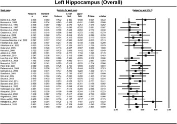
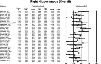
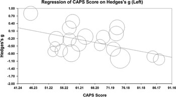
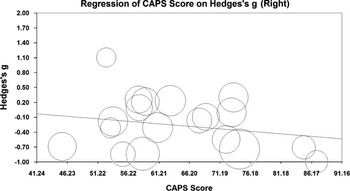
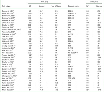
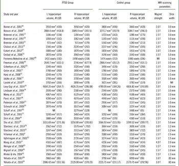
After the initial meta-analyses, two studiesReference Gurvits, Shenton and Hokama 34 , Reference Hedges, Allen and Tate 35 were identified as outliers, as they had very large effect sizes, high standard errors, and small sample sizes. Therefore, they were excluded from further consideration. Demographic information from the remaining 35 studies is provided in Table 1. Mean hippocampal volumes are given in Table 2. Total n for the left hippocampus was 1354 (PTSD=654, controls=700) and 1368 for the right hippocampus (PTSD=661, controls=707). The results of the overall meta-analysis showed significant volumetric reductions for both the left (g=–0.400; CI 95%=[–0.563, –0.237]; p<0.001; see Figure 1) and right (g=–0.462; CI 95%=[–0.621, –0.302]; p<0.001; see Figure 2) hippocampi in participants with PTSD compared to non-PTSD controls. These results correspond to a volumetric reduction of 5.24% for the left hippocampus and 5.23% for the right hippocampus. Secondary meta-analyses conducted only on those studies that reported CAPS scores again showed significant, but smaller, reductions in both the left (g=–0.307; CI 95%=[–0.546, –0.069]; p=0.012) and right hippocampus (g=–0.243; CI 95%=[–0.465, –0.022]; p=0.031). These results correspond to a volumetric reduction of 4.02% for the left hippocampus and 2.55% for the right hippocampus. However, only 19 studies reported CAPS scores (see Table 1). The meta-regression of CAPS scores on Hedges’ g for the left hippocampus (slope=–0.024; intercept=1.20; Z=–2.92, p<0.004; see Figure 3) was significant, but the meta-regression for the right hippocampus (slope=–0.010; intercept=0.38; Z=–1.26, p<0.21; see Figure 4) was not. In other words, increased symptom severity was significantly associated with decreased volume of the left, but not the right, hippocampus.
Forest plot showing overall left hippocampal volume effect sizes (Hedges’ g) and 95% confidence intervals (CI 95%) for comparisons of PTSD and control groups. Negative effect sizes (Hedges’ g) indicate smaller hippocampal volume in the PTSD participant group. Square size indicates relative study weight in the meta-analysis. Mean effect size and confidence interval across studies are indicated by the solid diamond.

Forest plot showing overall right hippocampal volume effect sizes (Hedges’ g) and 95% confidence intervals (CI 95%) for comparisons of PTSD and control groups. Negative effect sizes (Hedges’ g) indicate smaller hippocampal volume in the PTSD participant group. Square size indicates relative study weight in the meta-analysis. Mean effect size and confidence interval across studies is indicated by the solid diamond.

Meta-regression of participants’ CAPS scores on Hedges’ g for the left hippocampus. Circle size indicates relative study weight in the meta-regression. Slope=–0.024; intercept=1.20; Z=–2.92, p<0.004.

Meta-regression of participants’ CAPS scores on Hedges’ g for the right hippocampus. Circle size indicates relative study weight in the meta-regression. Slope=–0.010; intercept=0.38; Z=–1.26, p<0.21.

Demographic information for PTSD and control participants in studies that reported mean hippocampal volumes.

* Gender breakdown not reported. CAPS=Clinician-Administered PTSD Scale; DSM=Diagnostic and Statistical Manual of Mental Disorders; DTS=Davidson Trauma Scale; F=number of female participants; ICD=International Classification of Diseases; M=number of male participants; M–PTSD=Mississippi Scale for Combat-Related PTSD; NR=not reported; SCID=Structured Clinical Interview for DSM–IV; SI–PTSD=Structured Interview for Posttraumatic Stress Disorder.
Hippocampal means (M)/standard deviations (SD), and scanning parameters for studies included in the meta-analysis

Volumes not originally reported in mm3 have been converted to mm3 for ease of comparison, with the exception of one studyReference Fennema-Notestine, Stein, Kennedy, Archibald and Jernigan 48 for which this was not possible, as voxel dimensions were not reported. mm=millimeter; mm3=cubic millimeter; NR=not reported; T=tesla.
Discussion
This meta-analytic study produced two main findings. First, PTSD in adults was associated with significant bilateral volumetric reduction of the hippocampus, as has been reported in previous meta-analyses.Reference Kitayama, Vaccarino, Kutner, Weiss and Bremner 60 – Reference O’Doherty, Chitty, Saddiqui, Bennett and Lagopoulos 66 Second, increased PTSD symptom severity was associated with decreased left hippocampal volume. No association was found between symptom severity and right hippocampal volume. This conclusion, however, must be viewed with caution, as studies that reported CAPS scores had lower mean effect sizes than those in the overall meta-analysis. Therefore, the relationship between symptom severity and right hippocampal volume may have been underestimated.
Because of the cross-sectional nature of the studies reviewed here, the observed volume differences in the overall meta-analysis could either indicate that PTSD produces hippocampal volume deficits or that they are a predisposing factor. A recent model of PTSD suggests that a hyperresponsive amygdala and dorsal anterior cingulate cortex (dACC) are predisposing factors to the development of the disorder, while reduced hippocampal connectivity with the vmPFC is a consequence of it.Reference Admon, Milad and Hendler 69 Additional research demonstrates that current, but not lifetime, PTSD symptomatology predicts hippocampal volume deficits.Reference Apfel, Ross and Hlavin 70 If smaller hippocampal volume were a predisposing factor to PTSD, one would expect that both current and lifetime symptom severity would be associated with volumetric deficits.Reference Apfel, Ross and Hlavin 70 Furthermore, in an 18-month prospective study of Israeli Defense Force soldiers pre- and postcombat, increased PTSD symptomatology postcombat was associated with reduced hippocampal volume.Reference Admon, Leykin and Lubin 76 Additionally, in a study of veterans with brain lesions, the vmPFC and amygdala were found to be critical in the etiology of PTSD, while the hippocampus was not.Reference Koenigs, Huey and Raymont 77 These findings suggest that the hippocampal volume changes observed in PTSD are a result of the disease rather than its cause. However, a study of monozygotic twins (one with and one without PTSD) found that PTSD symptom severity in the affected twin was inversely associated with hippocampal volume in both PTSD and non-PTSD twins.Reference Gilbertson, Shenton and Ciszewski 50 This suggests that reduced hippocampal volume constitutes a predisposition to PTSD.Reference Gilbertson, Shenton and Ciszewski 50 A more recent study also suggested that left hippocampal volume reduction is a risk factor for the persistence of PTSD.Reference van Rooij, Kennis, Sjouwerman, van den Heuvel, Kahn and Geuze 78 Clearly, more prospective studies are necessary to determine the reasons for these hippocampal volume differences.
The finding that left hippocampal volume was significantly and inversely associated with symptom severity is consistent with the results of several previous studies. For example, total CAPS score and CAPS–B score significantly predicted left hippocampal volume deficits in a civilian PTSD sample, while time since trauma, illness duration, and age did not.Reference Villarreal, Hamilton and Petropoulus 2 Another study also found a significant correlation between CAPS total and CAPS–C score and left, but not right, hippocampal volume.Reference Shu, Xue and Liu 28 Additionally, CAPS–B score was inversely correlated with left hippocampal volume in a sample of Dutch police officers.Reference Lindauer, Vlieger and Jalink 36 , Reference Lindauer, Olff, van Meijel, Carlier and Gersons 68 Furthermore, in a study of survivors of a Chinese coal-mining disaster using both voxel-based morphometry (VBM) and region of interest (ROI) approaches, reduced gray matter volume in the left hippocampus and left parahippocampal gyrus were observed.Reference Zhang, Tan and Yin 79 There was also reduced gray matter density in the left hippocampus. While there was no correlation between left hippocampal volume and total CAPS score, a significant inverse correlation (r=–0.49) was found between left hippocampal gray matter density and total CAPS score.Reference Zhang, Tan and Yin 79 These findings suggest that increased PTSD symptom severity may be associated with hippocampal pathology in ways that are not always observable using traditional volumetric MRI techniques. Similarly, in a study of civilians with recent-onset PTSD, significant inverse correlations were found between left hippocampus NAA/Cr ratio and total CAPS (r=–0.939), CAPS–B (r=–0.829), CAPS–C (r=–0.743), and CAPS–D (r=–0.635) scores.Reference Shu, Xue and Liu 28 No such relationships between NAA/Cr ratio and symptom severity were observed for the right hippocampus.Reference Shu, Xue and Liu 28 However, other studies examining the effects of PTSD on the hippocampus have not found any effects of symptom severity on hippocampal volume.Reference Bremner, Randall and Scott 15 , Reference Schuff, Neylan and Lenoci 27 , Reference Bossini, Tavanti and Calossi 29 Taken together with the current results, these findings suggest that PTSD damages the left hippocampus more than the right hippocampus and that this relationship may manifest itself in additional ways other than those observable with MRI volumetry.
Study limitations
There are several factors that limited this study. Not all studies investigating hippocampal volume reduction in PTSD reported mean CAPS scores, and those that did had smaller effect sizes than those that did not. This may have led to the underestimation of the relationship between symptom severity and hippocampal volume, especially for the right hippocampus. Furthermore, we were unable to account for some potentially important moderator variables, such as illness duration (only seven studies in this sample reported illness duration), psychiatric comorbidity (which is high in this population), MRI scanning parameters, and substance abuse, all of which may affect reported hippocampal volume. There was also substantial variance in the anatomical boundaries used to delineate the hippocampus. This variance may at least partially explain the wide range in hippocampal volumes reported in the studies included in this meta-analysis. Finally, as the majority of studies examined were cross-sectional designs, rather than longitudinal, we cannot unequivocally state whether or not the observed differences in hippocampal volume were consequences of the disorder or predisposing factors.
Conclusions
In conclusion, the studies reviewed in this meta-analysis indicate that PTSD is associated with moderate bilateral volumetric reduction of the hippocampal formation in adults. Increased symptom severity appears to be associated with reduced left, but not right, hippocampal volume in this group of studies. More prospective studies are needed to clarify the etiology of the differences in hippocampal volume associated with PTSD.
Acknowledgments
The authors wish to thank Anna Marie Medina, PhD, and Gary L. Thorne, PhD, for helpful comments concerning this manuscript, and Norbert Schuff, PhD, for providing hippocampal volumes not included in a published article.
Disclosures
Michael Nelson and Alecia Tumpap hereby state that they have no conflicts of interest to disclose.