Ventricular septal defect is a common congenital cardiac malformation, and percutaneous closure of ventricular septal defect has emerged as a valuable alternative to surgical treatment for certain kinds of ventricular septal defect.Reference Lim, Forbes, Rothman, Lock and Landzberg1,Reference Carminati, Butera and Chessa2 Compared to the surgical approach, which has a 60-year history, transcatheter closure of ventricular septal defect is a newer procedure. As the transcatheter approach is associated with the benefit of shorter hospitalisation time and does not require cardiopulmonary bypass and sternotomy.Reference Butera, Carminati and Chessa3,Reference Xunmin, Shisen, Jianbin, Haidong and Lijun4 However, significant controversy exists with transcatheter closure of ventricular septal defect compared with open surgical techniques. During the development and clinical application of transcatheter closure, significant problems were encountered such as acute aortic insufficiency, haemolytic anaemia, tricuspid regurgitation, device embolisation, endocarditis, and complete heart block. Our previous large sample study shows that by selecting appropriate patients, using proper manoeuvring, and avoiding oversised devices, atrioventricular block is no longer an obstacle to transcatheter ventricular septal defect closure using symmetric devices.Reference Wang, Cao and Li5 Short-term and mid-term follow-ups have indicated that ventricular septal defect closure with the occlude is a safe and effective method for the management of medium to some large ventricular septal defects,Reference Yang, Yang and Yu6,Reference Zheng, Zhao and Zuo7 but the long-term outcome of this interventional approach remains unclear.
To the best of our knowledge, this is the first report of aortic regurgitation caused by occlude 9–12 years following transcatheter closure and regurgitation were gradually increased.
Case report
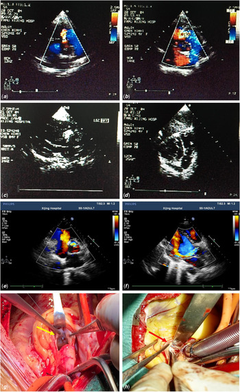
Four children were admitted to our department with severe aortic regurgitation. According to their medical histories, the patients all underwent transcatheter ventricular septal defect closure approximately 10 years ago. The details of each patient and ventricular septal defect type are listed in Table 1. The anatomy of four cases of ventricular septal defect were simple defects with no aneurysm. They all met the indications for transcatheter closure by then, which included 1) age > 3 years; 2) weight > 15 kg; 3) the size of ventricular septal defect between 3 to 10 mm; and 4) the length of subaotic rim more than 2 mm. All of them were discharged following the procedure without any complications, as control aortography after occluder placement did not show aortic regurgitation. All four patients underwent clinical examination, Holter or electrocardiographic monitoring, chest roentgenogram, and transesophageal echocardiography were performed at 1, 6, and 12 months after the procedure and yearly thereafter. Moderate aortic regurgitation was observed in 4 patients 9–12 years following transcatheter closure and regurgitation were gradually increased (Fig 1). All four patients need to undergo aortic valve replacement according to examination and indications.
Clinical characteristics of the four patients.

AR = Aortic regurgitation; VSD = ventricular septal defect.
Echocardiography and aortic valve replacement surgery of the patients. Panels (a, c, e and g) and (b, d, f and h) belong to two patients, respectively. Panels (a and b) show the perimembranous VSD before transcatheter closure. Panels (c and d) show the images after the occlusion procedure. Panels (e and f) show aortic regurgitation after the transcatheter closure of VSD. Panels (g and h) show that the aortic valve was encroached upon by the occluder, leading to severe aortic valve insufficiency. Yellow arrow shows the waist of occluder. Red arrow shows the margin of occluder disk. VSD = ventricular septal defect.

Surgery was performed through median sternotomy. Cardiopulmonary bypass was established via ascending aortic cannulation and right atrium drainage. A venting tube was placed in the left atrium through the right superior pulmonary vein. After cardiac arrest, aortotomy was performed. We found that the aortic valve was encroached upon by the occluder, which impaired mobility of left coronary cusp and leading to severe aortic valve insufficiency (Fig 1). A mechanical heart valve/biological heart valve was implanted after valve excision. In each case, the patient was then weaned off cardiopulmonary support uneventfully. All four patients recovered well following the operation.
Discussion
The standard treatment of ventricular septal defect is open heart surgery, which requires cardiopulmonary bypass and has minimal operative mortality and morbidity. However, surgical repair albeit with a high success rate, negligible mortality, and good long-term outcomes is associated with morbidity, discomfort, and thoracotomy scars. Thus, transcatheter closure has become an alternative way to treat selected kind of ventricular septal defect. Short-term and mid-term follow-ups have indicated that the transcatheter closure of ventricular septal defect is safe and effective.Reference Yang, Yang and Wan8 However, the implantation of a foreign body in the heart raises the possibility of heart block, valve problems, and occluder migration.Reference Pamukcu, Narin, Baykan, Sunkak, Tasci and Uzum9 Clinicians have certain doubts regarding transcatheter ventricular septal defect closure.
According to a review by Butera et al, the total number of percutaneous closures of ventricular septal defect in United States of America and European countries was less than 1000 cases.Reference Pamukcu, Narin, Baykan, Sunkak, Tasci and Uzum9 Compared with the United States of America and with European countries, China has a more open attitude towards the transcatheter closure of ventricular septal defect; Thus, the implementation of transcatheter closure occurred earlier and more widely in China than in other nations due to concerns of cardiopulmonary bypass risk and to cosmetic concerns related to the incision used in traditional surgery.
In our case, we first found that the occluder was encroached upon the aortic valve and leading to aortic valve insufficiency long time after transcatheter ventricular septal defect closure. A progressive aortic regurgitation could be a long-term complication of transcatheter closure of ventricular septal defect. In our department, the amount of transcatheter ventricular septal defect closure surgery was approximately 800 per year since 2000, and we mainly use symmetric ventricular septal occlude. We found four cases out of approximately 4800 patients which performed ventricular septal defect closure in 2000–2006, and the incidence of occluder leading aortic regurgitation was 0.1%. However, the number may be underestimated due to the lack of physical examination and follow-up. There is reporting that tricuspid regurgitation occurred 2 years after transcatheter closure of ventricular septal defect.Reference Butera, Chessa and Carminati10 Our findings raise an alarm for clinicians who are using occlusion to treat ventricular septal defect. These clinicians must account for the serious long-term complication of aortic regurgitation, and the patients and their families should also be informed of this serious complication before they chose the operation procedures. For those who received transcatheter closure should do regularly examination in order to early detection of complications such as aortic regurgitation.
Conclusions
Our findings may be representative of a series of cases in which patients may experience aortic regurgitation after undergoing the transcatheter closure of ventricular septal defect. Despite encouraging short- and mid-term follow-ups of transcatheter closure of ventricular septal defect, this technique is still investigational. Longer follow-up is necessary to determine the risk of bacterial endocarditis and to ascertain the safety of the procedure with regard to trauma to the conduction system and aortic and tricuspid valves. However, the absence of such complications in the short- and mid-term does not preclude their occurrence in the long-term follow-up. Multi-centre cooperation may be a good way to clarify this issue.
Acknowledgements
This work was supported by grants from the National key research and development program (2018YFC1002603 to Yuehu Han) and the Boost foundation (XJZT18MJ48 to Yuehu Han) and Natural science foundation of Shaanxi province (2019JQ-575 to Yuehu Han).
Financial Support
This research received no specific grant from any funding agency, commercial or not-for-profit sectors.
Conflicts of Interest
None.
Ethical Standards
Ethical approval: All procedures performed in studies involving human participants were in accordance with the ethical standards of the institutional.
Informed consent: Informed consent was obtained from all individual participants included in the study.