Introduction
Fasciola hepatica is a trematode parasite that establishes in the liver of the final host. It infects several animal species including humans, demonstrating its anthropozoonotic scope (Mas-Coma et al., Reference Mas-Coma, Valero and Bargues2009). Parasite prevalence has been reported to range from 0.12 to 100% in sheep from Argentina, USA, Ireland and elsewhere, contaminating more than 300 million cattle and water buffaloes worldwide (Byrne et al., Reference Byrne, McBride, Lahuerta-Marin, Guelbenzu, McNair, Skuce and McDowell2016; Mehmood et al., Reference Mehmood, Zhang and Sabir2017, Pritsch et al., Reference Pritsch, Stanula, dos Anjos, Bertot and Molento2019). The disease may cause production losses of US$90–210 million annually (Rahmeto et al., Reference Rahmeto, Fufa, Mulugeta, Solomon, Bekele and Alemayehu2010; Molento et al., Reference Molento, Bennema, Bertot, Pritsch and Arenal2018), and Dutra et al. (Reference Dutra, Molento, Naumann, Biondo, Fortes, Savio and Malone2010) reported that F. hepatica is endemic in Rio Grande do Sul (RS), a state located in the south of Brazil, with an average prevalence of 14.3%. The distribution of F. hepatica in Brazil is extensive in the southern region and its presence reaches up to the south border of the Amazon region (Bennema et al., Reference Bennema, Scholte, Molento, Medeiros and Carvalho2014, Reference Bennema, Molento, Scholte, Carvalho and Pritsch2017; Pritsch & Molento, Reference Pritsch and Molento2018).
Most of the economic losses from F. hepatica arise from a decrease in milk production and reduce weight gain. According to Arias-Pacheco et al. (Reference Arias-Pacheco, Lucas, Rodríguez, Córdoba and Lux-Hoppe2020), the losses due to liver condemnation may reach 55.72%, with a market value of US$35.080, and a direct loss by the reduction in weight gain of approximately US$354.570 in Peru. In acute or chronic infections, daily blood loss per animal was determined to be about 0.2 to 0.5 ml per worm (Khan et al., Reference Khan, Sajid, Khan, Iqbal, Arshad and Hussain2011). This is due to the invasion of a large number of migrating young flukes, and the posterior aggregation of inflammatory cells in the cicatricial tissue, causing parenchymal destruction, liver failure and bleeding into the peritoneal cavity, leading to haemorrhagic hepatitis. The parasite damage to the host tissue is seen by hepatobiliary injury, and the host response is determined by fibro-cellular reactions. Changes in the metabolic activity of rat hepatocytes exposed to the somatic proteins of F. hepatica revealed that the protein concentration, as well as the duration of their action, decreased the longevity of these cells (Trivilin et al., Reference Trivilin, Sousa, Carvalho Nunes, Sousa Rocha and Martins2014; Taibi et al., Reference Taibi, Aissi, Harhoura, Zenia, Zait and Hamrioui2019).
Analysis of economic losses due to fluke infection in livestock has a high degree of uncertainty, resulting from the numerous possible causes of injury attributed to infections. The multifactorial effects of F. hepatica on animals and human health suggest that modelling approaches may be useful to calculate the impact of the disease in cattle (Charlier et al., Reference Charlier, Levecke, Devleesschauwer, Vercruysse and Hogeveen2013). Disease-based simulation studies are being applied to study fasciolosis, providing additional support for diagnostic and control strategies in Europe and South America (Kantzoura et al., Reference Kantzoura, Kouam, Demiris, Feidas and Theodoropoulos2011; Silva et al., Reference Silva, Freitas, Dutra and Molento2016, Reference Silva, Freitas, Dutra and Molento2020; Molento et al., Reference Molento, Bennema, Bertot, Pritsch and Arenal2018). To overcome these difficulties, risk analysis simulations such as Monte Carlo have been used to control the variability of some parameters (Schweizer et al., Reference Schweizer, Braun, Deplazes and Torgerson2005). This technique is generally applied to situations where the probability of an event is difficult or impossible to calculate by algebraic methods. Additionally, a better understanding of the impact of fascioliasis in cattle may contribute to disease control programs and the improvement of animal welfare. The Susceptible-infected-recovered (SIR) epidemic model is made of ordinary equations based on the rate of susceptible (S), infected (I) and the recovery (R) of individuals from viral infection (Vynnycky & White, Reference Vynnycky and White2010). This model is being vastly used to determine Severe acute respiratory syndrome coronavirus 2 (SARS-CoV-2) infection, responsible for the coronavirus disease 2019 (COVID-19) pandemic, giving the possibility to predict the virus evolution and measure the success rate of sanitary protocols (social distance, use of mask, etc.).
The aim of this study was to characterize the injuries caused by F. hepatica in the liver and the impact in weight gain of infected and uninfected cattle. This research also focused on correlating the incidence of the disease (quantitative sampling) in slaughtered animals from an endemic region in Brazil and the economic impact of the parasite to the beef cattle industry in the region.
Material and methods
Experiment 1: area, animals and clinical measurements
The experiment was conducted in the city of Dom Pedrito (30°58′56″S, 54°40′91″W), RS, south of Brazil. The farm was located at an altitude of 300 m above sea level. For this experiment, 18 adult Hereford cows were used. This European breed is representative of the livestock population in the entire Pampa region. For the inclusion criteria, all animals were checked with faecal egg count (FEC) sedimentation method (Girão & Ueno, Reference Girão, Ueno, Ueno and Gonçalves1994), and were positive for F. hepatica. Nine animals were kept positive for F. hepatica (FhG group) and nine were treated with triclabendazole (TBZ) in four occasions (30, 60, 150 and 240 days) to avoid infection (TbG group). Fasciola hepatica infection was monitored individually by performing FEC examinations, which were carried out every 21 days, over ten months. The animals were weighed six times at bimonthly intervals, including day 0 (zero). All animals had similar weight (P < 0.05) at the start of the experiment and were subjected to similar care over the experimental period. Food and water were provided ad libitum.
Liver histopathology
At the end of the samplings, all animals were sent to a slaughterhouse approved by the Federal Service of Meat Inspection, from the Ministry of Agriculture. Samples from all livers were collected from FhG and TbG for histological analysis. The livers were inspected to determine the degree of injury in the tissues, using a ranking system (absent, mild, moderate and severe lesions), established by Bostelmann et al. (Reference Bostelmann, Luz, Thomaz-Soccol and Cirio2000). The histology data from Experiment 1 were used to determine and to correlate the degree of injuries in the livers of animals from Experiment 2 (see the following section). Tissue sections were fixed in 10% formalin, and, after fixation, the tissues were dehydrated, diaphanized and embedded in paraffin, after which 10–155-um slices were cut from each section (FhG and TbG). The tissues were stained with haematoxylin and eosin prior to the analysis. To determine the degree of fibrosis, the sections were stained with Masson's trichrome marker.
Experiment 2: quantification of parasites from a large liver sampling and disease distribution
Based on the description of Experiment 1 and our previous data (Dutra et al., Reference Dutra, Molento, Naumann, Biondo, Fortes, Savio and Malone2010), assuming an average of 5 to 15% of liver condemnation, a total of 12,236 cattle from multiple breeds (Red Angus, Hereford, Brangus, Charolais) were inspected. Liver was collected from all animals and parasite specimens were counted from livers with suggestive external lesions. The organs were open with approximately 10–15 1-inch sections close to the lesions. The content was passed through a 1 mm sieve under warm water. Parasites were identified by their morphological characteristics (Periago et al., Reference Periago, Valero, Panova and Mas-Coma2006). The level of infection was divided from 0 to 5, where a gross score of 0 represented liver lesions but no parasites; 1 represented 1–5 parasites; 2 represented 6–50 parasites; 3 represented 51–100 parasites; 4 represented 101–250 parasites; and a score of 5 represented more than 250 parasites. Individual carcass weight was obtained to correlate the score with the number of parasites, as well as the description of all macroscopic lesions.
On average, animals are slaughter at 30 months of age (Cucco et al., Reference Cucco, Abdalla, Chiocca, Sá, Zieher, Agostini and Theodorovicz2015) in RS, with approximately 400–600 kg. As calves are raised on pasture, they have the opportunity to get infected from early age (Molento, pers. obs.); therefore, it is reasonable to consider that the animals in RS have been infected for at least 20–22 months at slaughter.
Statistical analysis, Monte Carlo simulation and SIR modelling
For Experiment 1, the data were tested for normality using the Kolmogorov–Smirnov test. A descriptive analysis of the data from the 18 animals from the treated and untreated groups was performed using a generalized linear mixed model (GLMM). The analysis used weight as a dependent variable, and the independent variables comprised the groups, the individual animals and the time (month), using the following equation (Breslow & Clayton, Reference Breslow and Clayton1993).
Linear mixed model:

This model was used to determine the individual differences, assuming the random variables with an intercept to each animal. Intercepts of different values were generated to each animal, and the GLMM represents the estimation of these intercepts. The GLMM analysis was run using R version i386 4.0.0 (The R Foundation for Statistical Computing, Vienna, Austria), with the lm4 package. A Student's t-test was also performed between the two groups (n = 18).
For the risk analysis of the weight loss caused by the trematode, the weight of the animals, period and treatment group were determined by Monte Carlo simulation using R version i386 4.0.0 with P < 0.05 for the first set of data (Experiment 1). For Experiment 2, a descriptive analysis was made to characterize the distribution of the disease by breed, regions of the state and carcass weight. The results from the weight differences (Experiment 1) and the quantified proportion of infected livers (Experiment 2) were used to estimate the economic impact of F. hepatica to the beef cattle industry of RS. For the economic extrapolation, we used the total slaughter data (total carcass weight) of cattle under the State and Federal Meat Inspection (IBGE, 2016). The average cost of meat in 2015 (IEPE, 2016) and the contribution of the beef industry to the gross domestic product were based on the Brazilian Livestock Industry Input using the CEPEA/USP/Brazil platform (https://cepea.esalq.usp.br/br) (CEPEA, 2016).
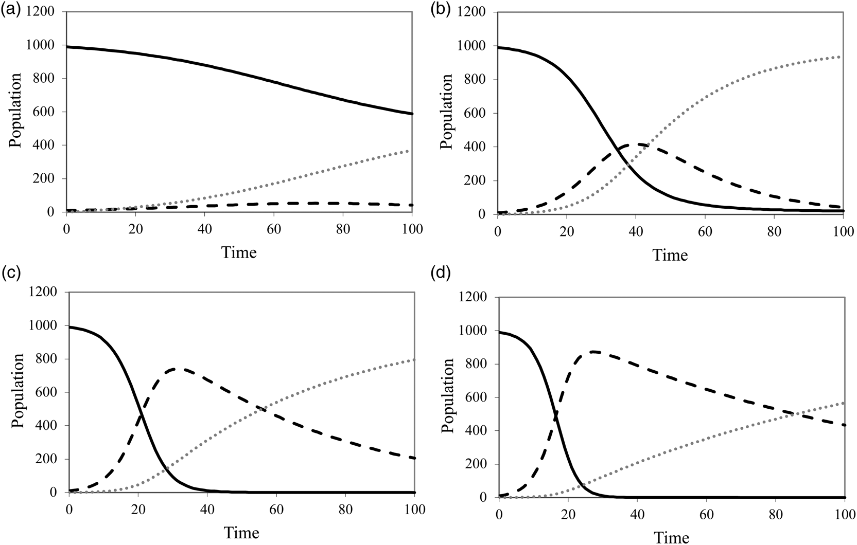
The SIR model was used to determine the present level of infectivity and also other distinctive scenarios, using a fixed number of animals (n = 1000), interaction level (0.001), infected at the start (10) and time interval (1). The assumptions at the start were: time equals to zero plus one (t = 0 + 1), susceptible equals to negative (S = −N), as infection had to start with one infected individual (I = 1), and different recovery rates (R = 0.10 to 0.01) (table 1). The four specific conditions were established on the different disease prevalence in the state of RS (low, mild, moderate, severe), based on the positivity index described by Silva et al. (Reference Silva, Freitas, Dutra and Molento2016, Reference Silva, Freitas, Dutra and Molento2020). Condition A represents, an optimal situation with minimal clinical cases and a high recuperation level; condition B represents a mild level of prevalence in an area with a low level of recuperation; condition C has a moderate number of infections, with a decreasing number of recuperations; and condition D is a severe situation, with a high number of infectivity and a very low number of recuperations. The following equation was used for the model:

where S is the number of susceptible individuals, a is a constant for the level of disease contact (dispersal/host–parasite interaction), I is the number of infected individuals; t is a unit of time; (–) minus assumes that the number of healthy individuals will diminish; and u is a parameter representing the rate of loss of infection in infected animals and is proportional to the inverse of the life expectancy of the parasite. The term uI represents the increase in susceptible individuals caused by the loss of parasites in infected animals. The data from experiments 1 and 2 were used to run the SIR model based on the disease condition of cattle in RS.
Established conditions for the SIR model for Fasciola hepatica infection in cattle in Rio Grande do Sul, Brazil.

Results
Experiment 1
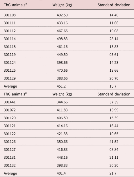
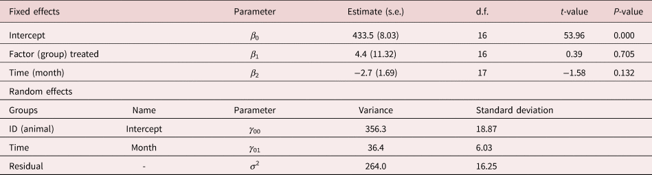
The Kolmogorov–Smirnov test showed that the data had a normal distribution. The control untreated group had a Decimal reduction time/number (D-value) equal to 0.164 and P = 0.937, and the TBZ group had D = 0.244 and P = 0.573. Therefore, the t-test was used for comparison. We determined a change in the weight of both groups along the experimental period, with a statistically significant difference (P = 0.0098) between the groups. The weight gain of the animals had a reduction in both groups during the third and fourth periods, probably due to the occurrence of a short drought, and a decrease in pasture availability. The temporal average weight gain for the fluke-free cattle was constant, showing a strong positive correlation with carcass weight (R 2 = 0.82). The FhG showed a ratio of R 2 = 0.74, with a strong tendency to lose weight (table 2). Therefore, considering the average weight of TbG and FhG of 451.2 kg and 401.4 kg, respectively, at the end of Experiment 1, there was a difference in weight of about 11% (49.8 kg) between groups. In table 3, it can be seen that the estimated values forβ 0, β 1 andβ 2 for the fixed effects refer to the 18 animals. Therefore, the estimate β 0 = 433.48 kg (intercept) is relative to the average weight of the animals; β 1 = 4.36 corresponds to the group 2 (treated animal) factor, indicating that group 1 (control animals) is the reference (control group = intercept). Both groups had similar F. hepatica egg counts (P > 0.05) at the start of the experiment, when TbG received the first treatment. We observed no statistical differences (P > 0.05) from the FEC among infected animals, ranging from 150 to 2025 during the entire period. TbG had consistently low FEC, with one animal presenting a maximum of three eggs in one count. The overall efficacy of TBZ was above 98%.
Final individual weight and standard deviation after six measurements from nine naturally infected cattle with Fasciola hepatica (FhG) and nine treated with triclabendazole (TbG) in Brazil.

a Animal numbers refer to their individual tag identification, as all animals were part of the National Program of Cattle Traceability of Brazil.
Estimated parameters and fit statistics of the model that corresponds to a t-test fit to the Fasciola hepatica groups.

s.e., standard error associated to each parameter; d.f., degrees of freedom of each model.
Liver histology
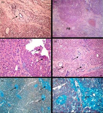
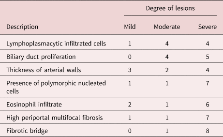
The histological analysis showed the presence of lesions that were suggestive of chronic fascioliasis with angiocholitis, thickening of the portal wall and eosinophil infiltration, in addition to the presence of widespread fibrotic tissue. Figure 1 shows the histopathological findings in FhG livers with varying degrees of infection. Table 3 describes the degree of these lesions in the infected organs. In this study, liver fibrosis was observed in three animals (fig. 1a and table 4). The presence of numerous biliary ducts with similar injuries were evident, with differences only in the location of acinar formations, which were arranged along the middle third of the walls with lymphocyte, plasma cell and eosinophils infiltration (fig. 1b, c and table 4). Calcification was observed in the duct walls of two livers (fig. 1d and table 4). The lesions were moderately correlated with clinical sings (0.467), as we observed three animals showing apathy and anaemia (haematocrit <18%) in the FhG. For the TbG, the lesions (recovering after five treatments) were not correlated with clinical sings (−0.071), as only one animal showed signs of apathy and anaemia (haematocrit: 25%) after 150 days of the first treatment. These data were used for liver evaluation in Experiment 2.
Histological images of Fasciola hepatica-infected cattle livers. The arrows indicate: (a) moderate thickness of the wall of the portal area; (b) fibrotic bridge (FB); (c) nucleated polymorphic infiltrated cells/eosinophils; (d) excessive multifocal periportal fibrosis; (e) Masson stain with trichrome, showing connective tissue (CT), obj. 10×; (f) Masson stain with trichrome showing connective tissue (CT), obj. 40×.

Histopathological findings from cattle livers infected with Fasciola hepatica and the degree of lesions at necropsy.

Monte Carlo simulation
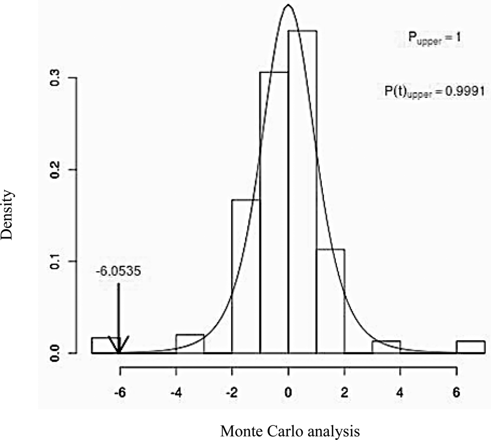
For the Monte Carlo simulation, a bootstrap analysis of 1000 replicates were run using the weight data in random drawings. The confidence interval of 95% was between 0.312 and 74.866 (fig. 2). The output demonstrated solid evidence that the differences between groups were due to the presence of the infection (FhG) and the treatment (TbG) (P < 0.0001). The results from the analysis suggest that although the month and climate variations may have had an impact in the production parameters, the differences in weight between groups were attributed to F. hepatica infection.
Histogram of the Monte Carlo analysis referring to the calculation of the total weight for the Fasciola hepatica and control groups. Value of the test: −6.0535, corresponding to P = 0.0001. The confidence interval of 95% was between 0.312 and 74.866.

Experiment 2
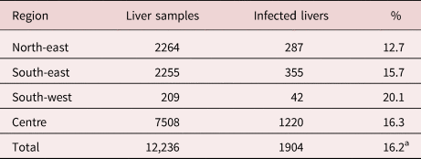
The incidence of infected livers from different regions of RS was above 16% (table 5). Animals from the south-west region had the highest percentage (20.1%), possibly because of the extensive farm management and the low altitude of the terrain. The incidence of hepatic hypertrophy (24%), biliary duct calcification (71%), purulent calcification (20%), dark brown-coloured thick exudate (52%) and duct thickness (90%) were the main external and internal organ lesions observed in the infected livers. From the 1904 positive livers, the distribution was 30.31, 35.03, 31.88, 2.36, 0.39 and 0% for categories 0, 1, 2, 3, 4 and 5, respectively. We did not find a statistically significant correlation between parasite incidence and cattle breed (data not shown), and no clinical signs were observed before slaughter. Although several factors (i.e. treatment history, nutrition, farm management, acute/chronic infections, parasite aggregation) could be responsible for animal performance, we have determined a low correlation value (R 2 = 0.354) between carcass weight and parasite numbers. These findings reveal that field animals would benefit from diagnostic and treatment of the disease, as also reported in Experiment 1. It was also confirmed that animals deteriorate when disease conditions are maintained with no diagnosis.
Incidence (%) of Fasciola hepatica in infected livers of cattle from the main cattle-raising regions of Rio Grande do Sul, Brazil.

a Average of percentages.
The total weight difference was determined by calculating the initial and final weight of each group, being −15.7 kg for the FhG and 21.7 kg for the TbG. As a consequence, the estimated loss caused by fascioliasis was US$56.60/animal (retail price) deducted from the carcass weight of each animal at slaughter. Based on the Monte Carlo simulation (Experiment 1), it may be considered that this value can be used as the maximum projection loss (worse-case scenario), serving as the basis for the extrapolation of economic estimates to other F. hepatica endemic regions. The animals also had a poor welfare condition, measured by the differences in weight, as well as liver damage and some signs of anaemia.
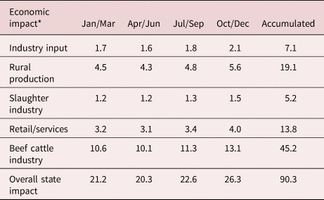
The data revealed that F. hepatica had an important economic impact on the beef cattle industry, as calculations were based on the Industry Input using the CEPEA/USP/Brazil platform (CEPEA, 2016). The overall estimated State loss was about US$90.3 million during 2015 alone, where rural production (US$19.1 million) and meat sales (US$45.1 million) represented the most affected segments (table 6).
Estimate of the economic impact of Fasciola hepatica on the beef cattle industry of Rio Grande do Sul, Brazil, in 2015.

* US$ millions.
SIR model for disease infectivity and recovery
From the Monte Carlo simulation, we confirmed the damage caused by F. hepatica between the groups (Experiment 1). The different disease prevalence based on Experiment 2 was also determined (table 5). From these experiments, the SIR model was used to run different infection scenarios that occur in RS, also based on Silva et al. (Reference Silva, Freitas, Dutra and Molento2016). Using the SIR model, it was possible to demonstrate the progression of F. hepatica infectivity using low to high infectivity rates (fig. 3a–d). The SIR model demonstrated that once the infection level is high, as in Experiment 2 (up to 20%) (fig. 3d), recovery would take longer to be achieved, compared to lower levels (fig. 3a–c).
SIR model for four simulated conditions based on the level of infectivity of Fasciola hepatica in cattle (refer to table 1). Lines represent susceptible (solid line), recovered (dotted line) and infected (dashed line) individuals.

Discussion
The histological analysis showed the presence of F. hepatica specimens in the bile duct collected from the FhG, with lesions suggesting the damage of a chronic infection. These findings have shown a high incidence of duct thickness, biliary duct calcification and thick exudate in slaughtered cattle. Similar findings have also been reported by Bostelmann et al. (Reference Bostelmann, Luz, Thomaz-Soccol and Cirio2000), where they determined chronic cholangitis and obstruction that lead to stenosis of the bile ducts and peritubular fibrosis. Ross (Reference Ross1970) reported the injury to the high-calibre bile ducts of bovine due to F. hepatica, nine weeks after artificially infecting animals, accompanied by an increase in thickness of the liver wall, acinar formation with irregular structures on the inner portion and eosinophilic infiltration beyond the outer middle portion. Supporting the current study, Hussain & Zghair (Reference Hussain and Zghair2017) and Trivilin et al. (Reference Trivilin, Sousa, Carvalho Nunes, Sousa Rocha and Martins2014) reported haemorrhage points on the parietal surface in chronically infected livers, representing the points of entrance of the immature parasites into the liver structure, and enlarged/distended ducts, caused by immunological reaction of macrophages and lymphocytes infiltration during the later stage of fasciolosis. Oyarzún-Ruiz et al. (Reference Oyarzún-Ruiz, Alvelo, Vera and Moroni2019) reported that biliary ducts hyperplasia and hypertrophy with fibroblastic formation, as also observed in infected wild rabbits in Chile. Some of the external lesions were observed in the livers from Experiment 2.
The Monte Carlo simulation analysis randomly pulled out the weight data from each group and calculated the differences between them. This procedure was performed to find the average weight for the groups, and the bootstrap analysis revealed a highly significant probability value (Manly, Reference Manly2006), confirming that the differences in weight were caused by the disease. It was also found that the differences were progressing as the experiment continued, being less expressive at the beginning of the ten-month period. Torgerson & Claxton (Reference Torgerson, Claxton and Dalton1999) reported that the greatest weight loss in beef cattle occurred within the first 16 weeks of infection; however, Monteiro Noel et al. (Reference Monteiro Noel, Fontes-Pereira, Castillo, Fernández, Fonseca and Percedo2013) found that even after the animals were considered free from infection, their lower initial performance continued until slaughter, when compared to non-infected animals.
Fasciola hepatica was found in a high prevalence (up to 20%) across RS, indicating room for improvement in animal health and the adoption of more strict control strategies. Although we did not correlate any welfare indicators, mostly referred as chronic clinical sings from the disease, the data demonstrated that TBZ treatment was efficient in controlling the present infection, even though TBZ resistance has already been reported in the south of Brazil (Oliveira et al., Reference Oliveira, Ferreira, Stival, Romero, Cavagnolli, Kloss, Araújo and Molento2008). The results also showed that 60% of the animals had a parasite burden of between one and 50 flukes. Even though we have examined the liver of cattle of difference ages, previous studies revealed that moderate fluke infections can significantly decrease performance of cattle, reducing their weight gain by 10% (Swai & Ulicky, Reference Swai and Ulicky2009). A low degree of infection may result in only a few clinical signs of the disease, as observed in the present experiment. From this, we hypothesize that the treated animals had better welfare conditions, represented by a significant increase in weight, which may have been reflected in a higher predisposition for foraging and a more efficient immune response (Aleri et al., Reference Aleri, Hine, Pyman, Mansell, Wales, Mallard and Fisher2016). Follow-up experiments should look into cell-mediated responses, plasma cortisol and antibody levels between groups.
In the present study, it was evident that the use of direct (faecal measurements and histology) and indirect (weight gain) diagnostics (Experiment 1) were important health measurements that could be used for disease simulations. Although a small group of animals were used in Experiment 1, these variables were analysed together with the data from RS (Experiment 2) to estimate the impact of the incidence of F. hepatica in cattle on the entire industry, supported by the use of Monte Carlo, SIR and GLMM analysis. Malone & Craig (Reference Malone and Craig1994) reported that herds consisting of more than 25% of infected animals had a significant economic loss, and when animals were experimentally infected with 40–140 cercariae, they showed a reduction in weight gain of up to 28% when compared to non-infected animals. More recently, in a large-scale UK abattoir study, Mazeri et al. (Reference Mazeri, Rydevik, Handel, Bronsvoort and Sargison2017) showed a significant relationship between the presence of liver fluke infection and slaughter age, using parasite burden as an indicator. The impact of fasciolosis was also determined in Cajamarca, Peru, associating the downgrading of the carcass and its value paid to farmers (Raunelli & Gonzalez, Reference Raunelli and Gonzalez2009). This is consistent with the results of Gavinho et al. (Reference Gavinho, Kulek and Molento2008), in a study conducted in Parana, Brazil, where infected cattle (average of 15 parasites/liver) were compared to healthy animals. The groups had a difference of 12.3 kg, which represented 4.9% in carcass weight and a loss of US$30/infected animal. Schweizer et al. (Reference Schweizer, Braun, Deplazes and Torgerson2005) reported an average annual loss of approximately US$1515.00 for a herd of 31 dairy cows in Switzerland.
In the SIR model, a susceptible individual could eventually get infected, and this situation would be maintained for as long as the animal lives (approximately 30 months) with continuous reinfection, or when it receives an antiparasitic treatment. As cattle have little or no immunity to the disease (Graham-Brown et al., Reference Graham-Brown, Hartley, Clough, Kadioglu, Baylis and Williams2018), the second option is more plausible for F. hepatica when determining the rate of recovery. The SIR model also considers that an infected individual will be cured or recover after some time. In the case of fascioliasis, infectivity would not mean that an individual could acquire full immune protection, as animals can be infected by another parasite or even get re-infected, after the temporary benefit from the chemical protection. Full fascioliasis recovery can only be achieved by the use of a vaccine or the use of an effective drug (i.e. TBZ), and this condition can only be verified through the diagnostic of 100% of the animals.
Some other limitations in the use of the SIR model are also important to note. The first and most important is the diagnostic of F. hepatica, which would be used to determine when an individual passes from being susceptible to infected, based on the presence of parasites in the liver (slaughter houses), FEC or clinical examination. As the two last methods are hardly used, the damage to the animals is very pronounced, as seen by the histological slides, when infection is caused by more than 20 parasites. The model also assumes that a recovered individual will never be susceptible again, due to immune protection (i.e. some viral infections), but in the case of F. hepatica, adults and young animals will keep getting infected as long they have contact to the metacercaria, the parasite infective stage.
The four different infection scenarios established for F. hepatica demonstrate the complexity of the disease. The possibility to have a low infection rate, as in the north of RS (Silva et al., Reference Silva, Freitas, Dutra and Molento2016), illustrated in condition A, would reflect a low environmental contamination with a low level of disease transmission. Unfortunately, fascioliasis is highly endemic in cattle and buffaloes in RS and other areas of South America, so conditions C and D are more frequent, reflecting the great economic impact of the disease. Silva et al. (Reference Silva, Freitas, Dutra and Molento2016) reported that rainfall volume was strongly correlated with a higher occurrence of the disease in southern Brazil, and that a La Nina event affected the positivity index of bovine fascioliasis through significant changes in air temperature and rainfall regimens. Aleixo et al. (Reference Aleixo, Freitas, Dutra, Malone, Martins and Molento2015) and Dutra et al. (Reference Dutra, Molento, Naumann, Biondo, Fortes, Savio and Malone2010) reported that the prevalence of F. hepatica had no year effect, measuring low, medium and high infection levels distributed in RS. Silva et al. (Reference Silva, Freitas, Dutra and Molento2020) have recently determined that fascioliasis is a disease that is epidemiologically constant, requiring year-long monitoring. Therefore, and considering that F. hepatica is a chronic infection that may last for up to two years in infected animals (Ross, Reference Ross1968) with significant clinical damage, the SIR model may be used to assess regional control programs once a diagnostic of infectivity is implemented. As mentioned above, we believe that the effect of parasite mortality may have little impact on the disease dynamic in endemic areas that have a low rate of parasite replacement. This condition would not affect the worse-case scenarios, as the return to susceptibility would be very low.
In the present study, the infection caused by F. hepatica, especially in its chronic form, was responsible for considerable damage to the infected group of animals, confirming the relation between reduced weight and the presence of infection in adult cows. It was possible to calculate the weight difference between groups, which was used to estimate regional economic losses. Forecasting disease incidence and prevalence studies have focused on measuring their spatial distribution and the potential risk to the animals and local communities (Bennema et al., Reference Bennema, Molento, Scholte, Carvalho and Pritsch2017). Thus, the present data reinforce the importance of continuous diagnostic (clinical, liver inspection) of F. hepatica, and also validate the use of modelling to determine disease incidence, as well as to calculate the economic impact on local communities. Although modelling can facilitate the broad view of the disease impact, the use of genomic information with transcriptome and proteome data should also be integrated to better determine the processes involved in host invasion and future drug and vaccine targets (Cwiklinski et al., Reference Cwiklinski, Jewhurst and McVeigh2018).
Conclusion
Fasciola hepatica had a high prevalence in the state of RS, causing a chronic inflammation in the biliary ducts of adult cows. The most prevalent histopathological lesions were the presence of polymorphic nucleated cells, eosinophil infiltrate, high periportal multifocal fibrosis and fibrotic bridges. Based on Monte Carlo simulations and the prevalence of 16% of infection, it was estimated that fascioliasis can cause US$90.3 million in losses to the state of RS, due to poor health conditions. Fascioliasis is hard to control in endemic areas as infectivity is constant, with a low rate of recovery, as determined by the SIR model.
Acknowledgement
The authors wish to thank Coordenação de Aperfeiçoamento de Pessoal de Ensino Superior/CAPES for offering an MSc scholarship to L. Dutra.
Financial support
This research received no specific grant from any funding agency, commercial or not-for-profit sectors.
Conflicts of interest
None.
Ethical standards
The authors assert that all procedures contributing to this work comply with the ethical standards of the relevant national and institutional guides on the care and use of laboratory animals.