Troponin levels are often obtained when evaluating patients with chest pain. When obtaining troponin levels, it is critical to understand the cardiac and the non-cardiac causes of troponin elevation, particularly in younger patients.
The case involves an 18-year-old young man presented to the emergency department with elevated troponin levels, and we discuss the causes of troponin elevation and a diagnostic framework to evaluate these patients.
Case report
An 18-year-old male presented to the emergency department with stabbing, 8/10, central chest pain that radiated through his left arm without associated palpitations or shortness of breath. He denied any recent fevers, cough, runny nose or sick contacts or trauma. He reported normal urine output and stools. He endorsed having decreased appetite and some nausea.
The patient had a previous history of similar chest pain 2 years ago and was admitted for 5 days at that time. He denied any other history of illness. He took no medications and denied nutritional supplements or illicit drugs. He recently started playing basketball with college-level athletes. He did not report any other change in his exercise regimen. The remainder of his social and family history was unremarkable.
On presentation, his heart rate was 51 bpm, blood pressure 139/87 mmHg, and respiratory rate of 18 rpm. He was in visible pain, without obvious aggravation by palpation of the chest wall or by movement of his arm. He had unlaboured breathing with clear breath sounds and good air movement throughout his lung fields. He had normal heart sounds without murmurs, gallops, or rubs and he had good distal pulses. He did not have jugular venous distension. His abdomen was soft and non-tender.
Laboratory evaluation began in the emergency department and demonstrated a normal complete blood count. Troponin I (TnI) was 11 ng/ml and N-terminal-β-natriuretic peptide was 14 pg/ml (lab normal less than 126).
An electrocardiogram (ECG) was obtained, which demonstrated sinus bradycardia with normal chamber size and normal intervals. T-wave inversions were observed in the inferior and lateral leads. Serial ECGs throughout his admission showed a transition to upright T waves in the inferolateral leads, with new T-wave inversions in the anterior leads. At the time of discharge, his ECG showed upright T waves in the anterior and inferior leads, with T-wave inversions in the lateral leads, similar to his most recent pre-admission ECG from 2017.
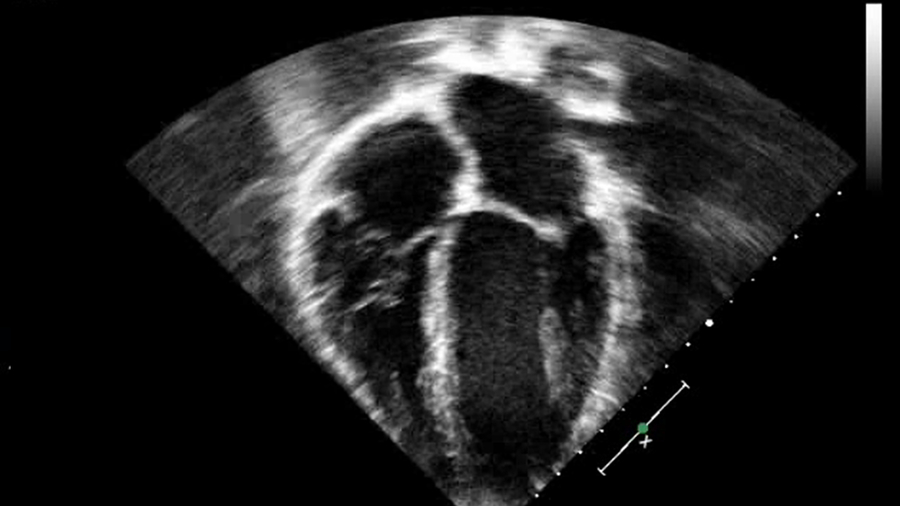
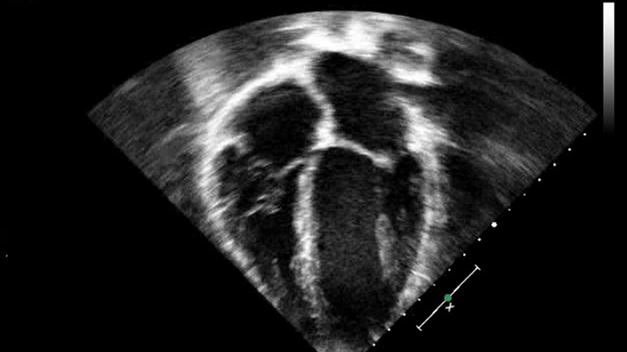
An echocardiogram performed in the emergency department demonstrated normal cardiac anatomy and biventricular size, thickness and function without regional wall motion abnormalities. The remainder of the examination was normal including the proximal coronaries. Figure 1 is a representative four-chamber view from the initial echocardiogram.
Four-chamber echocardiographic image demonstrating normal-sized atria and ventricles. The ventricles are neither hypertrophied nor dilated.
The patient was admitted to the paediatric intensive care unit where he continued to have normal vital signs and perfusion. Venous blood gases demonstrated a normal arteriovenous saturation difference and renal near infrared spectroscopy values were in the 70s, further indicating adequate cardiac output. Additional testing demonstrated an elevated creatine kinase (CK) with a level of 1102 IU (lab normal less than 300). Intravenous hydration was started at 150% of maintenance in the setting of CK levels consistent with rhabdomyolysis. Creatinine levels were normal and urine output remained adequate.
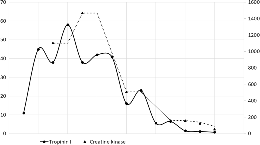
The TnI levels increased through the 2nd day of admission, peaking at 58.0 ng/ml and then downtrended. Initial CK levels started at 1102 IU, peaking at 1469 IU on the 2nd day of admission. On the 8th day of admission, the TnI level was 0.49 and CK was 94. Figure 2 graphically depicts the TnI and CK levels. It is interesting to note that the TnI and CK levels trended together. Ebstein-Barr virus testing, including heterophile antibody testing, was negative. Urine myoglobin demonstrated a value of less than 1.0.
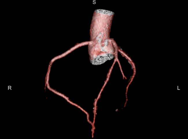
Three-dimensional reconstruction of coronary computed tomographic angiography demonstrating normal coronary arteries. The coronary arteries arise from the proper location and course distally without any occlusion, stenosis, or myocardial bridging.
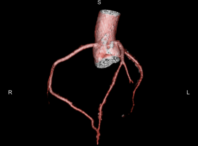
Repeat echocardiograms during the admission also demonstrated normal biventricular function. A computed tomography angiography study was obtained to evaluate the entire coronary system. This demonstrated normal coronary anatomy without any luminal narrowing and without any evidence of myocardial bridging.
The admission was complicated by two episodes of asymptomatic ventricular tachycardia. Electrolyte levels were normal during the admission except for some relative hypomagnesaemia (1.9 mg/dl). The patient was given supplemental magnesium and was started on metoprolol, which was titrated until the ventricular tachycardia was controlled after he experienced a sustained 25-beat episode of asymptomatic ventricular tachycardia. No further arrhythmias was observed; however, he continued to have single and couplet premature ventricular contractions.
The patient was eventually discharged on the 10th day of admission on metoprolol with a diagnosis of rhabdomyolysis with cardiac involvement secondary to strenuous exercise and inadequate hydration. Repeat cardiac magnetic resonance imaging was obtained and demonstrated normal biventricular size and systolic function without regional wall motion abnormalities. Mid-myocardial and epicardial delayed gadolinium enhancement was noted, suggesting the presence of inflammation, infiltration, or fibrosis as well as early enhancement and abnormal STIR, suggesting ongoing or incompletely resolved acute inflammation and oedema in a similar distribution when compared to the previous study from 2017. A 30-day event monitor was placed prior to discharge, which showed single premature ventricular contractions, one ventricular triplet at 100 bpm, and a six-beat run of atrial tachycardia. A previous Holter Monitor from 2018 showed sinus rhythm with rare supraventricular (0.07%) and ventricular ectopy (0.01% composed of single, monomorphic premature ventricular contractions).
During the admission, the patient’s parents and the patient shared details regarding the initial hospitalisation that was 18 months prior. The patient was admitted for chest pain and elevated TnI. He was discharged after 4 days with a diagnosis of myocarditis on a medication that he could not recall. He saw a cardiologist as an outpatient and had a cardiac magnetic resonance imaging study done as well as cardiopulmonary stress test, though the details of neither were shared.
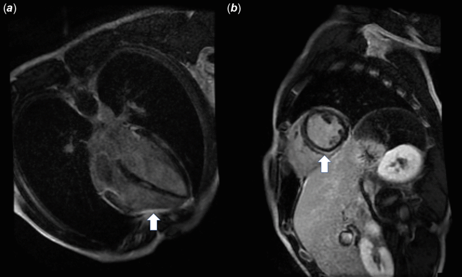
Review of the previous records demonstrated that the patient presented with chest pain and had a TnI of 155.3 ng/ml at the time of presentation during his first admission. At that time, he had endorsed having an episode of increased exercise intensity a few days prior to the episode and limited hydration for the days preceding his admission. He was admitted to the paediatric intensive care unit where his initial TnI represented the peak level which decreased subsequently. On the 5th day of admission, the day he was discharged, the TnI level was 5.2 ng/ml. He had three echocardiograms done during that admission, all of which demonstrated normal cardiac anatomy and normal biventricular function. He also had episodes of asymptomatic ventricular tachycardia and was discharged on metoprolol. Outpatient follow-up included a cardiac magnetic resonance imaging study that demonstrated normal biventricular function but some delayed gadolinium enhancement that was felt to be in a distribution consistent with myocarditis. The delayed enhancement was limited to the epicardium primarily in the inferior and anterior walls. Figure 3 demonstrates a representative slice from the cardiac magnetic resonance imaging study demonstrating this delayed gadolinium enhancement. A cardiopulmonary exercise test was also done, which demonstrated excellent athletic endurance, normal pulmonary function, and no arrhythmia. Due to patient non-compliance, he did not continue metoprolol as an outpatient. Figure 4 is a graph demonstrating the TnI level and the simultaneous CK level.
Magnetic resonance imaging demonstrating epicardial-delayed gadolinium enhancement primarily in the anterior and inferior walls. Panel (a) is in the axial plane, while panel (b) is in the sagittal plane. The arrows point to the areas of delayed gadolinium enhancement.

Discussion
This is a unique case of an athletic teenager who presented with chest pain and troponin elevation. He also had elevated CK levels. These findings led to a diagnosis of rhabdomyolysis with cardiac involvement.
A similar case has been reported before. Westermeyer and colleagues reported a case of a 24-year-old male athlete who presented with a syncopal episode after running 25 miles. This patient had significant TnI elevation for 3 days. Admission TnI was 6.3 ng/ml, with TnI peaking at 16.0 ng/ml on the 1st day of admission. On the 7th day of admission, the TnI level normalised. CK level on admission was 1074 IU and peaked at 23,563 IU on the 5th day of admission, after which it began to decrease. This patient underwent a multiple gated acquisition scan, which was normal. Echocardiogram in this patient had also demonstrated relatively normal biventricular function. The patient was discharged on the 9th day of admission without a cardiac catheterisation, as no regional wall motion abnormalities had been noted earlier and no additional concerns regarding coronary anatomy was observed.Reference Westermeyer and Eilbert 1
Our case is similar to the case reported by Westermeyer and colleagues with respect to the elevations in TnI and CK in the setting of normal biventricular function. Our case differs from that of Westermeyer and colleagues with regard to higher TnI levels, the presence of ventricular tachycardia, and repeat episode.
These cases highlight the need to understand the source of TnI. Korff and colleagues reviewed the differential diagnosis of elevated TnI.Reference Korff, Katus and Giannitsis 2 The following are the cardiac causes of TnI elevation: acute myocardial infarction, post-cardiac catheterisation, open-heart surgery, pericarditis, myocarditis, congestive heart failure, and defibrillator shocks.Reference Alpert, Thygesen, Antman and Bassand 3 – Reference Missov and Mair 7 Non-cardiac causes of TnI elevations include acute pulmonary embolism, end-stage renal disease, aortic dissection, strenuous exercise, endurance activities, chemotherapy, chest wall trauma, amyloidosis, and rhabdomyolysis.Reference Giannitsis, Muller-Bardorff and Kurowski 8 – Reference Lavoinne and Hue 12
In our patient, normal biventricular function with no regional wall motion abnormality and normal computed tomographic angiography of the coronary arteries was not consistent with acute myocardial infarction or congestive heart failure. No history of cardiac catheterisation or cardiac surgery was reported in our patient. Echocardiogram did not demonstrate a pericardial effusion. Additionally, the history of a low C-reactive protein and previously mentioned echocardiogram findings were not consistent with infectious myocarditis. The patient did not receive defibrillator shocks. No tachycardia, tachypnoea, or desaturation was observed to suggest pulmonary embolism in our patient. Renal function was normal and so end-stage renal disease was ruled out. No evidence of aortic dissection nor history of trauma, chemotherapy, or amyloidosis was observed. Both Strenuous exercise and rhabdomyolysis were present in our patient, and rhabdomyolysis is the most common non-cardiac cause of TnI elevation. Thus, after adequately excluding the all other potential diagnoses, a diagnosis of rhabdomyolysis with cardiac involvement in the form of TnI leak was made.
Several studies demonstrated elevation in TnI with strenuous exercise.Reference Westermeyer and Eilbert 1 , Reference Neumayr, Gaenzer and Pfister 13 , Reference Koller 14 Neither the precise mechanism of this nor the true significance of this finding is understood. Most of these studies, however, have noted only mild elevations in TnI levels after strenuous exercise, and our current case represents the highest TnI levels in such a situation.Reference Neumayr, Gaenzer and Pfister 13 Koller and colleagues hypothesised that both reversible and irreversible myocyte degeneration may occur with strenuous exercise but that the area of myocardium involved in both situations is likely quite small. Despite a small affected area, the TnI elevation is detectable.Reference Koller 14 Other studies have proposed that such myocyte degeneration may be due to elevated catecholamines inducing some degree of coronary vasospasm.Reference Rowe 15 , Reference Laslett, Eisenbud and Lind 16 Only limited studies are available to actually confirm these proposed mechanisms.
Rhabdomyolysis is the most common non-cardiac cause of TnI elevation.Reference Punukollu, Gowda and Khan 17 It is unlikely that rhabdomyolysis is the direct cause of TnI elevation in patients. It is more likely that the underlying cause of rhabdomyolysis leads to TnI elevation. In our patient, strenuous exercise with limited hydration can potentially explain both the rhabdomyolysis and the TnI elevation. A temporal relationship was found in the increase and decrease of both TnI and CK levels in our patient. Interestingly, the degree of TnI elevation does not seem to be related to CK levels in patients with rhabdomyolysis and TnI elevation. In adult studies of rhabdomyolysis, 50–75% patients will have TnI elevations.Reference Wongrakpanich, Kallis and Prasad 18 , Reference Li, Zapata and Tillem 19 Most of these individuals will have normal biventricular function. The mechanism of the TnI elevation remains unclear in these studies, as stated before. Other investigations have demonstrated that skeletal TnI may cross-react in different TnI assays meant to be cardiac specific, leading to significantly high cardiac TnI values.Reference Hyytia, Heikkila, Hedberg, Puolakanaho and Pettersson 20
Repeat episodes without cardiac abnormalities identified should lead to consideration of defects in β-oxidation of fatty acids or an underlying myositis such as polymyositis or dermatomyositis. Defects in β-oxidation of fatty acids can be screened by with a spot lactate check and/or by an acylcarnitine profile. Suspicion for myositis can be screened for by C-reactive protein, erythrocyte sedimentation rate, and/or anti-nuclear antibody. In our patient, a normal C-reactive protein and lack of muscle tenderness on physical examination made polymyositis less likely. The rapid normalisation of CK also made polymyositis less likely.
Graphical representation of troponin I and creatine kinase levels as a function of time. The left axis is the troponin I level in ng/ml, while the right axis is the creatine kinase level in International Units (IU).

Conclusion
Troponin elevations in young athletic patients may not be cardiac in nature. Troponin elevation may be present in the setting of rhabdomyolysis and may not be in proportion to CK. Elimination of cardiac causes of troponin elevation is necessary before making a diagnosis of rhabdomyolysis-induced troponin elevation.
Acknowledgements
None.
Financial Support
This research received no specific grant from any funding agency, commercial or not-for-profit sectors.
Conflicts of Interest
None.