

Introduction
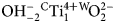
Our knowledge of oxo amphiboles, i.e. amphiboles with O2– as the dominant W anion, has increased rapidly in the last 25 years, particularly as secondary ion mass spectrometry analysis and structure refinement (SREF) has allowed accurate quantification of the OH component. Indeed, the latest report on amphibole classification and nomenclature (Hawthorne et al., Reference Hawthorne, Oberti, Harlow, Maresch, Martin, Schumacher and Welch2012) has classified oxo amphiboles as a separate group of the amphibole supergroup. Oxo amphiboles may derive from the different amphibole rootnames either via the coupled exchange C ${\rm (Mg,Fe)}_{{\rm \ndash 1}}^{{\rm 2 +}} $W
 ${\rm OH}_{{\rm \ndash 2}}^\ndash $ C
 ${\rm Ti}_1^{{\rm 4 +}} {} ^{\rm W}\!{\rm O}_2^{2\ndash} $ or via iron oxidation according to C
 ${\rm Fe}_{{\rm \ndash 2}}^{{\rm 2 +}} $W
 ${\rm OH}_{{\rm \ndash 2}}^\ndash $ C
 ${\rm Fe}_2^{{\rm 3 +}} $W
 ${\rm O}_2^{2\ndash} $ In the first case, the occurrence of the oxo amphibole is restricted by the bulk chemistry; in the second case, it depends on the T, P and
 $f_{\rm O_2}$ conditions, so that oxidation and deprotonation can occur easily by experimental annealing even during SREF investigation. In both cases, high-charged cations order mainly at the M(1) site and possibly at the M(3) site (Oberti et al., Reference Oberti, Hawthorne, Cannillo, Cámara, Hawthorne, Oberti, Della Ventura and Mottana2007, Reference Oberti, Boiocchi, Zema, Hawthorne, Redhammer, Susta and Della Ventura2018a).
In accord with the current amphibole nomenclature (Hawthorne et al., Reference Hawthorne, Oberti, Harlow, Maresch, Martin, Schumacher and Welch2012), oxo amphiboles with CTi contents higher than 0.5 atoms per formula unit (apfu) require a distinct rootname, whereas oxo amphiboles with Ti contents ≤ 0.5 apfu maintain the proper rootname in the amphibole group and assume the prefix oxo.
Among CTi–rich oxo amphiboles, the most petrologically important and best known compositions are those related to the calcium group, which assume the rootname kaersutite, NaCa2(Mg3AlTi4+)(Si6Al2)O22O2. The two other rootnames known with CTi4+ in their formula derive from the sodium group: obertiite, ideally, NaNa2(Mg3AlTi4+)Si8O22O2 and dellaventuraite, NaNa2(MgAl2Ti4+Li)Si8O22O2. For both of these, only the Mn3+ or Fe3+ varieties have been described so far (Hawthorne et al., Reference Hawthorne, Cooper, Grice and Ottolini2000, Reference Hawthorne, Ball and Czamanske2010; Oberti et al., Reference Oberti, Boiocchi, Hawthorne, Ball and Blass2017a; Tait et al., Reference Tait, Hawthorne, Grice, Ottolini and Nayak2005; names corrected after Hawthorne et al., Reference Hawthorne, Oberti, Harlow, Maresch, Martin, Schumacher and Welch2012). The other oxo amphibole related to the sodium group is mangano-mangani-ungarettiite, NaNa2( ${\rm Mn}_2^{2 +} {\rm Mn}_3^{3 +} $)Si8O22O2, where the presence of WO2– is allowed by an inverse ordering of the C cations (Mn2+ at M(2) and Mn3+ at M(1,3)), which restricts its occurrence to the end-member formula (Hawthorne et al., Reference Hawthorne, Oberti, Cannillo, Sardone, Zanetti, Grice and Ashley1995a; Oberti et al., Reference Oberti, Della Ventura, Boiocchi, Zanetti and Hawthorne2017b).
Here, we present the complete characterisation of potassic-jeanlouisite, ideally K(NaCa)(Mg4Ti)Si8O22, a new species of the oxo amphibole group, the first that is related to the sodium–calcium subgroup and in particular to the richterite composition. As CTi is higher than 0.5 apfu, a new rootname was required for this composition. Jean-Louis Robert (1948–2017) dedicated much effort (first at the Centre de Recherches sur la Synthèse et la Chimie des Minéraux (CRSCM) at Orleans and then at the Institut de Minéralogie, de Physique des Matériaux et de Cosmochimie (IMPMC) in Paris) to the crystal-chemical characterisation of micas and amphiboles, with a particular focus on natural and synthetic richterites with significant Ti content. Because the rootname ‘robertite’ was too close to the already used mineral name robertsite, and the name ‘jeanlouisrobertite’ was thought too cumbersome, the rootname ‘jeanlouisite’ was dedicated to Jean-Louis Robert.
The mineral and its name were approved by the International Mineralogical Association Commission on New Minerals, Nomenclature and Classification, vote 2018-050 (Oberti et al., Reference Oberti, Boiocchi, Hawthorne, Della Ventura and Färber2018b). The holotype material of potassic-jeanlouisite is deposited in the mineral collection of the Dipartimento di Scienze della Terra e dell'Ambiente, Università di Pavia, Italy, catalogue number 2018-01.
Occurrence and optical properties
Potassic-jeanlouisite was discovered in a rock specimen collected some years ago by Gunnar Färber. The specimen is a pale pinkish-brown leucite, which is found in the lava layers, ~7 cm × 7 cm in size, and was collected in an active gravel quarry on Zirkle Mesa (41°46′54.58″N, 108°53′23.82″W), in particular in a small zone with faulted layers. The leucite-lava layer was ~20 cm thick. Zirkle Mesa is part of an area ~10 km × 10 km known as Leucite Hills, close to the town of Superior, Wyoming, USA. In the past, the locality was mined as a source of fertiliser due to the high potassium contents of lamproites, and there are still several gravel quarries active.
The type specimen (Fig. 1) is a brown ochre porous block, a couple of centimetres across, in which the matrix consists of microcrystalline amphibole and leucite (as determined by powder X-ray diffraction analysis), with acicular potassic-jeanlouisite crystals (smaller than 200 μm × 20 μm × 20 μm) in small vugs. In the type specimen, potassic-jeanlouisite occurs as acicular crystals with good {110} cleavage. It is pale-yellow to colourless with a white streak, vitreous lustre, and does not fluoresce in short-wave or long-wave ultraviolet light.
The type specimen studied in this work (catalogue number 2018-01). The acicular potassic-jeanlouisite crystals shown in the micro-photo are ~200 μm × 20 μm × 20 μm in size.

In transmitted plane-polarised light (λ = 590 nm), potassic-jeanlouisite is non-pleochroic. A spindle stage was used to orient a crystal for measurement of refractive indices and 2V by extinction curves (Bartelmehs et al., Reference Bartelmehs, Bloss, Downs and Birch1992). The optical orientation was determined by transferring the crystal from the spindle stage to a single-crystal diffractometer and measuring the relative axial relations by X-ray diffraction. It is biaxial (–), α = 1.674(2), β = 1.688(2), γ = 1.698(2), 2Vmeas = 79(1) and 2Vcalc = 79.8°. The orientation is: X ^ a = 17o (in β obtuse), Y || b, Z ^ c = 3o (in β acute).
Single-crystal and powder diffraction analysis
During this work, several crystals were characterised by single-crystal structure refinement and electron microprobe (EMP) analysis. Most of them are strongly zoned. In crystal 1290, compositional zoning is less prominent and hence the agreement between the two analyses is best. The crystal code is that in the amphibole database at the CNR-Istituto di Geoscienze e Georisorse (CNR-IGG) in Pavia.
Diffraction data were collected in the θ range 2–30° with a Bruker-AXS CCD diffractometer using graphite-monochromatised MoKα X-radiation (λ = 0.71069 Å). Omega rotation frames (scan width = 0.3°, scan time = 20 s and sample-to-detector distance = 50 mm) were processed with the SAINT software (Bruker, Reference Bruker2003) and intensities were corrected for Lorentz and polarisation effects; absorption effects were empirically evaluated with the SADABS software (Krause et al., Reference Krause, Herbst–Irmer, Sheldrick and Stalkeand2015) and an absorption correction was applied to the data. Unit-cell parameters were calculated by a least-squared procedure based on the positions of 720 measured reflections with I > 10 σI in the 2θ range 4°–60°. The collected reflections were reduced to unique reflections (mean redundancy = 5). No twinning was observed.
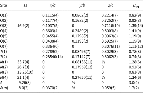
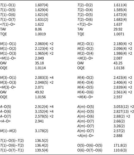
Following the procedures and terminology described in Hawthorne et al. (Reference Hawthorne, Ungaretti and Oberti1995b) and Oberti et al. (Reference Oberti, Ungaretti, Cannillo and Hawthorne1992), scattering curves for fully ionised chemical species were used at sites where chemical substitutions occur; neutral vs. ionised scattering curves were used at the T and anion sites [except O(3)]. Full-matrix unweighted least-squares refinements on the (observed) reflections with I > 3 σI were done with a program locally written to handle complex solid-solutions (Cannillo et al., Reference Cannillo, Germani and Mazzi1983). Crystallographic details are given in Table 1, refined atom coordinates and equivalent displacement parameters, in Table 2, and selected bond lengths and angles are given in Table 3. Observed structure factors are deposited with the crystallographic information file as Supplementary materials (see below). The a:b:c ratio calculated from the unit-cell parameters is 0.5518:1:0.2932.
Data and experimental details for potassic-jeanlouisite crystal 1290.

Refined site-scattering (ss, epfu), atom coordinates and equivalent-displacement parameters (B eq, Å2) for potassic-jeanlouisite crystal 1290.

Selected interatomic distances (Å) and angles (°), tetrahedral and octahedral angle variances (°^2) and quadratic elongations (Robinson et al., Reference Robinson, Gibbs and Ribbe1971) in potassic-jeanlouisite crystal 1290.

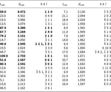
Powder X-ray diffraction data (CuKα, λ = 1.54178 Å) were obtained using the XPREP utility of SAINT (Bruker, Reference Bruker2003), which generates a 2D powder diffractogram (Debye-Scherrer technique) starting from the F 2obs collected on the single crystal and taking into account solely the information concerning the unit-cell dimensions and the Laue symmetry. No Lorentz and polarisation corrections were applied. The strongest eight reflections in the powder X-ray pattern [d values (in Å), (I) (hkl)] are: 2.703 (100) (151); 3.380 (87) (131); 2.541 (80) ( $\bar 2$02); 3.151(70) (310); 3.284 (68) (240); 8.472 (59), (110); 2.587 (52), (061); 2.945 (50) (221,
 $\bar 1$51). The whole data are given in Table 4.
Powder X-ray diffraction data for potassic-jeanlouisite 1290.

Note: the strongest eight reflections are in bold. Only reflections with I rel ≥ 5 are reported.
Infrared spectroscopy
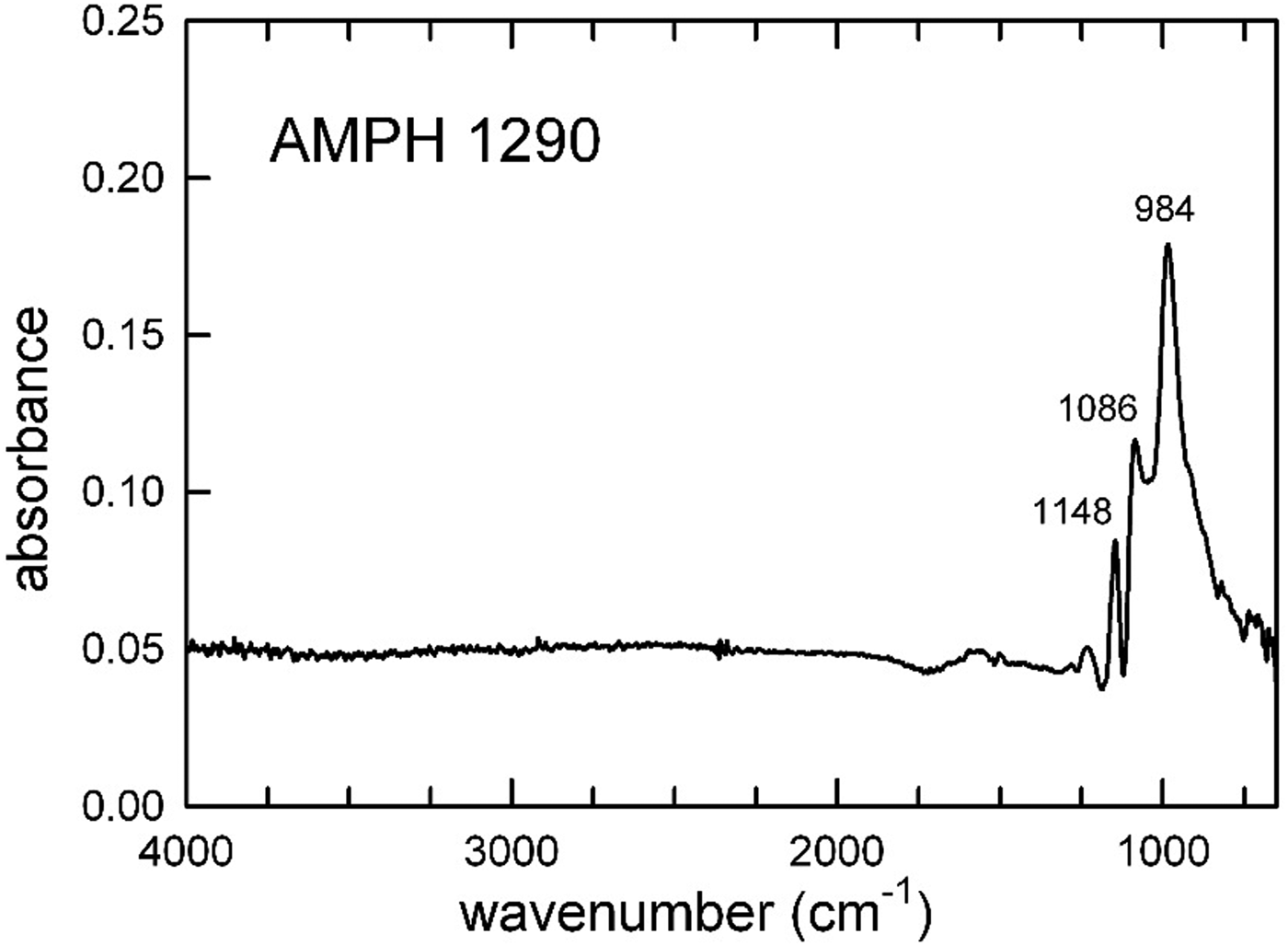
Because of possible inhomogeneity, Fourier-transform infrared (FTIR) analysis was undertaken on two potassic-jeanlouisite crystals used for single-crystal structure refinement and EMP analysis (#1289 and #1290 in the amphibole database of the CNR-IGG), and thus already embedded into the epoxy resin and polished. Hence, the FTIR data were collected in reflection mode with a Bruker Hyperion 3000 microscope equipped with a N2-cooled MCT detector at the INFN-LNF (Istituto Nazionale di Fisica Nucleare, Laboratori Nazionali di Frascati, Rome). The microscope was attached to a Vertex V70 optical bench, equipped with a KBr broadband beamsplitter and a Globar IR source. The beam covered the complete surface of the embedded crystals, i.e. ~20 μm × 80 μm so as to maximise the signal from the amphibole; the nominal resolution was 4 cm–1 and 128 scans were accumulated for both spectrum and background. The patterns collected for both amphiboles are virtually identical; a number of peaks typical of an [4]Al-free amphibole structure are observed in the low-frequency region <1200 cm–1 (Fig. 2), while no signal due to the presence of hydroxyl is observed in the OH-stretching region (3700–3600 cm–1). Based on our data, we cannot exclude beyond any doubt the presence of a very small amount of hydroxyl in the crystals examined because of the extremely small dimensions of the sample analysed and the fact that, in reflection mode, we probe only a small volume of the sample under the crystal surface.
The FTIR spectrum of potassic-jeanlouisite, crystal 1290.

Chemical analysis
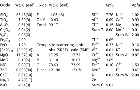
Chemical analysis (8 points) on the crystals used for structure refinement were performed using a CAMECA SX–100 electron microprobe (WDS mode, 15 kV, 10 nA, 5 μm beam diameter). The standards used were as follows: Si: diopside (TAP); Ti: titanite (LPET); Al: andalusite (TAP); Cr: chromite (LPET); V: VP2O7 (LLiF); Fe: fayalite (LLiF); Mn: spessartine (LLiF); Mg: forsterite (LTAP); Zn: gahnite (LLiF); Ni: pentlandite (LLiF); Ca: diopside (LPET); Na: albite (TAP); K: orthoclase (LPET); F: fluoro–riebeckite (TAP); and Cl: tugtupite (LPET). Chlorine was below the detection limit. An image of crystal 1290 is shown in Fig. 3. Fluorine is lower at the rim (0.70 and 0.93 wt.% at points 9 and 16, respectively) and increases from 1.08 wt.% at point 10 to 2.15 wt.% in point 15. Titanium is higher at the rim (5.01 and 4.96 wt.% at points 9 and 16, respectively) and decreases from 4.78 wt.% at point 10 to 3.23 wt.% at point 15. Magnesium is slightly lower and Fe is slightly higher at the rim. Thus the only significant (inverse) correlation is between F and Ti, which is in agreement with the absence (or minor amount) of OH. Hence, and because the FTIR analysis showed no signal for OH, the empirical formula was calculated on the basis of 24 anions pfu, 16 cations pfu and no H2O, giving: A(K0.84Na0.16)Σ1.00B(Ca0.93Na1.02Mg0.04  ${\rm Mn}_{{\rm 0}{\rm. 01}}^{2 +} $)Σ2.00C(Mg3.85
 ${\rm Fe}_{{\rm 0}{\rm. 16}}^{2 +} $Ni0.01
 ${\rm Fe}_{{\rm 0}{\rm. 33}}^{2 +} {\rm V}_{{\rm 0}{\rm. 01}}^{\,\,\,\,\,\,\,\,\,3 +} $Ti0.65)Σ5.01T(Si7.76Al0.09Ti0.15)Σ8.00O22 W[O1.53F0.47]Σ2.00 (where the dominant cations/anions are in bold). The chemical composition and calculated formula are reported in Table 5.
The crystal of potassic-jeanlouisite studied in this work (code 1290). EMP analysis points are shown in red.

Chemical composition (average of 10 points) and unit formula* for potassic-jeanlouisite 1290, and a comparison between observed and calculated site-scattering values.

* Calculated on the basis of 16 cations and 24 (O,F)
esd – estimated standard deviation
However, FTIR analysis was undertaken in reflection mode, and thus we could not explore the inner part of the crystal. For the sake of completeness, a second calculation was made assuming the minimum amount of the oxo component compatible with the analysis (allowing for no Fe2O3, i.e. H2Omax wt.% = 0.34, wt.% sum = 99.22). The resulting unit formula is: A(K0.84Na0.16)Σ1.00B(Ca0.93Na1.02Mg0.04 ${\rm Mn}_{{\rm 0}{\rm. 01}}^{2 +} $)Σ2.00C(Mg3.85
 ${\rm Fe}_{{\rm 0}{\rm. 49}}^{2 +} $Ni0.01
 ${\rm V}_{{\rm 0}{\rm. 01}}^{3 +} $Ti0.65)Σ5.01T(Si7.76Al0.09Ti0.15)Σ8.00O22W[O1.20(OH)0.33F0.47]Σ2.00, which is still well within the compositional field of potassic-jeanlouisite.
The simplified end-member formula of potassic-jeanlouisite is AKB(NaCa)C(Mg4Ti)TSi8O22WO2, which requires SiO2 56.16, TiO2 9.33, MgO 18.83, CaO 6.55, Na2O 3.62, K2O 5.50, total 100.00 wt.%. Based on the empirical formula, the calculated density of potassic-jeanlouisite is 3.146 g/cm3. The compatibility index (1 – (K P/K C)) is 0.009 (superior; Mandarino, Reference Mandarino1981).
Comments on the crystal-chemistry of potassic-jeanlouisite
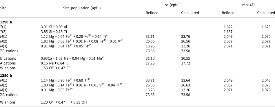
Oberti et al. (Reference Oberti, Hawthorne, Cannillo, Cámara, Hawthorne, Oberti, Della Ventura and Mottana2007) had reported crystal-chemical relationships suitable to calculate the oxo component starting from the refined M(1)–M(2) distance in calcium and sodium–calcium amphiboles. This method would provide 1.14WO2– apfu for crystal 1290, supporting the unit–formula with the lowest oxo content (1290b in Table 6). However, recent (still unpublished) studies done at CNR-IGG on calcium amphiboles with a significant to dominant oxo component and significant WF contents have shown that the presence of WF does affect the calculation, and leads to lower estimates of the oxo component. Because a further modelling, which takes into account WF, is still underway (these compositions are rare and very difficult to characterise), we haven't used the crystal-chemical approach in the case of potassic-jeanlouisite.
Site populations for potassic-jeanlouisite crystal 1290 calculated (a) with the option of no (OH)– present as a W anion (oxo max, as in Table 5), and (b) of oxo min (see text, only for C cations and W anions). There is a good agreement (see text for more detail) between the refined values of site scattering (ss, epfu) and those calculated based on the proposed site-populations. The agreement for mean bond lengths (mbl, Å) at the M(1,2) sites is less close, indicating significant relaxation.

Site populations were derived for crystal 1290 from the unit formula and the results of the structure refinement, and are given in Table 6. It contains a large amount of Ti (0.80 apfu) which is partitioned between the C- and T-group cations, where it orders at the M(1) and T(2) sites, respectively. The presence of Ti at the T(2) site is a common feature of richterites crystallised at high-T and low P conditions (cf. Oberti et al., Reference Oberti, Ungaretti, Cannillo and Hawthorne1992, Della Ventura et al., Reference Della Ventura, Robert and Bény1991), and is detectable by the refined value of the angular variance (Robinson et al., Reference Robinson, Gibbs and Ribbe1971) at that site (29.9°^2, Table 3). The complete ordering of CTi at the M(1) site is coherent with the reaction M(1)Mg2+ + 2 O(3)OH– → M(1)Ti4+ + 2 O(3)O2–, and is detectable also via the high value refined for the equivalent atomic-displacement parameter (B eq) at the M(1) site (1.28 Å2) with respect to the M(2) and M(3) sites (Tiepolo et al., Reference Tiepolo, Zanetti and Oberti1999; Oberti et al., Reference Oberti, Hawthorne, Cannillo, Cámara, Hawthorne, Oberti, Della Ventura and Mottana2007). This reaction locally implies short-range order of M(1)(Ti4+, Fe3+) and O(3)O2– versus M(1)Mg and O(3)(F,OH)–, which could not be confirmed in this composition by FTIR spectroscopy due to the absence of any OH– signal.
The relaxation observed for the M(2) octahedron (<M(2)–O> obs = 2.087 Å to be compared with <M(2)–O> calc = 2.077 Å), is also typical of natural and synthetic richterites with significant oxo component (Oberti et al., Reference Oberti, Ungaretti, Cannillo and Hawthorne1992).
Because the presence of Ti at the T(2) site also implies a significant increase in volume of that tetrahedron, these crystal-chemical peculiarities are in agreement with low-P conditions of crystallisation in an oxidised environment. The inverse relation between the F and Ti contents is particularly interesting for amphibole crystal-chemistry and is being investigated further using natural and synthetic samples.
Relation with other species and a review of related compositions
Potassic-jeanlouisite is the first sodium–calcium amphibole with WO2– >1.0 apfu. The formula of jeanlouisite can be derived from that of richterite via the chemical substitution CMg–1 W ${\rm OH}_{{\rm \ndash 2}}^\ndash $ C
 ${\rm Ti}_1^{{\rm 4 +}} {} ^{\rm W}\!O_2^{2\ndash} $. Table 7 reports a comparison between potassic-jeanlouisite, holotype potassic-richterite from the Pajsberg ore field, Värmland, Sweden (Holtstam et al., Reference Holtstam, Cámara, Skogby, Karlsson and Langhof2019) and a TTi-bearing potassic-fluoro-richterite found in lamproites from the West Kimberley region, Australia (Oberti et al., Reference Oberti, Ungaretti, Cannillo and Hawthorne1992). The unit-cell volume of potassic-jeanlouisite is smaller than those of the other samples, in agreement with the presence of significant CTi. The largest difference in the unit-cell edges is in the length of the a edge, also in agreement with the (relatively) high WF (Della Ventura et al., Reference Della Ventura, Robert, Bény, Raudsepp and Hawthorne1993); in contrast, the c edge is longer than expected based on the modelling of the unit-cell parameters reported by Hawthorne and Oberti (Reference Hawthorne, Oberti, Hawthorne, Oberti, Della Ventura and Mottana2007), which is in agreement with the presence of TTi (Oberti et al., Reference Oberti, Ungaretti, Cannillo and Hawthorne1992).
Comparison of the mineral data available for potassic-jeanlouisite from Leucite Hills (this work), for its holotype W(OH)-dominant counterpart (Pjsberg ore field, Värmland, Sweden; Holtstam et al., Reference Holtstam, Cámara, Skogby, Karlsson and Langhof2019), and for a TTi-bearing potassic-fluoro-richterite from lamproites in the West Kimberley region, Australia (no. 9 in Oberti et al., Reference Oberti, Ungaretti, Cannillo and Hawthorne1992).

[1] A(K0.84Na0.16)Σ1.00B(Ca0.93Na1.02Mg0.04 ${\rm Mn}_{{\rm 0}{\rm. 01}}^{2 +} $)Σ2.00C(Mg3.85
 ${\rm Fe}_{{\rm 0}{\rm. 16}}^{2 +} $Ni0.01
 ${\rm Fe}_{{\rm 0}{\rm. 33}}^{2 +} {\rm V}_{{\rm 0}{\rm. 01}}^{3 +} $Ti0.65)Σ5.01T(Si7.76Al0.09Ti0.15)Σ8.00O22W[O1.53F0.47]Σ2.00
[2] A(K0.61Na0.30Pb0.02)Σ0.93B(Na1.14Ca0.79Mn0.07)Σ2.00C(Mg4.31Mn0.47 ${\rm Fe}_{{\rm 0}{\rm. 20}}^{2 +} $)Σ5.00T(Si7.95Al0.04
 ${\rm Fe}_{{\rm 0}{\rm. 01}}^{3 +} $)Σ8.00O22W(OH1.82F0.18)Σ2.00
[3] AK1.00B(Ca1.03Na0.94)Σ1.99C(Mg4.65 ${\rm Fe}_{{\rm 0}{\rm. 30}}^{2 +} {\rm Mn}_{{\rm 0}{\rm. 01}}^{2 +} $Ti0.04)Σ5.00T(Si7.54Al0.07Ti0.39)Σ8.00O22W[(OH)0.53F1.47]Σ2.00
Acknowledgements
Careful and constructive reviews by Fernando Cámara, Peter Leverett and the Editorial handling of Anthony Kampf and Stuart Mills are gratefully acknowledged.
Supplementary material
To view supplementary material for this article, please visit https://doi.org/10.1180/mgm.2019.38.