


Pulmonary embolism (PE) is a serious condition with an annual incidence rate of 0.50 to 0.69 per 1,000 persons in the general population Reference Skaf, Stein, Beemath, Sanchez and Olson1 and is associated with mortality as high as 24%. Patients with ischemic stroke are at risk of PE, both as a concurrent presentation due to shared risk factors of hypercoagulability and causal pathway of paradoxical embolism, and as a subsequent consequence of their immobility.
Previous work has shown PE rates of nearly 0.3–1% in the hospitalized ischemic stroke population. Reference Pongmoragot, Rabinstein, Nilanont, Swartz, Zhou and Saposnik2 These studies are mostly limited to symptomatic cases during the hospitalization period and show a three-month mortality between 8.6 and 17% in ischemic stroke patients with PE.
In contrast, the literature on concurrent PE at the time of hyperacute stroke is sparse and limited to retrospective case series. Reference Chen, Yang and Bhaskar3 In these studies, CT technique only included the proximal pulmonary arteries and upper lobes. Additionally, subspecialty chest radiologist reporting was not implemented. These factors may have possibly led to underreporting of the true rates of concomitant PE in the hyperacute stroke population.
The objective of this study was to investigate the rate of concomitant PE in hyperacute stroke patients. We performed a retrospective analysis of a previous prospective cohort study, whose methods were previously described Reference Lee, Dhillon and Pourafkari4 and followed the Strengthening the Reporting of Observational Studies in Epidemiology Statement. In brief, suspected hyperacute stroke patients presenting to a Canadian comprehensive stroke center underwent modified Code Stroke imaging with a 64-slice CT scanner, where the longitudinal axis of CT angiography was extended from the carina to the diaphragm as part of neurovascular imaging. This allowed imaging of the full cardiac structures, as well as extended view of the lungs and pulmonary vasculature. The cardiac and chest component of the imaging was reported after acquisition by experienced fellowship-trained chest and cardiothoracic radiologists. The primary outcome in this analysis was rate of concomitant PE. Participants presenting on Code Stroke were enrolled through deferral of consent during daytime working hours only, due to requirement for timely reporting of in-house cardiothoracic radiologists.
Microsoft Excel (Version16.61.1, 2019) was used to calculate percentages, means, medians and interquartile ranges. Interrater reliability of PE findings was assessed using kappa score. P-values are reported as per t-tests for continuous data and chi-squared tests for categorical data.
Ethics approval was obtained from the Queen’s University Health Sciences Research Ethics Board (HSREB; #6030510).
Among 301 patients, 208 were diagnosed with stroke. PE was found in 12/208 (5.8%, CI 95% 3.02 – 9.86%) of stroke and 1/93 (1.1%, 95% CI 0.03– 5.83) of mimic patients (see Figure 1A, p = 0.06). Interrater reliability kappa score was 1.00 for detection of PE based on two readers (G.D. and M.P.) blindly reviewing imaging. All PEs were clinically asymptomatic at presentation, with no evidence of hypoxia or tachycardia.
(A) Rates of pulmonary embolism (PE) in patients undergoing extended CT angiography during hyperacute stroke imaging. PE was found in 12/208 (5.8%, 5.8%, CI 95%, 3.02% – 9.86%) of stroke patients and 1/93 (1.1%, 95% CI 0.03– 5.83) of stroke mimics. (B) Most PE (10/12; 83%) found in stroke patients were low risk as per AHA/ESC classification, while 2/12 cases (17%) were of intermediate risk. All PEs were clinically asymptomatic.

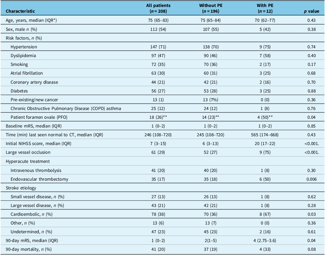
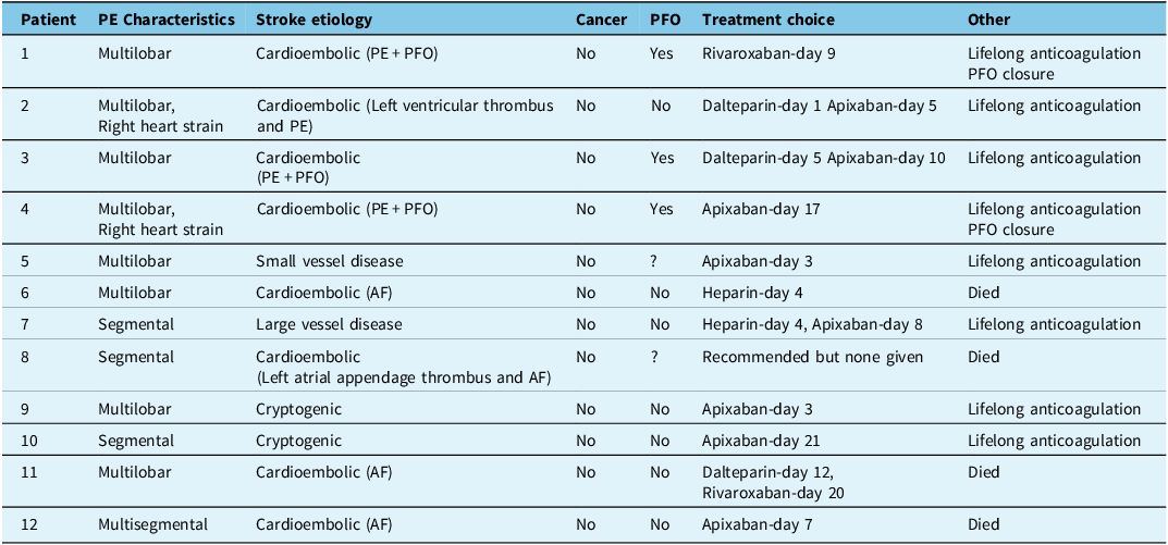
Stroke patients with and without PE were matched with respect to age, sex, pre-existing functional status and major risk factors, including cancer or underlying lung disease (see Table 1). PE was associated with severe strokes, presence of large vessel occlusion, cardioembolism and worse functional outcomes (see Table 1). Most cases of PE were low risk, as per American Heart Association (AHA) and European Society of Cardiology (ESC) classification, with the exception of two cases that were submassive (AHA)/ intermediate risk (ESC; see Figure 1B). PEs were often multilobar or segmental (Table 2). Lifelong anticoagulation was recommended by subspecialists for all patients. Initial treatment included heparin and oral anticoagulants starting at day 1 to day 21 and no treatment complications occurred.
Stroke patients with and without pulmonary embolism

* Interquartile range.
** 8/12 stroke patient with PE and 62/196 stroke patient without PE had test for PFO.
PE = pulmonary embolism.
Characteristics of patients with pulmonary embolism

PE = pulmonary embolism; AF = atrial fibrillation.
Patent foramen ovale (PFO) rate in patients with PE versus without PE was 50% versus 23%, respectively. Although this difference did technically reach statistical significance (p = 0.04), it should not be interpreted as true comparative rates between the two groups, as 8/12 (66.7%) of stroke patients with PE versus 62/196 (31.6%) without PE had testing for PFO with a transthoracic echocardiogram with bubble study. Median time from stroke onset to stroke CT imaging was not significantly higher for patients with PE compared to those without.
In summary, in this cohort study of hyperacute stroke patients who underwent extended CT Angiography (CTA), we found 1) concomitant asymptomatic PE is associated with increased stroke severity, Large vessel occlusion (LVO), cardioembolic etiology and worse clinical outcomes, 2) PE findings change clinical management, specifically anticoagulation, as recommended by subspecialists.
No clinical trials or observational studies have evaluated the incidence or prevalence of concomitant stroke and PE and reported data are based on case reports and case series. Reference Saleh Velez and Ortiz Garcia5 Our series is the largest to highlight these conditions using extended CTA.
There are no specific guidelines on treatment of incidental PE in non-cancer patients. Management decisions are usually individualized based on emboli size, location, patient factors and institutional protocols. Generally, subsegmental PE may not need treatment Reference Kearon, Akl and Ornelas6 but there is general trend in treating asymptomatic PEs that are not subsegmental to prevent chronic thromboembolic pulmonary hypertension or post-PE syndrome. All PEs in this study were segmental or lobar and treated with lifelong anticoagulation.
Patients with PE had more severe stroke, higher frequency of LVO and stroke secondary to cardioembolism. Potential mechanisms could include paradoxical embolism via PFO and atrial fibrillation (AF), both of which were present in some of our patients. The Tromsø study Reference Enga, Rye-Holmboe and Hald7 concluded that AF was associated with increased risk of PE. This relationship is thought to be bidirectional Reference Lutsey, Norby and Alonso8, such that 1) increased pulmonary vascular resistance, right ventricular afterload and cytokine release can lead to elevated right atrial pressure and strain, leading to AF, and 2) AF which in turn could lead to PE through right atrial thrombosis. Reference Watson, Shantsila and Lip9 Therefore, anticoagulation for stroke prevention in AF may be beneficial for PE prophylaxis, while anticoagulation to prevent venous thromboembolic disease (VTE) recurrence can provide protection for AF. Reference Miao, Shi and Yu10
Our study has notable limitations. Given that our enrollment was non-consecutive, findings do not represent incidence or prevalence of PE in hyperacute stroke. Additionally, no multivariate analysis was performed. These results should therefore be viewed as possible suggestion of a signal that require further evaluation with properly designed studies.
Acknowledgements
This study was supported by the Translational Institute of Medicine at Queen’s University, Kingston, Ontario.
Author contribution
HA assisted in the data collection and drafted the initial copy of the manuscript. JD and IA collected demographic data. DD, GD, and ARN assisted in the interpretation of the imaging. AJ and JGB assisted with the idea development and interpretation. SJ suggested the initial idea, coordinated the project, and completed the final draft of the manuscript.
Funding statement
Shirin Jalini received funding from the Translational Institute of Medicine at Queen’s University for this work.
Competing interests
Authors have no other disclosures relevant to this work.


 Open access

