Introduction
Ground beetles of the genus Calosoma Weber, 1801 (Coleoptera: Carabidae) are large, conspicuous predatory beetles common throughout North America and most of the Northern Hemisphere. At least three species of the genus were introduced in the United States of America over the last century as biological control agents for caterpillars, although only C. (Calosoma) sycophanta (Linnaeus, 1758) appears to have become established (Burgess Reference Burgess1911; Bousquet Reference Bousquet2012). The only major analyses covering the Nearctic fauna (Gidaspow Reference Gidaspow1959; Lindroth Reference Lindroth1961) and the description of a single new species (Dajoz Reference Dajoz1997) resulted in the recognition of 41 valid native North American species and subspecies and a single introduced species (Bousquet Reference Bousquet2012). Six species of Calosoma occur in central Texas, United States of America: Calosoma (Callitropa) externum Say, 1823, C. (Callitropa) macrum LeConte, 1826, C. (Calodrepa) scrutator (Fabricius, 1775), C. (Calodrepa) wilcoxi LeConte, 1848, C. (Camegonia) marginale Casey, 1897, and C. (Castrida) sayi Dejean, 1826.
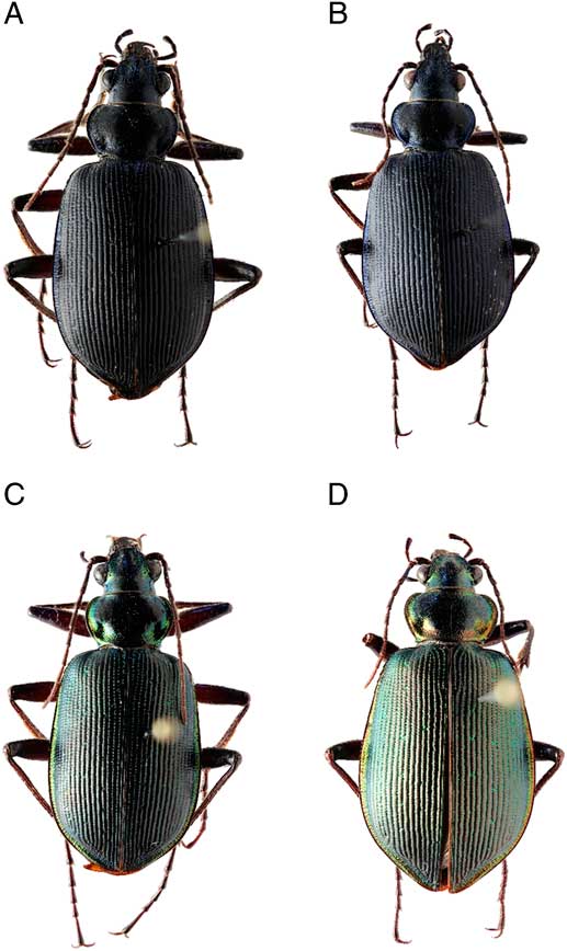
In April of 2011, an aberrant specimen of this genus was collected in Waco, Texas. The specimen was similar in size and habitus to C. wilcoxi, but was dark blue-black and not the typical green (Fig. 1). Only one specimen was known until 2013, when a second was found at the same location. In order to properly identify these specimens, a combined morphological and genetic approach, using DNA barcoding methods, was applied.
Habitus photographs of the two colour morphs of Calosoma wilcoxi. (A) Male and (B) female of blue morph. (C) Male and (D) female of the green morph.
DNA barcoding has enriched the study of biodiversity by providing quick and cost effective methods for the identification of distinct mitochondrial lineages and robust species-level identification (Hebert et al. Reference Hebert, Penton, Burns, Janzen and Hallwachs2004). For animals, a section of the cytochrome oxidase subunit I (COI) gene is the primary marker for DNA barcoding. By comparing morphological variation to DNA barcoding results, increased confidence in the identification of similar or cryptic taxa can be achieved.
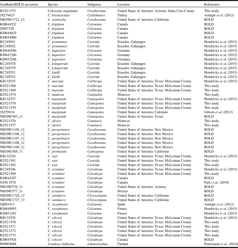
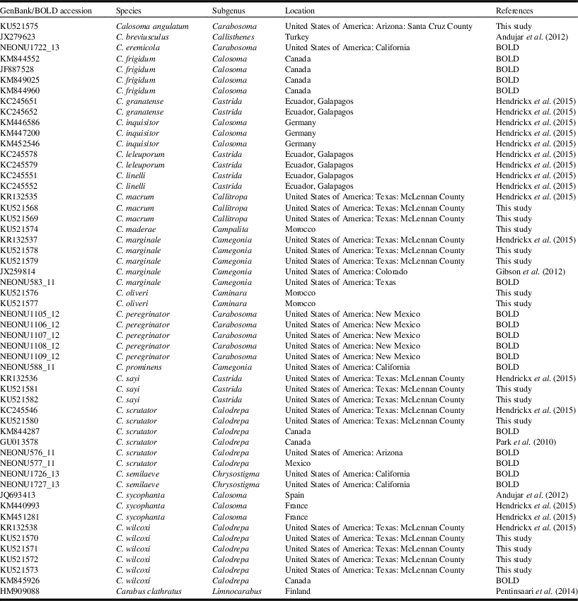
Here we genetically compared the two aberrant Calosoma specimens to specimens of all other local species in order to determine whether these specimens represented a separate lineage, or phenotypic plasticity within C. wilcoxi. We sequenced the COI gene of the aberrant specimens, as well as the five species sympatric at the sampling location, C. (Carabosoma) angulatum Chevrolat, 1834 from Arizona, United States of America, and two northern African species (C. (Caminara) oliveri Dejean, 1831 and C. (Campalita) maderae (Fabricius, 1775)) as outgroups. In addition, we incorporated previously published sequences as available on GenBank (www.ncbi.nlm.nih.gov/genbank) and the Barcode of Life Data Systems (BOLD) (www.boldsystems.org), providing further coverage of North American, South American, and European species (Table 1). Combined with our newly generated sequences, we generated a preliminary phylogeny covering 20 species of Calosoma from four continents. We discuss the results of our phylogenetic reconstruction and compare them to previously established relationships in the genus.
GenBank/BOLD accession numbers for sequences used in this study.
BOLD, Barcode of Life Data System.
Materials and methods
Laboratory methods
We extracted total DNA of 15 specimens belonging to eight species, of which two specimens belonged to the new phenotype (Cal14, Cal15; specimens preserved in the collection of the Centrum für Naturkunde (CeNak) of the University of Hamburg, Hamburg, Germany). Extracts were prepared from a single mesothoracic leg of dried or ethanol stored material. DNA extractions were carried out according to Paxton et al. (Reference Paxton, Thorén, Tengö, Estoup and Pamilo1996). Polymerase chain reactions were performed using standard protocols and barcoding primers provided by Folmer et al. (Reference Folmer, Black, Hoeh, Lutz and Vrijenhoek1994): LCO1490 (5'-GGTCAACAAATCATAAAGATATTGG-3') and HCO2198 (5'-TAAACTTCAGGGTGACCAAAAAATCA-3'). Polymerase chain reactions (total volume 10 μL) contained 0.1 µL GoTaq (5 u/µL) (Promega, Madison, Wisconsin, United States of America), 2 µL GoTaq-Buffer (5×) (Promega), 0.2 µL MgCl2 (25 mM), 0.3 μL of both primers (10 µM), 0.2 µL dNTPs (10 mM), 1.0 μL DNA template, and 5.9 μL H2O. Polymerase chain reaction cycling conditions were as follows: initial denaturation 94 °C for 3.0 minutes, followed by 35 cycles of 94 °C for 30 seconds, 48 °C for 45 seconds, 72 °C for one minute, and final elongation 72 °C for eight minutes. Polymerase chain reactions products were purified using EXOSAP-it (Affymetrix, Santa Clara, California, United States of America) following the manufacturer’s instructions. Sanger sequencing was performed by GATC-Biotech (Konstanz, Germany).
Genetic analyses
Sequences were inspected and aligned using the MAFFT (Multiple Alignment using Fast Fourier Transform) algorithm as implemented in Geneious v.6 (Kearse et al. Reference Kearse, Moir, Wilson, Stones-Havas, Cheung and Sturrock2012). The most suitable substitution model was determined to be GTR+I+G using jModelTest2 (Guindon and Gascuel Reference Guindon and Gascuel2003; Darriba et al. Reference Darriba, Taboada, Doallo and Posada2012). We then used the Bayesian approach implemented in MrBayes v.3.2.5 (Ronquist et al. Reference Ronquist, Teslenko, Van Der Mark, Ayres, Darling and Hohna2012) to reconstruct the phylogeny of Calosoma. A sequence of Carabus clathratus Linnaeus, 1761 (HM909088) was defined as the outgroup. We ran MrBayes for 20 million generations sampling every 2000 generations resulting in 10 000 sampled trees. Convergence was confirmed by the average split frequencies being consistently below 0.02. A burn-in of 1000 trees was discarded before summarising the results in a consensus tree. The tree was visualised with FigTree v. 1.3.1 (http://tree.bio.ed.ac.uk/software/figtree/).
Results
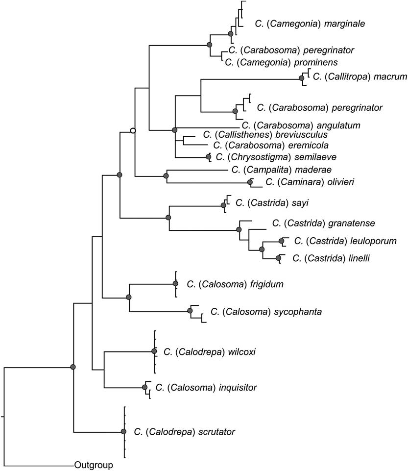
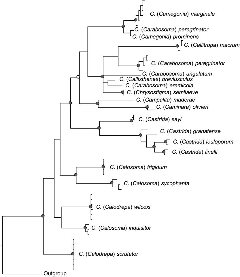
A total of 15 new COI sequences were generated for this study. After trimming, each sequence was 620 base pairs in length (Table 1). An additional 38 sequences from previous studies were incorporated into our phylogenetic analyses (Table 1). In total, our phylogenetic analyses included 53 sequences belonging to 20 species from the genus Calosoma, representing lineages from North America, South America, North Africa, and Europe and one sequence of Carabus clathratus as outgroup. The resulting tree (Fig. 2) indicates that C. (Calodrepa) wilcoxi is closely related to the European C. (Calosoma) inquisitor (Linnaeus, 1758), which along with C. (Calodrepa) scrutator are the most basal species in the genus of those investigated in this study.
Bayesian phylogenetic tree reconstructed with MrBayes; grey circles represent posterior probabilities above 0.95, white circles represent posterior probabilities above 0.90.
Discussion
Our genetic analyses confirm that the two aberrant Calosoma specimens collected in Texas represent black-blue morphological variants of the widespread, typically green C. wilcoxi. Melanism is known to occur in a wide variety of Coleoptera, including the families Cantharidae (Brakefield Reference Brakefield1985); Carabidae (Davis et al. Reference Davis, Bouvet and Vernon2007), and the subfamily Cicindelinae (Carabidae) (Pearson et al. Reference Pearson, Knisley, Duran and Kazilek2015); Chrysomelidae (Tower Reference Tower1906); Coccinellidae (Mikkola and Albrecht Reference Mikkola and Albrecht1988; Jong et al. Reference Jong, Gussekloo and Brakefield1996); Scarabaeidae (Ratcliffe and Mico, Reference Ratcliffe and Mico2001); and Tenebrionidae (Harris Reference Harris1988). Our results confirm the first recorded case of melanism in the genus Calosoma. Beetles of the genus, in particular the common North American species C. scrutator and C. wilcoxi, are well represented in collections; yet, we could find no evidence of previously reported melanism in either of these species, both of which are abundant, widespread, and typically green.
Our phylogenetic analysis, while limited due to our taxonomic and geographic coverage of the genus, does provide some useful information for understanding relationships within the genus. Our study strongly supports the monophyly of the subgenus Castrida Motschulsky, 1866 (0.98 posterior probability), based on the four species incorporated into this study (C. sayi, C. granatense Géhin, 1885, C. linelli Mutchler, 1925, and C. leuloporum Basilewsky, 1968). However, we did not recover the nominate subgenus Calosoma as monophyletic, based on three species incorporated into our analysis (C. frigidum Kirby, 1837, C. sycophanta, and C. inquisitor). Similar results were reported in a previous genetic analysis based on the mitochondrial ND5 marker (Su et al. Reference Su, Imura and Osawa2005). One specimen of C. (Carabosoma) peregrinator (BOLD ID NEONU1109-12.COI-5P) grouped within a cluster otherwise only consisting of Camegonia species (C. prominens and C. marginale). This is likely based on a misidentification of the specimen.
Our results indicate that C. wilcoxi is closely related to the European C. inquisitor, and the subgenera of Calosoma and Calodrepa Motschulsky, 1865 are the most basal of the genus. This does not conform with previous phylogenetic analyses, which indicated that subgenus Castrida occupied a basal position (Su et al. Reference Su, Imura and Osawa2005). However, this may partially be a result of different outgroup choice and taxon sampling. Based on morphological analyses, the subgenus Calosoma was originally identified as an ancestral lineage within the subfamily Carabinae (Jeannel Reference Jeannel1940); hence, it is not surprising that based on our choice of outgroup (Carabus) the analyses suggest a basal position of Calosoma. While any inference based on the results of our phylogenetic analyses are limited, given the sampling performed to identify the aberrant specimens, our results do indicate that the currently established subgeneric classifications within the genus are likely in need of revision.
Acknowledgements
The authors would like to thank Dr. Jeff Back of Baylor University for help collecting, as well as comments on a previous version of the manuscript. They further thank Carl Vangestel for providing sequences and comments. They thank Dr. Robert Paxton for providing the laboratory and sequencing facilities that supported this study. The authors would also like to thank Panagiotis Theodorou, Anja Miertsch, and Kerstin Goessel for support in the laboratory.