

INTRODUCTION
The large extinct rhinoceros Stephanorhinus kirchbergensis, also known in Russia as Merck’s rhinoceros, as well as other members of the genus (S. hemitoechus, S. etruscus, and S. hundsheimensis) lived in Eurasia during the Pleistocene. Stephanorhinus remains are known from Pliocene-Pleistocene deposits in western Europe (Gorjanovich-Kramberger, Reference Gorjanovich-Kramberger1913; Belyaeva, Reference Belyaeva1935, Reference Belyaeva1939; Gromova, Reference Gromova1935; Guérin, Reference Guérin1980, Reference Guérin1982; Ziegler, Reference Ziegler1995; Van der Made, Reference Van der Made2010; Persico et al., Reference Persico, Billia, Ravara and Sala2015; Rivals et al., Reference Rivals, Julien, Kuitems, Kolfschoten, van, Serangeli, Drucker, Bocherens and Conard2015) and eastern Europe (Beljaeva and David, Reference Beljaeva and David1975; Alekseeva, Reference Alekseeva1980; Danukalova et al., Reference Danukalova, Yakovlev, Alimbekova, Yakovleva, Morozova, Eremeev and Kosintsev2008). In Asia, Stephanorhinus remains have been found in Kazakhstan, Russia (southern Siberia), and in China (Brandt, Reference Brandt and von1877; Chersky, Reference Chersky1891; Dubrovo, Reference Dubrovo1957; Billia, Reference Billia2007, Reference Billia2014; Tong and Wu, Reference Tong and Wu2010; Tong, Reference Tong2012; Vasiliev et al., Reference Vasiliev, Serednyov, Milyutin, Slyusarenko, Kozlikin and Chekha2015). In Europe, the species S. kirchbergensis, hereafter Merck’s rhino, is associated with interglacial periods and woody (forest) vegetation (Flerov, Reference Flerov1989; Van der Made, Reference Van der Made2010) and has been recovered from a large territory across the middle part of the Eurasian continent (Billia and Zervanová, Reference Billia and Zervanová2015, fig. 1).
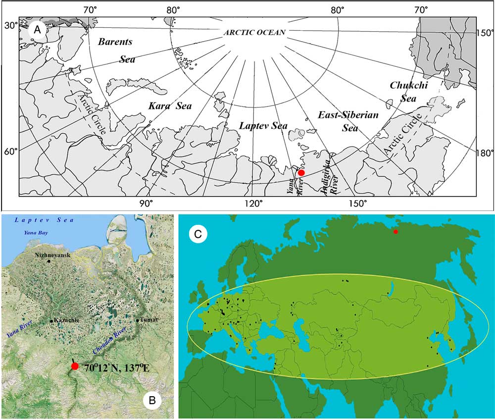
(color online) Site of the Stephanorhinus skull discovery. (A) Map of the Arctic territories. (B) Chondon River map (Google Earth). (C) Restored area of the rhinoceros (according to Billia and Zervanova, Reference Billia and Zervanová2015).

Merck’s rhino is well known from central Europe, where it is traditionally considered an element of the interglacial faunas. Its refugia during glacial periods have not been established. Remains from Spain, once thought to have been from Merck’s rhino, were reclassified as S. hemitoechus (Cerdeño, Reference Cerdeño1990). As a result, Spain does not seem to have been a refugium (Van der Made, Reference Van der Made2010). Other potential refugia include Italy, the Balkans, or southwest Asia, but no true records of glacial chronology containing Merck’s rhino are known from any of these locations. There is a long record of Merck’s rhino and similar species in China (Tong and Wu, Reference Tong and Wu2010), which led to the suggestion that Merck’s rhino may have arisen in China and persisted there during glacial times, at lower latitudes than Europe. If true, dispersal from China into Europe is likely to have been through Siberia, where fossils of Merck’s rhino are rare (Billia, Reference Billia2007, Reference Billia2008; Billia and Zervanová, Reference Billia and Zervanová2015), in particular compared with the widespread woolly rhinoceros (Coelodonta antiquitatis). In Russia, the few known fossils of Merck’s rhinoceros include the well-preserved but toothless “Irkutsk skull” (Brandt, Reference Brandt and von1877; Loose, Reference Loose1975; Billia, Reference Billia2008, Reference Billia2010).
In the summer of 2014, a skull of a rhinoceros of the genus Stephanorhinus was discovered in the middle reaches of the Chondon River (Yakutia, Russia; 70°12′N, 137°E; Fig. 1) near the bottom of a bank exposure 12 m high. It probably originated from the middle part of the outcrop. In this study, we used genetic, dietary, and morphological analyses to understand the taxonomic position, season and age of death, and diet of this Stephanorhinus individual.
GEOLOGIC, CLIMATIC, AND ENVIRONMENTAL SETTING
The Chondon River region is located in the northern part of the Primorskaya (Yano-Indigirskaya) lowland, which abuts the Laptev Sea. The geologic age of the sediments at the coast of the Laptev Sea and exposed by the river range from the middle Pleistocene to the Holocene. The general type of loose Pleistocene sediment in this region is an ice complex (so-called Edoma and other formations) comprising icy silts and large syngenetic polygonal ice wedges of mainly lake-alluvial origin (Baranova and Biske, Reference Baranova and Biske1964; Plakht, Reference Plakht1979; Sher, Reference Sher1981, Reference Sher1984). At this site, the sediments include the remains of the mammoth fauna that characterized the Zyryanskoe (early Würm, or early Wisconsinan) periods (Lomachenkov, Reference Lomachenkov1957), suggesting an age between 70,000 and 10,000 yr ago.
Today’s climate in the region is severe and cold because of its high latitude, the influence of the winter anticyclone, and the proximity of the Arctic Ocean. Long-term observations by meteorological stations in the Ust’-Yansk and Kazachie (Cossack) settlements describe an 8- to 9-month winter, in which average daily temperatures during the coldest months of December and January are −31°C to −32°C, and a 2- to 4-month summer from June to September, during which average daily temperatures range between 11°C and 24°C. The amount of precipitation in the region is small: up to 113 mm of precipitation in the summer and only 45 mm in the winter (Lomachenkov, Reference Lomachenkov1957).
The river network is poorly developed in the region. Steep-sided depressions resulting from permafrost thaw (= alasses in local terminology) are widespread in the watershed, with depths reaching 10–15 m. These alasses tend to be covered with meadow vegetation dominated by sedges (carex), dwarf willow (salix), and birch (betula). The tundra itself tends to include mosslike vegetation dominated by grasses and dwarf shrubs. The northern boundary of the forest vegetation (larch; larix) reaches Orotko Lake, near the lower Chondon River (Lomachenkov, Reference Lomachenkov1957; Dylis, Reference Dylis1981). Larch trees grow in the river valley but are sparse in the upper reaches of the Chondon River, where taiga is widespread.
MATERIALS
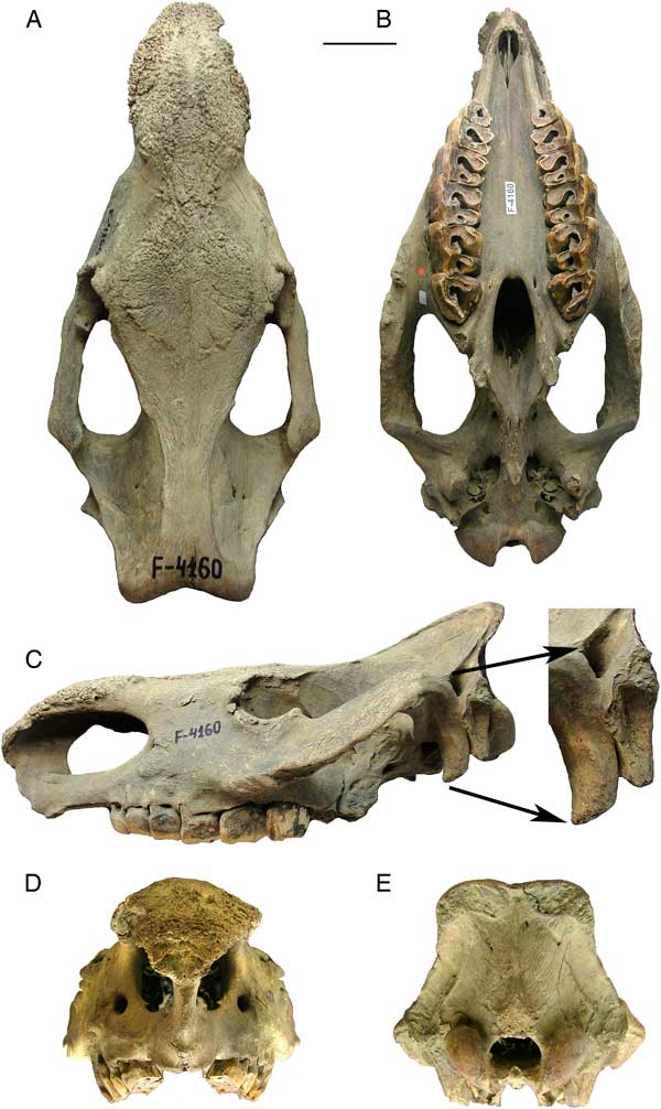
We investigated a well-preserved skull of Stephanorhinus sp. from the middle of the Chondon River valley. The specimen, which was a yellow-grayish color, retained the complete tooth rows (Fig. 2) but is missing the mandible. Within the cranial cavities, we found large, rolled and flattened gravel and small pebbles (up to 1.8 cm in diameter) with a dark-gray color, along with fine-grained sand with a gray-yellow color. The presence of this material indicates that the skull had been in an active aquatic environment, though this did not cause significant erosion. A greenish coating (the traces of microbial activity) on some sections of the skull suggests that the skull remained in subaerial conditions for some time, presumably when the perennially frozen sediments were thawed.
(color online) Skull of the Chondon rhino, specimen F-4160. (А) View from above. (В) Bottom view. (С) Left side view. (D) Front view. (E) Occipital view. Scale bar: 100 mm.

We found that stratification of different substances in the Chondon rhino (hereafter referred to as ChR) teeth cavities was the following. Sediments and other preserved materials formed “layers” within the teeth fossae: fine-grained dust and sand (with thickness 0.05–0.01 mm and 0.25–0.1 mm, respectively) was found at the bottom and was overlain by a vegetation “plug” comprising chewed branches of larch. Proceeding from this, we suppose that the skull lay “teeth up” in the river stream, which carried away the mud and larger particles, but the fine-grained sand passed through the plug and settled at the bottom of the cavities. Otherwise, we would have to assume that ChR at first ate fine-grained and well-sorted sand and then began to eat the plants.
The skull of the ChR is stored at present in the National Alliance of Shidlovskiy “Ice Age,” Ice Age Museum, Moscow, as accession number F-4160.
METHODS
Metric data
All measurements were made following the methodology for working with rhinoceroses by Guérin (Reference Guérin1980) and Van der Made (Reference Van der Made2010). All measurements are provided in millimeters.
Determining the age of ChR
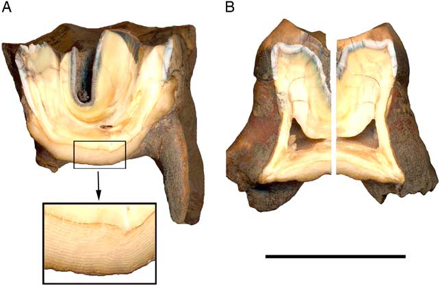
The individual age at death of large herbivorous mammals is usually determined by assessing the condition of the sutures between the cranial bones and the state of eruption and wear of the teeth with respect to annual increments of cementum. However, because tooth wear depends on the abrasiveness of the food, we did not use the degree of eruption and wear of the teeth (Goddard, Reference Goddard1970; Hitchins, Reference Hitchins1978; Hillman-Smith et al., Reference Hillman-Smith, Owen-Smith, Anderson, Hall-Martin and Selaladi1986; Garutt, Reference Garutt1994). Instead, we used a method developed for present-day rhinoceroses to estimate age based on annual cementum layers (Goddard, Reference Goddard1970; Hitchins, Reference Hitchins1978; Klevezal, Reference Klevezal1996, Reference Klevezal2007). We cut the М1 cementum pad vertically (Fig. 3) in transversal (external-internal) and longitudinal (antero-posterior) directions, and then the number of layers of annual increment was estimated. We used М1 because it erupts earlier than the other molar teeth and has longer life history.
(color online) Vertical cuts of М1. (A) Anteroposteriorly; inset shows the annual growth layers in the cement pad. (B) Second half of the tooth buccal-labial cut. Scale bar: 50 mm.

Microstructure patterns
We scanned the masticatory surface of the facets and the buccal and lingual lateral sides of four teeth with an Epson Perfection 2580 PHOTO scanner (Seikо Epson Corporation, Japan) with a resolution of 800 dpi. We measured the maximum height of the crown and the width of its base and estimated the square of buccal and lingual walls of each tooth (Supplementary Fig. 1). We examined microstructure patterns optically using a Keyence Digital Microscope VHX-1000 (Keyence Corporation, Japan) with a 5–50× and 20–200× lens. Four well-preserved enamel sections were selected, including those located on the dorsal and lateral sides of the crowns (Supplementary Fig. 1C, see arrows). To standardize the description of our results, we classified microstructure patterns into categories depending on their configuration, size, and orientation. Our classification is based on previously proposed “microwear” patterns (Solounias and Semprebon, Reference Solounias and Semprebon2002; Semprebon et al., Reference Semprebon, Godfrey, Solounias, Sutherland and Jugers2004). We distinguished the following categories: (1) large pits and small pits; (2) parallel coarse scratches and cross fine strokes; and (3) cleavages. We estimated the total number of patterns per 4 mm2 by superposition on a photomicrograph square frame with a 2-mm thickness.
Selection and identification of plant remains
We collected materials for analysis from the brain cavity and from the surface of several teeth with a chip-blower and a soft brush. We collected the content of tooth cavities with dental tools: first, we recovered plugs of large plant remains in the fossae of the cheek teeth, and then the fine-grained sand beneath.
The specimens examined were as follows: (1) grayish aleurite from the skull brain cavity, weighing 60 g; (2) yellowish-gray fine-grained sand from under the vegetation plug, total weight 0.6 g; and (3) the plug of plant detritus in the fossae of the teeth, total weight 4.5 g.
We sieved each sample using meshes of 0.5 mm and 0.25 mm. We treated the large fraction, up to 0.25 mm (twigs, bark) of (3) with 38% H2O2. A smaller fraction, less than 0.25 mm, was macerated, without adding alkali or sodium pyrophosphate. We performed low-density biogenic fractionation to separate phytoliths, detritus, and the remains of tissue and epidermis using a heavy liquid (KI+СdI with specific gravity 2.2 g/cm3). We examined these preparations using a Carl Zeiss Axiostar microscope at a magnification of 200–400× and a Vega 3 Tescan scanning electron microscope under high and variable vacuum, using the analyzers backscattered electron detector (BSE) and secondary electrons detector (SE). The unprocessed fraction (0.5–0.25 mm) of specimen (3) was examined under a binocular MBS-9.
To analyze wood remains, we used the methods of A.A. Yatsenko-Khmelevsky (Reference Yatsenko-Khmelevsky1954) and V.E. Benkova and F.H. Schweingruber (Reference Benkova and Schweingruber2004). We embedded 10 well-preserved shoots in a silicone sealer and examined these in cross sections of 12–16 μm thick, prepared with a sliding microtome, stained with safranin, and identified using transmitted and reflected light.
Mitochondrial phylogenetic analysis
We extracted DNA from an M1 tooth root of ChR following Dabney et al. (Reference Dabney, Knapp, Glocke, Gansauge, Weihmann, Nickel and Valdiosera2013). We converted the DNA extract to an Illumina-compatible library following Meyer and Kircher (Reference Meyer and Kircher2010) as modified by Heintzman et al. (Reference Heintzman, Zazula, Cahill, Reyes, MacPhee and Shapiro2015). We then enriched the library for rhinoceros mitochondrial DNA using a novel mammalian mitochondrial RNA bait set, adapted from Slon et al. (Reference Slon, Glocke, Barkai, Gopher, Hershkovitz and Meyer2016), which included bait from all four extant rhinoceros genera and Coelodonta (Supplementary Table 1). For enrichment, we followed the MyBaits v. 3 protocol (MYcroarray, Ann Arbor, MI). We sequenced ~350,000 reads of the enriched library on the Illumina MiSeq platform, using paired-end chemistry for 2×75 cycles. We merged reads and removed sequencing adapters and reads shorter than 25 base pairs (bp), using SeqPrep (https://github.com/jstjohn/SeqPrep).
We aligned the merged and remaining unmerged readings to three mitochondrial reference genomes (white rhinoceros, Ceratotherium simum, GenBank: NC_001808.1; Sumatran rhinoceros, Dicerorhinus sumatrensis, GenBank: NC_012684.1; and woolly rhinoceros, Coelodonta antiquitatis, GenBank: NC_012681.1) using the Mapping Iterative Assembler (MIA) (Briggs et al., Reference Briggs, Good, Green, Krause, Maricic, Stenzel and Lalueza-Fox2009), and named mitochondrial consensus sequences following Heintzman et al. (Reference Heintzman, Froese, Ives, Soares, Zazula, Letts and Andrews2016). We combined the three consensus sequences and filled in any remaining unnamed sequence using a manual primer walking approach, following Heintzman et al. (Reference Heintzman, Zazula, Cahill, Reyes, MacPhee and Shapiro2015). We then remapped the merged and remaining unmerged reads to the draft ChR mitochondrial genome, using MIA, and manually inspected the alignment to ensure sequence accuracy. Because of the uncertainty of mapping short reads to the tandem repeat section of the control region, we coded this section as missing data, following Heintzman et al. (Reference Heintzman, Zazula, Cahill, Reyes, MacPhee and Shapiro2015). The ChR mitochondrial genome has been accessioned in GenBank (KX646743).
To infer the phylogenetic placement of ChR, we conducted two analyses using: (1) full mitochondrial genomes and (2) a 751 bp portion of the mitochondrial control region. The latter was conducted to demonstrate that ChR falls outside of previously sampled intraspecific diversity for other rhinoceros taxa for which complete mitochondrial genome data are not available. We aligned the ChR mitochondrial genome to previously published rhinoceros data (Supplementary Table 2) using the MUSCLE algorithm in SeaView v. 4.6 (Gouy et al., Reference Gouy, Guindon and Gascuel2010) and used the Bayesian information criterion in jModelTest v. 2.1.6 (Darriba et al., Reference Darriba, Taboada, Doallo and Posada2012) to select the (1) GTR+G and (2) HKY+G models of molecular evolution for the two data sets. We analyzed the alignments using both Bayesian and maximum likelihood (ML) phylogenetic methods. Bayesian analyses were run in MrBayes v. 3.2.2 (Ronquist et al., Reference Ronquist, Teslenko, van der Mark, Ayres, Darling, Hohna, Larget, Liu, Suchard and Huelsenbeck2012) for 10 million generations, with the first 25% discarded as burn-in. We ran ML analyses in RAxML v. 8.2.4 (Stamatakis, Reference Stamatakis2014), which included 500 bootstrap replicates. For both data sets, we used the Malayan tapir (Tapirus indicus, GenBank: NC_023838.1) as outgroup.
14С dating and stable isotopes (13C, 15N)
We performed accelerator mass spectrometry (AMS) radiocarbon dating on a root of the М1 tooth (4.5 g) in Groningen. The sample underwent standard chemical cleaning and collagen extraction (Mook and Streurman, Reference Mook and Streurman1983) based on the method developed originally by Longin (Reference Longin1971). The collagen was combusted into CO2 by an elemental analyzer (EA), coupled online with a stable isotope mass spectrometer (MS), which enables precise measurements of the stable isotope ratios δ13C and δ15N. The EA/MS system is an Elementar Isocube/Isoprime 100 combination.
The EA was also used to purify the CO2, which was then cryogenically trapped using an automatic device and transformed into graphite by reacting under excess H2 gas (Aerts-Bijma et al., Reference Aerts-Bijma, van der Plicht and Meijer2001). The 14C/12C and 13C/12C isotope ratios of the graphite were measured by AMS (Van der Plicht et al., Reference Van der Plicht, Wijma, Aerts, Pertuisot and Meijer2000). The AMS is a 2.5 MV Tandetron system made by High Voltage Engineering Europa, the Netherlands. The isotope ratios are converted to conventional 14C ages, reported in BP (Mook and van der Plicht, Reference Mook and van der Plicht1999).
The stable isotope ratios are expressed as deviations (in per mil) from reference materials:
 $$\eqalignno{ & \delta {}^{{13}}{\rm C}\, {\equals}\, {{\left[ {{}^{{13}}{\rm C}/{}^{{12}}{\rm C}} \right]_{{\rm sample}} } \over {\left[ {{}^{{13}}{\rm C}/{}^{{12}}{\rm C}} \right]_{{\rm reference}} }}{\minus}1\left( \!{{\times}1000{%\permil }} \right)\,{\rm and}\, \cr & { \delta {}^{{15}}{\rm N}\, {\equals}\, {{\left[ {{}^{{15}}{\rm N}/{}^{{14}}{\rm N}} \right]_{{\rm sample}} } \over {\left[ {{}^{{15}}{\rm N}/{}^{{14}}{\rm N}} \right]_{{\rm reference}} }}{\minus}1\left( {{\!\times}1000{%\permil}} \right)$$
For 13C, the reference is a belemnite called Pee Dee belemnite; for 15N, the reference is ambient air (Mook, Reference Mook2006).
RESULTS
The discovery of the skull under the geologic cross section, without the context of the sediment, required very careful research.
Taxonomic description and comparison of the skull of the ChR
Only three extinct genera of Quaternary Eurasian rhinoceroses are known from the territory of Russia: Elasmotherium, Coelodonta, and Stephanorhinus. In the latter genus, four species, S. etruscus, S. hundsheimensis, S. hemitoechus, and S. kirchbergensis, are recognized (Fortelius et al., Reference Fortelius, Mazza and Sala1993; Lacombat, Reference Lacombat2006; Van der Made, Reference Van der Made2010). Though the type material of S. kirchbergensis, Merck’s rhino, consists of only a few teeth, the species is well known from other well-preserved material, including skulls (Loose, Reference Loose1975; Van der Made, Reference Van der Made2010). Fossil material that is most similar to Merck’s rhino includes the Chinese middle and late Pleistocene S. choukoutienensis and the early Pleistocene S. yunchuchunensis and S. lantienensis. The first of these was initially considered to be S. kirchbergensis, and, recently, this has again been suggested, while the other early Pleistocene species are considered valid (Tong and Wu, Reference Tong and Wu2010).
The skull of ChR is large (Supplementary Table 3), and the morphology of both the skull and teeth are typical of the Rhinocerotidae. The incisors and canines are absent, which distinguishes ChR from the living genera Rhinoceros and Dicerorhinus that both have two upper incisors. ChR also differs from the extinct genus Elasmotherium in that it lacks a large domelike outgrowth of the frontal bone and has cauliflower structures on the frontals and on the nasals, indicating that there were two horns; lacks the intense lamellar folding of the enamel; and has teeth with roots.
An ossified bony septum is absent in all four living rhinoceros genera but is present in Stephanorhinus and Coelodonta (Guérin, Reference Guérin1980). In ChR, an ossified nasal septum is preserved only partially in the anterior portion of the skull. The trail from the ossified nasal septum is clearly traced from the nasal tip dorsally at least to the level of the eye orbits, and, ventrally, it is preserved by a narrow band about 13 mm wide, reaching the level of P2 (further broken). Perhaps it reached the level of the palatine bone; at least, there is no reason to believe that it ended in this fragment. The latter has a spongy structure and is not fused with skull, like the front and upper parts. Apparently, in this part the ossification of the septum has not yet occurred and is connected with palatine bone by cartilage or soft tissues.
ChR has some characteristics that indicate affinity with Stephanorhinus. Seen from behind, the occipital crest of ChR is narrow and the skull becomes markedly wider at the level of the zygomatic arcs, giving this part of the skull a trapezoidal outline. This is as in Stephanorhinus, whereas in Coelodonta the occipital crest is much wider and the size increase at the level of the zygomatic arcs is much less, resulting in a squarer outline (Van der Made, Reference Van der Made2010). In side view, the occipital crest does not extend much more posteriorly than the occipital condyles, which differs from both Coelodonta and S. hemitoechus (Zeuner, Reference Zeuner1934; Van der Made and Grube, Reference Van der Made and Grube2010, fig. 5). Seen from above, the occipital crest has an open V shape, which is typical only for S. kirchbergensis, but not for the other species of Stephanorhinus nor for Coelodonta, where the crest is straight.
In ChR, the processus postglenoideus and the processus paroccipitalis are massive, not very divergent, and placed close together. The processus posttympanicus is not really developed as a separate process but is covering the posterior side of the processus postglenoideus. This configuration is as in S. kirchbergensis, but it differs from that in S. hemitoechus and S. hundsheimensis, where the two massive processes are placed at a greater distance, and from that in the living rhinoceroses, where the posttympanic is more of an independent process (Loose, Reference Loose1975, fig. 5, plates 12 and 13; Billia, Reference Billia2008).
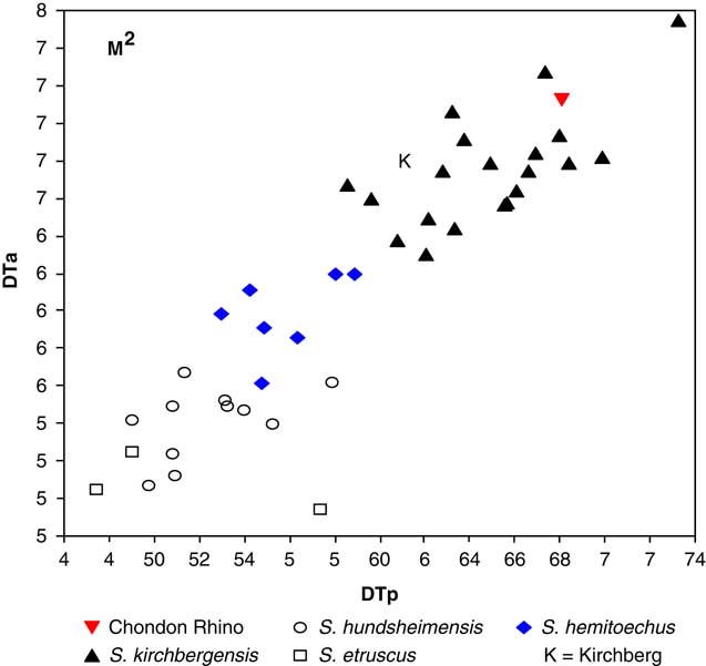
The teeth of ChR have low crowns and very finely crenellated enamel, which again differs from Coelodonta and S. hemitoechus (Kurtén, Reference Kurtén1968; Guérin, Reference Guérin1973; Fortelius et al., Reference Fortelius, Mazza and Sala1993; Billia, Reference Billia2008). The size of the teeth indicates affinity to S. kirchbergensis (Fig. 4 and Supplementary Fig. 2). ChR’s teeth are very large (Supplementary Table 4), and much larger than in S. hemitoechus, S. hundsheimensis, and S. etruscus. The P2 is not disproportionately large, as in S. hundsheimensis (compare Van der Made, Reference Van der Made2010, fig. 12; see also Van der Made, Reference Van der Made2014). The teeth are much larger than in S. hemitoechus, but the skull is not larger (Supplementary Table 3). Relatively large teeth compared with the skull were noted as a feature of S. kirchbergensis (Van der Made, Reference Van der Made2010).
(color online) The widths of the anterior (DTa) and posterior (DTp) lobes of the second upper molar (M2) of the Chondon rhinoceros compared with those of other species of Stephanorhinus: S. kirchbergensis from type locality Kirchberg, Mosbach, Neumark Nord, Ehringsdorf, Bilzingsleben, Rhinegravels; S. hemitoechus from Murr, Neumark Nord, Rhinegravels; S. hundsheimensis from Voigtstedt, Süssenborn, Mosbach, Soleilhac, Mauer; and S. etruscus and S. aff. etruscus from the Upper Valdarno and Atapuerca TD4. All teeth from tooth rows (so their identity as M2 is clear), save for one of the type specimens of S. kirchbergensis from Kirchberg (marked as “K”). For comparisons of further cheek teeth and provenance of the data, see the Supplementary Materials.

The ectolophs of the cheek teeth have gently undulating surfaces, which is common in Stephanorhinus, while in Coelodonta, the buccal relieve is much more marked. The buccal fold of the paracone is more marked, and there is a clear vertical depression in the middle of the ectoloph. The crochet does not completely separate a small fossa from the transverse valley. This is normal in Stephanorhinus, but in Coelodonta a separate fossa is very typical.
The orbits are small, somewhat protruding, and high set. The jugal arches under the eye socket are massive, and their lower portion beyond the orbit has no pronounced fold. The middle portion of the jugal arch is very wide but thin.
Taken together, these morphological characteristics indicate that the ChR skull belongs to S. kirchbergensis.
Genetic classification of the ChR skull
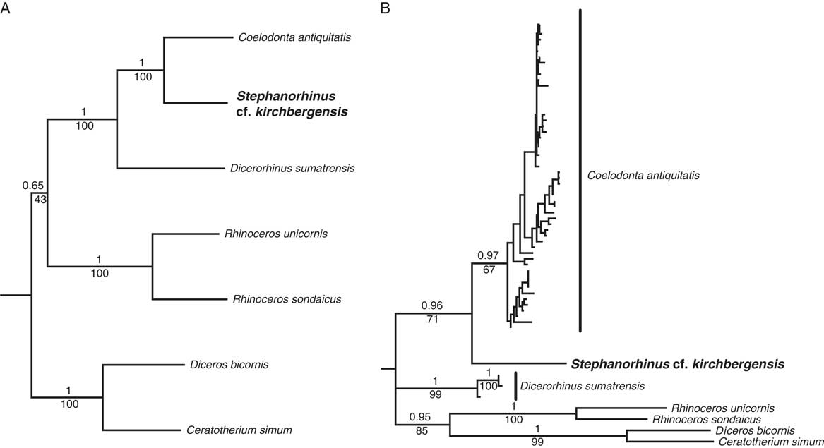
Phylogenetic analysis of mitochondrial genomes isolated from ChR and other rhinoceroses indicate that ChR is sister to the woolly rhinoceros (C. antiquitatis), with strong statistical support (Fig. 5A). Analysis of the short fragment of the control region confirms that ChR is distinct and does not fall within the sampled diversity of C. antiquitatis (Fig. 5B and Supplementary Table 2), which is consistent with ChR belonging to S. kirchbergensis. These results are also consistent with the morphological data, in that they preclude ChR from belonging to an extant rhinoceros genus.
The phylogenetic placement of the Chondon rhino within crown group Rhinocerotidae, based on full mitochondrial genomes (A) and a 751 bp section of the mitochondrial control region (B). The Chondon rhino is found to be sister to Coelodonta antiquitatis in both analyses. Support values above branches are Bayesian posterior probability values, whereas those below branches are percentage support from 500 maximum likelihood bootstrap replicates. The outgroup (Tapirus indicus) is not shown.

Determination of the age of the ChR individual at the time of death
Several lines of evidence suggest that the skull belonged to a mature individual. Most prominently, we observed an overgrowth of the sutures between the cranial bones and the erupted and worn M3, indicating maturity. In both living Diceros bicornis (Hitchins, Reference Hitchins1978) and Ceratotherium simum (Hillman-Smith et al., Reference Hillman-Smith, Owen-Smith, Anderson, Hall-Martin and Selaladi1986), the premolars are completely replaced by 8 yr of age, and the last molar, аn М3, erupts at an age of 8–16 yr. In addition, the roots of all of the teeth are closed and filled with dentin. Seven accretions are observed in the antero-external root of Р4, presumably reflecting annual accumulation. The cut in the lingual portion of М1 shows a small cavity that is not completely filled, with small denticles within it (Fig. 3A). All of the premolars, in particular Р3 and Р4, show a pronounced expansion of the root in the middle portion, corresponding to intensive growth for closure of the apical portion. The interroot cementum is very thick. The outer cementum is found at the base of the crown at the sides of each tooth and on the crown itself at the front and rear of the molars. The largest cementum increment is in М1, which is the first molar to erupt and is responsible for the major masticatory load before the other two molars erupt and the molar predecessors are replaced by the permanent teeth. The traces of initial resorption are visible on the Р2 roots and, to a much lesser extent, on the roots of other teeth. This indicates an intensive load on the chewing parts of the animal, most likely associated with eating roughage, but it also indicates that time has passed since the teeth erupted.
In “forest” S. kirchbergensis individuals, age can be inferred by adding 3 to the number of annual layers in the М1 pad (Goddard, Reference Goddard1970). In ChR, the cross section of М1 has 16–17 layers (Fig. 3А, inset) suggesting a minimum of 20 yr of age. The last layer in ChR appears to be dark in reflected light, which has been associated with periods of no growth (a cold season).
Microstructure patterns
The crowns of all teeth are heavily worn, particularly on the buccal side (Supplementary Fig. 1). The length of the base of the teeth along the left upper jaw increases from premolars to molars, reaching a maximum size of 63 mm in М2. However, the crown height shows no linear trend, changing from 22.5 mm in P2 to 40 mm in М2, reaching a maximum in М2 (Supplementary Table 5). On the lingual side, the crowns also increase in size from premolars to molars, but compared with those on the posterior side, they are lower, (i.e., the tooth slopes lingually) (Supplementary Table 5). This is consistent with the natural wear of the teeth.
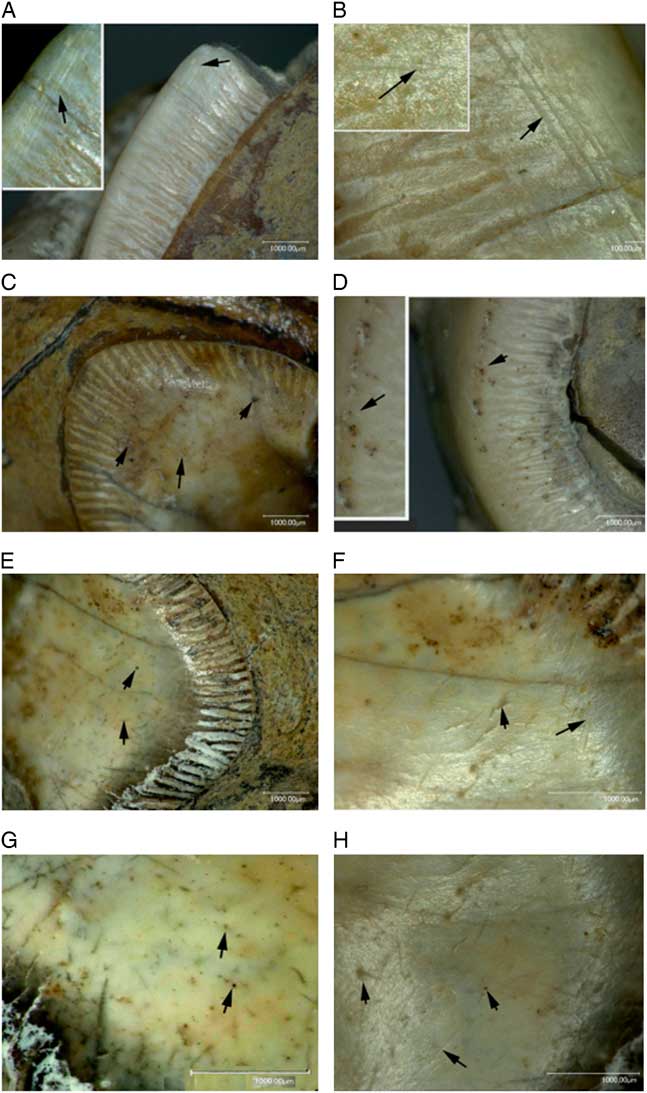
The amount, type, and distribution of microstructure patterns vary among teeth in ChR (Supplementary Table 6). The masticatory surface is damaged, probably because of the friction of the teeth of the upper and lower jaws and particles of the soil being ingested together with the food. We observe large, straight, long, and wide (9 to 30 μm) scratches, sometimes grouped in a parallel pattern (Fig. 6A [inset] and B, arrows). In addition, we observe short and thin strokes oriented in different directions that are frequently crossed (Fig. 6B [inset], F, and H, arrows) and numerous pits that vary in size from 1 to 20 μm and in configurations including round, oblong, scalloping, and with or without a pronounced central cavity. These pits are sometimes situated in random ways (Fig. 6С, E–H, arrows) or in the form of an arrow (Fig. 6D) and are found mainly on the lateral sides of the teeth. Of particular interest is the arch-shaped arrangement of large pits on the apex of the enamel lateral surface (Fig. 6D). The long and rough scratches arranged in a parallel cluster are the rarest. Fine, short strokes are also situated locally.
(color online) Microstructure enamel patterns (indicated by arrows) in dorsal (А, B) and lateral (C–H) sides of the left cheek teeth of the Chondon rhino. (А [inset], B) Wide parallel scratches. (В [inset], F, H) Small overlapping scratches. (C, E–H) Small and large pits located chaotically. (D [inset]) Large pits grouped in arch. The scale bar represents 1000mμ.

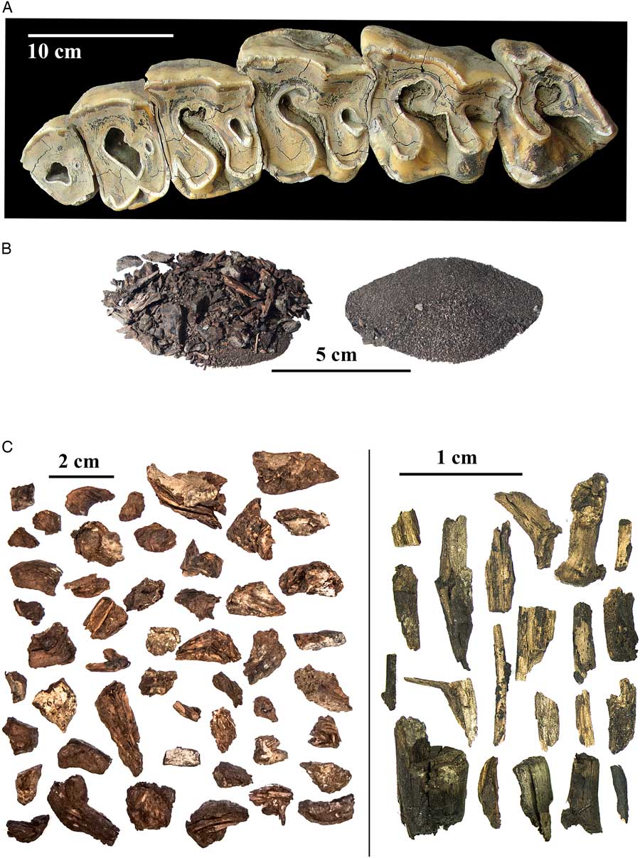
(color online) The remains of food from upper cheek teeth of Chondon rhino. (А) Left tooth row with cavities, hammered by “plug.” (В) Plant detritus (left) and fine-grained sand (right) from the bottom of teeth cavities. (С) Remains of woody plants: coniferous bark fragments (left) and fragments of sprouts (right).

Paleobotanical studies
We identified pollen and spores in samples taken from the brain cavity and the teeth. In addition, all specimens of diatomic algae are found, but algae were most numerous and well preserved in the brain cavity. Among these, the dominant species was Fragilaria ulna (Nitzsch) Lange-Bertalot by thin, long, and almost intact sections—this is a cosmopolitan species that is found commonly in freshwater bodies. We also observed in all specimens the epidermis of grasses and herbs, mosses of the genera Aulacomnium and Polytrichum, and vascular tissues of herbs.
The bulk of the plant remains came from the fossae of the teeth (Fig. 7В, left). In the unprocessed fraction, we found several twigs of Salix sp. and Betula sp., but the most highly represented remain was bark and shoots of Larix sp.
The macerate of the microremains comprised more than 150–200 μm of plant detritus, including the epidermis and the tissues of herbs, lignified tissues, and the conductive elements of branches/wood and moss tissues (Supplementary Table 7). Plant tissues retained a well-preserved cell structure and stomatal complexes. We also identified fragments of the epidermis of Ericaceae with stomatal complexes. Up to 20% of tissue remains are mosses. No remains of Cyperaceae were found.
Phytoliths were not abundant in the tooth cavities. We found fewer than 10 morphotypes of phytoliths, represented by elongated (rectangular and cylindrical) smooth sticks and trapezoid or occasionally conical and round forms. Polylobate and cubic shapes were rare. Elongated sticks with a spinous surface form, which are typical in dry conditions, were absent. The relative uniformity of phytolith forms is presumably associated with a predominance of tissues and macroremains of wood and shrub vegetation, which are better preserved than herbaceous remains.
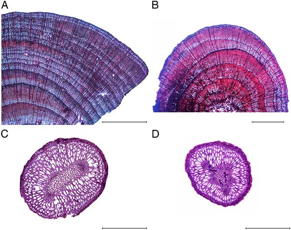
We identified numerous remains of woody plants with an average size of 0.6 mm3. These were mainly fragments of tree bark. Fragments of shoots ranged from 2 to 12 mm long and from 0.5 to 5 mm in diameter. Occasionally, these fragments were covered with bark (Fig. 8), with a thickness reaching 4–5 mm (i.e., the consumed shoots were 5 to 12 mm in diameter). We identified 25 woody remains as belonging to the families Pinaceae (23) and Ericaceae (2). The division of the genera Larix and Picea is problematic even for present-day specimens; nevertheless, 7 of the recovered remains were definitely attributed to Larix. The age of these remains ranges from 14 to 15 yr, based on the counting of annual rings. In some of the specimens, the last annual ring ends with late wood, which indicates that death (consumption) of the plant occurred in the middle of the vegetation season or during winter (Fig. 8А). In others, there is only early wood in the last (external) annual ring (Fig. 8B), indicating the beginning of seasonal growth.
(color online) Cross sections of sprouts from the dental cavities of the Chondon rhino. (A) Larix sp. (B) Larix/Picea. (C, D) (?) Vaccinium sp. Scale bars: 1 mm.

The width of the annual rings in the larch remains varied between 0.05 and 0.2 mm. The low radial increment indicates unfavorable conditions for tree growth, in particular a short and cold vegetation season (Naurzbaev and Vaganov, Reference Naurzbaev and Vaganov2000).
Two remains of shrubs (Fig. 8) with ages of 2–3 yr belong to Vaccinium. They died (or were consumed) between the late summer and early spring of the following year.
AMS dating and stable isotopes (13C, 15N)
The ChR sample provided excellent quality collagen. For the collagen, the measured isotope values and parameters are shown in Supplementary Table 8. The stable isotope values for the bone collagen (δ13C and δ15N) are consistent with a herbivorous diet.
The 14C measurement results returned infinite ages, >45,000 14C yr BP (see also Van der Plicht and Palstra, Reference Van der Plicht and Palstra2016). This corresponds to about 48,000 cal yr BP (Reimer et al., Reference Reimer, Bard, Bayliss, Beck, Blackwell, Bronk Ramsey and Buck2013).
The collagen yield of the bone was 12%. The collagen quality parameters show excellent values. The C %, N %, and C/N values are 43.4%, 16.4%, and 3.1, respectively, all within the range that is required (DeNiro, Reference DeNiro1985; Ambrose, Reference Ambrose1990).
DISCUSSION
Significance of the ChR
S. kirchbergensis is traditionally considered to be typical of the interglacial faunas of Europe, where it is not known from glacial faunas. A possible refugium may have been China, where the species is known from lower latitudes than in Europe (Tong and Wu, Reference Tong and Wu2010). If this is correct, the species must have dispersed each interglacial period from China across Siberia and into Europe. Its fossil record in Siberia is rare but is becoming better known (Billia, Reference Billia2007, Reference Billia2008; Billia and Zervanová, Reference Billia and Zervanová2015). The ChR skull is the first discovery of a rhinoceros from the genus Stephanorhinus north of the Arctic Circle, farther north than indicated by previous discoveries. Given that S. kirchbergensis in Europe is a typical indicator of interglacial (warm) conditions, this finding is surprising and sheds new light on its potential environmental constraints.
The mitochondrial genetic data are consistent with this taxonomic identity. We established strong statistical support for a sister relationship between ChR and Coelodonta antiquitatis. Given the morphological identification of ChR as Stephanorhinus, this result has systematic implications for understanding the relationships between rhinoceros species. Some taxonomists have suggested previously that, based on morphological features, the genus Stephanorhinus was closely related to, if not synonymous with, Dicerorhinus (e.g., Billia, Reference Billia2014; Persico et al., Reference Persico, Billia, Ravara and Sala2015). However, limited cladistic analyses using morphological data suggest a closer relationship to Coelodonta (Cerdeño, Reference Cerdeño1995; Prothero et al., Reference Prothero, Manning and Hanson1986), in line with the views of other taxonomists (e.g., Groves, Reference Groves1983) and the results presented herein. We caution, however, that genetic testing of additional individuals will be required to determine whether morphologically defined Stephanorhinus represents a monophyletic clade sister to Coelodonta.
According to the radiocarbon result, the ChR skull is from an individual that lived more than 48,000 yr ago. Based on geologic and other paleontological data from the site (Lomachenkov, Reference Lomachenkov1957), we conclude that ChR lived between 48,000 and around 70,000 yr ago. This corresponds to the beginning of the Karginsky interglacial period (i.e., the Marine Oxygen Isotope Stage [MIS] 4/MIS 3 boundary).
Reconstruction of the diet of fossil rhinoceroses
The diet of the most widespread Eurasian Pleistocene rhinoceros, the woolly rhinoceros C. antiquitatis, has been reconstructed from preserved plant remains in the folds of preserved cheek teeth (Brandt, Reference Brandt and von1849; Schmalhausen, Reference Schmalhausen1876; Garutt et al., Reference Garutt, Meteltseva and Tikhomirov1970) and from the stomach contents of preserved mummies (Nоwak et al., Reference Nоwak, Panоw, Tokarski, Szafer and Stасh1930; Kubiak, Reference Kubiak1969; Lаzarev and Tirskaya, Reference Lazarev and Тirskaya1975; Boeskorov, Reference Boeskorov2012). These plant remains are preserved because of the conservation effects of the enclosing sediments (perennially frozen sediments) or, in the case of the rhinoceros from Starunia, by mineral wax (Nоwak et al., Reference Nоwak, Panоw, Tokarski, Szafer and Stасh1930). The taxonomic composition of plants from these remains indicates a diet that consisted mainly of herbaceous plants (primarily grasses, sedges, and Artemisia) but also included branches of shrubs and trees. This combination points to a grassland or steppe tundra habitat across the range of C. antiquitatis. This reconstructed diet and also its body size and proportion and skull structure suggest that C. antiquitatis is most similar to the present-day white rhinoceros, Ceratotherium simum (Burchely) (Garutt et al., Reference Garutt, Meteltseva and Tikhomirov1970), and the Javan rhinoceros, Rhinoceros sondaicus (Boeskorov, Reference Boeskorov2012).
In contrast, our reconstructed diet of S. kirchbergensis is most similar to that of the present-day black rhinoceros, Diceros bicornis, which largely feeds on shrub branches and also has a narrow snout. For western Europe, a plausible biotope for this species appears to be the deciduous forests and a relatively closed environment (Fortelius, Reference Fortelius1982). Previous work indicated that the diet of the Neumark-Nord interglacial S. kirchbergensis included Populus, Quercus, Crataegus, Pyracantha, Urtica, Nymphaea, and also Betulaceae, Rosaceae, and Poaceae (Van der Made and Grube, Reference Van der Made and Grube2010).
The great difference between western European and ChR diet is likely because of the significant geographic and temporal differences between the two sites, reflecting the range of the possible diets despite the fact that both rhinoceroses lived during interglacial times. Even during the relative warming in the second half of the Pleistocene, climate in the northeast of present-day Russia was significantly colder than in western Europe. We note, however, that woody plants are the main part of the rhinoceros’s diet in both locations.
Identification of natural conditions, regional range, and diet of the ChR based on plant remains
The sample taken from within the brain cavity of the ChR skull can be used to characterize the conditions in which the skull was buried. An abundance of well-preserved diatomic shells indicates an aquatic environment that is confirmed by numerous inclusions in the skull cavities of waterworn gravel, small pebbles, and fine-grained sand. The sample is also free of clay, which is readily removed by water. This is indicative of a rapid water flow passing over the skull. The absence of plant pollen in all the specimens suggests either a long presence of the skull in the water or the cold season of death and burial of the rhinoceros, or both.
The plant remains accumulated as plugs in the fossae of the cheek teeth are the most informative source for a reliable reconstruction of the diet and habitat of ChR. Phytoliths provide good indications on the environmental conditions (Sangster and Parry, Reference Sangster and Parry1969; Twiss et al., Reference Twiss, Suess and Smith1969; Twiss, Reference Twiss1987; Gol’eva, Reference Gol’eva2001). In ChR, the majority of phytoliths have smooth walls and lack spinules or processes, indicating a moist habitat. Dicotyledonous herbs, grasses, and mosses are indicative of grassland communities. Bricaceae and Aulacomnium are suggestive of habitats with acidic soils and abundant moisture.
Although the observed plant remains indicate that ChR lived in a moist habitat, we do not observe remains of Cyperaceae (sedges), which are typical in such habitats. Sedges are nutritious and readily consumed and account for a considerable portion of the diet (occasionally more than 90%, particularly in autumn and in spring) both in present-day forest bison (Bison bison) (Larter and Gates, Reference Larter and Gates1991) and reindeer (Rangifer tarandus) (Andreev, Reference Andreev1934) and are observed in the diet of ancient herbivores (Garutt et al., Reference Garutt, Meteltseva and Tikhomirov1970; Tomskaya, Reference Tomskaya2000). It is unlikely that ChR should have neglected sedge, which is nutritionally valuable, in favor of mosses, which are nutritionally poor. The abundance of mosses in the tooth plugs most likely indicates a lack of food. Even reindeer in the tundra zone consume mosses only when there is lack of food (Andreev, Reference Andreev1934). Sedges, in comparison with grasses, contain more protein and less fiber and can be digested better (Tomme, Reference Tomme1964).
However, obligate browsers such as moose usually avoid consumption of sedge, despite its nutritional value (Filonov, Reference Filonov1983). Their diet depends on the season of the year. In a vast region (the Kola Peninsula, the Arkhangelsk region, the Komi region, the Altai, Yakutia), mainly in the spring and early summer, moose consume some species of sedges. In the spring, after the final snowmelt and before the appearance of the first foliage, there is a sharp change in diet—almost complete switching to herbaceous plants, including grasses, sedges, and grasses of forest glades (Timofeeva, Reference Timofeeva1974).
A predominance of conifer and shrub species (Betula, Vaccinium) in the recovered plant remains indicates a considerable proportion of twig fodder in ChR’s diet. The presence of shoots that died in different seasons of the year suggests that ChR fed on coniferous species throughout the year. Significant variability in the width of annual rings in coniferous shoots indicates considerable variation of climate conditions throughout the life of the trees (Fritts, Reference Fritts1976; Vaganov et al., Reference Vaganov, Shiyatov and Mazepa1996). Judging by the bark thickness (up to 5 mm), some of the consumed shoots may have been older than 14 yr.
The remains of the larch that sealed the tooth cavities of ChR may have been the last meal of the animal. Although we cannot conclude whether larch was common in ChR’s diet, we can conclude that larch was available. Today, larch forests are common in the northeast of Russia. Based on the composition of the plant community of these remains, we conclude that during the life of ChR, larch forests or larch trees with shrubs and meadow herbaceous associations, including grasses and mosses, were also common in the region.
For ChR, polymorphic pits serve as indices for feeding on plants in open areas (Semprebon and Rivals, Reference Semprebon and Rivals2007). There are numerous arch-shaped pits, which indicate the diet of a browser. The presence of large scratches and cleavages indicates some woody plants in the diet, as it is known that different microstructure patterns are associated with the type of feeding, and large scars are an indicator for browsers (Walker et al., Reference Walker, Hoek and Perez1978; Rivals et al., Reference Rivals, Julien, Kuitems, Kolfschoten, van, Serangeli, Drucker, Bocherens and Conard2015). However, the presence of thin strokes confirms that, in addition to the rough twig fodder, ChR consumed foliage and herbaceous plants. This is consistent with our paleobotanical observations. Possibly, variability of microscopic patterns found in ChR reflects seasonal differences in diet, similar to those shown for Pleistocene horses (Rivals et al., Reference Rivals, Julien, Kuitems, Kolfschoten, van, Serangeli, Drucker, Bocherens and Conard2015).
Comparison of teeth between S. kirchbergensis and S. hemitoechus from the Pleistocene of western Europe has demonstrated that it is incorrect to consider S. kirchbergensis a purely forest species, and S. hemitoechus a purely steppe species, although the feeding specialization in these species is more pronounced than in S. hundsheimensis (Kahlke and Kaiser, Reference Kahlke and Kaiser2011; Asperen and Kahlke, Reference Asperen and van, Kahlke2015). Nevertheless, both are preferably classified as mixed-feeding species.
Determining the season of the death of the ChR
The nutritional value of rangelands varies seasonally. When plants are soft and juicy, green fodders have the best nutritional value for herbivorous animals. Before blossom time, herbaceous plants, including Cyperaceae and grasses, contain significant amounts of protein. The protein content declines as these plants develop.
In summer, the potential diet for herbivorous animals includes forbs (dicotyledonous herbs), aquatic and juicy vegetation near water, plus branches of shrubs and trees for browsers. During the second half of the warm season, the most nutritious forms are deciduous shrubs. By the end of the summer, the most nutritious plants include some grasses, forbs, willows near water, and birches. Finally, during the cold season, nutrition comes from shrubs, retained clusters of dry grasses and sedges, and larch branches. Characterization of the plants preserved in the teeth indicates that ChR died in a cold season (for this latitude, it lasts now from October to April), which is consistent with data on recording structures in teeth layers. Its last meal consisted of branches of larch.
Ecological aspect
Given the geographic position of the find, one can hypothesize that the load on the front horn was not limited to that incurred during tournament battles but probably included moving snow or raking the ground. Similar uses have been proposed for the horns of the woolly rhinoceros (Haase, Reference Haase1914; Garutt et al., Reference Garutt, Meteltseva and Tikhomirov1970; Fortelius, Reference Fortelius1983). Finally, although we cannot test this hypothesis directly using the available data, it is possible that, given a life in the far north and other similarities to the woolly rhinoceros, perhaps Merck’s rhinoceros had a woolly coat.
CONCLUSIONS
We report the first discovery of the extinct rhinoceros S. kirchbergensis from above the Arctic Circle, which we identify using both morphological and genetic data. The skull, which is between 48,000 and 70,000 yr old, expands both the habitat region of this rhinoceros far to the north of Asia and knowledge of its biology, in particular the ability to live in the extremely harsh conditions of northern Siberia.
Analyses of macro- and microfossils preserved within the skull and of patterns of tooth wear reveal aspects of both the behavior and natural history of the extinct species, as well as and some details of the habitat in which it lived. Our results show that the pastures of the ChR included meadow habitats with grass-forb, moss associations and larch forests or detached larches. Based on identification of plants preserved in the teeth and the patterns of tooth wear, we conclude that it was a mixed feeder, with a diet consisting of herbaceous leaf and woody plants. The preserved plant material also suggests that the animal lived under harsh climate conditions, similar to the modern climate of the Chondon River region. The death of the animal probably occurred during the cold season, with the last meal comprised mainly of larch branches.
New is the fact that the “interglacial” rhinoceros lived in conditions far from warm regions together with the woolly rhinoceros, in the same places and conditions, without competition but occupying another ecological niche.
This find from the Chondon River is the first, but probably not the last, Merck’s rhinoceros remnant in the region.
ACKNOWLEDGMENTS
We are grateful to the following persons who allowed access to material used for comparison: F.X. Amprimoz, J.M. Bermúdez de Castro, E. Cioppi, E. Frey, R.D. Kahlke, H. Lutz, D. Mania, W. Munk, J. Rodríguez, B. Sánchez Chillón, and R. Ziegler. We express our gratitude to Z.V. Pushina for the identification of diatoms; G.A. Klevezal for the discussion of recording structures; and senior editor Derek Booth, associate editor Jeff Pigati, and both anonymous reviewers for very useful comments and positive attitude toward our research, which made it possible to significantly improve and complete our manuscript. We thank Petr Aleinikov for the text translation, and Joshua Kapp and Galina Goncharova for technical assistance. The study was supported by the Russian Foundation for Basic Research (Grant No. 15-04-08552).
Supplementary Material
To view supplementary material for this article, please visit https://doi.org/10.1017/qua.2017.53