INTRODUCTION
Adaptation to extreme environments is an important subject of biological research, hitherto taken up mainly in the search for life on other planets. For this reason, attention has concentrated on primitive organisms (Rotschild & Mancinelli, Reference Rotschild and Mancinelli2001; Rotschild, Reference Rotschild, Pudritz, Higgs and Stone2007 with references). Only a few marine sponges have been studied from habitats subjected to strong variation in environmental parameters such as salinity and alkalinity, and not in great detail. Examples include sponges from hypersaline Shark Bay (Australia) (Hentschel, Reference Hentschel, Michaelsen and Hartmeyer1909), without information on their environmental distribution; and sponges from the anchihaline lakes Kakaban and Maratua (Indonesia) (de Voogd et al., Reference Voogd de, Weerdt de, van Soest, Custódio, Lôbo-Hajdu, Hajdu and Muricy2006) and from saline lakes in New South Wales (Australia) (Barnes, Reference Barnes, Custódio, Lôbo-Hadju, Hadju and Muricy2006), without many details of the physical parameters. Previous reports of sponges from the hydrochemically unique Satonda crater lake (Indonesia) (Kempe & Kaz'mierczak, Reference Kempe and Kaz'mierczak1993; Reitner et al., Reference Reitner, Wörheide, Arp, Hooper and Kempe1999; Arp et al., Reference Arp, Reimer and Reitner2003, Reference Arp, Reimer and Reitner2004) lack descriptions and illustrations. We have since been able to study the morphological characteristics of the Satonda sponges, thus assembling a basis for future comparisons with related species from similarly restricted or open-ocean, unstressed environments. With more material available, it will be possible to determine systematic relationships and special adaptations for tolerating stress in an extreme hydrochemical environment. Sponges, associated biota, and the unusual hydrochemistry of the Satonda Lake can provide some insight into the conditions in this unique setting.
MATERIALS AND METHODS
Study location
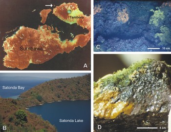
Satonda (8°7′S 117°45′E), Indonesia, is a small volcano of Holocene age, forming a circular island with a crater lake at its center (Kempe & Kaz'mierczak, Reference Kempe, Kaz'mierczak, Ittekkott, Kempe, Michaelis and Spitzy1990a, Reference Kempe and Kaz'mierczakb, Reference Kempe and Kaz'mierczak1993; Kempe et al., Reference Kempe, Kaz'mierczak, Reimer, Landmann, Reitner, Reitner, Neuweiler and Gunkel1996, Reference Kempe, Kaz'mierczak, Reimer, Landmann, Reitner, Tomascik, Mah and Moosa1997; Kaz'mierczak & Kempe, Reference Kaz'mierczak and Kempe2004) (Figure 1). It is situated north of the Tambora volcano on Sumbawa, famous for the historic eruption of 15 April 1815. Satonda was then blanketed with about 1 m of ash, disrupting life on the island for years. When Satonda itself last erupted is unknown. Its central lake fills two nested craters and measures 0.77 km2 on the surface, is 69.5 m deep, and has a volume of 0.034 km3. Its surface is almost at sea level, with a >1 m seasonal fluctuation. No tides are measurable in the lake, suggesting no appreciable subsurface infiltration of seawater at present.
(A) Location of Satonda Island (arrow); GeoSat image; (B) general view of the Satonda crater lake; (C and D) macroscopic morphology of the suberitid sponges at a depth of ~20 m (C—Suberites sp.) and close to the lake surface (D—Protosuberites lacustris).

14C dating of peat underneath the biocarbonate deposits along the lake's shores shows that the lake was filled with freshwater until about 3000 years ago. It then suddenly filled with seawater, an event that may have been triggered by the partial collapse of the crater wall to the south-west, the scar of which forms ‘Satonda Bay’ today (Figure 1). This collapse left a 13-m-high remnant of wall, creating the only breach in the otherwise steep, up to 300-m-high crater wall. Alternatively, the regionally rising sea level may have caused seawater to filter in through the much older collapse scar. Whatever its cause, the seawater ingression allowed a few marine bivalves and gastropods to populate the lake, while serpulid polychaete fringing reefs started to develop along the shore. Since that time, regional sea level has dropped by about 1 to 1.5 m, and the receding sea has left beach walls in Satonda Bay that eventually isolated the lake from water exchange with the ocean. During the dry season, the lake now stands about 80 cm higher than at spring tide and may push water out slowly to the ocean through the volcanic edifice (Kempe & Kaz'mierczak, Reference Kempe and Kaz'mierczak1993).
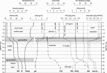
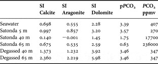
The lake has three layers, separated by density interfaces (pycnoclines). The top layer is aerobic and has salinity less than seawater, indicating dilution by rain. The lower two layers have turned anaerobic: salinities there are greater than seawater and indicate prolonged evaporation. As a consequence, the water of Satonda Lake has deteriorated from its original marine composition to a quasi-marine, alkaline state. Alkalinity is produced in the lower layers by sulphate reduction and exported to the upper layer (‘alkalinity pump’) by diffusion and/or mixing events. Such an event probably occurred between the two sampling periods, as is evident from the differences between the 1986 and 1993 pH values and alkalinity curves in the upper pycnocline. Table 1a lists the main hydrophysical and hydrochemical parameters and Table 1b the calculated mineral saturations (PHREEQUE) and the CO2-pressure (PCO2) from three samples of the three layers, in comparison with a seawater sample from Satonda Bay. Figure 2 plots some of these parameters and the remaining samples in order to illustrate the stratification of Lake Satonda and its chemistry (for details of hydrochemical evolution of the lake, see Kempe & Kaz'mierczak, Reference Kempe and Kaz'mierczak1993; Kempe et al., Reference Kempe, Kaz'mierczak, Reimer, Landmann, Reitner, Reitner, Neuweiler and Gunkel1996).
Hydrochemical profiles of the water column of Satonda Crater Lake sampled during 1986 (black triangles) and 1993 (open circles) expeditions (after Kempe & Kaz'mierczak, 1997 and Kempe et al., Reference Kempe, Kaz'mierczak, Reimer, Landmann, Reitner, Tomascik, Mah and Moosa1997, modified)

Water composition for water samples taken at various depths, Satonda crater lake (Indonesia), and seawater for comparison (J.K. and S.K. own data; for details see Kempe & Kaz'mierczak, Reference Kempe, Kaz'mierczak and Seckbach2007).

Mineral saturation indices (log (IAP/Kmineral) of some carbonate, CO2-pressure (pPCO2 = − log PCO2). Degassed, PCO2 degassed to 347 ppmv. Calculation by PHREEQUE (Parkhurst et al., Reference Parkhurst, Thorstenson and Plummer1990) (J.K. and S.K. own data; for details see Kempe & Kaz'mierczak, Reference Kempe, Kaz'mierczak and Seckbach2007).

Sampling
Water samples have been taken from the water column with a 1.5 l Nansen bottle mounted on a hand-held nylon line. Immediately after recovery of the samples the following parameters were determined: temperature, conductivity, pH and Eh. Oxygen was preserved with MnSO4 and KI-KOH solution for later titration in a Winkler bottle. A limited set of samples for δ18O and δ13C determination was taken. At the field camp aliquots of the samples were acidified with HCl to pH 2 for later determinations of cations concentrations (Na, K, Mg, Ca, Fe and Mn) by standard atomic absorption methods. Glass fibre filters (GF) were used in the analysis of suspended matter for particulate organic carbon (POC). The GF filtered matter was poisoned with a few drops of saturated HgCl2 solution for later determination of the dissolved organic carbon (DOC). Alkalinity was determined with a HACH hand-held titrator in the field camp. Nutrient concentrations (NO3, NO2, NH4+, PO43- and SiO2) were analysed for 18 samples aboard the RV ‘Sonne’ a few hours after the samples had been taken according to standard methods. Some results of the chemical determinations are given in Table 1a, b. Sediment and biota samples were taken both at the water surface and at depth by SCUBA diving. The hard rock (reef) samples were dried in the sun before packaging. Biosamples, sponges included, were divided into two groups: one group was air-dried and transported in plastic bags; the other was preserved in ~60% ethanol diluted with lake water. Sponge spicules, skeletal organization, and morphology were studied using both light and scanning electron microscopy. For detailed information on the survey and sampling and analytical methods used by the German–Indonesian Satonda Island Expedition of 1986 that collected these suberitid sponges, see Kempe & Kaz'mierczak (Reference Kempe and Kaz'mierczak1993).
The investigated material is housed in the Institute of Paleobiology, Polish Academy of Sciences, Warszawa, under the designation ZPAL Pf.20
RESULTS
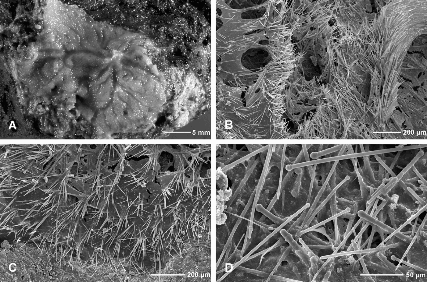
Sponges are among the main groups of macroorganisms in Satonda Lake. Typically, they grow in a patchy manner over the cyanobacterial–red algal reef surface from the lake shallows down to almost the chemocline (Figure 1C). Occasionally, irregular bunches overgrow the bottom-attached and free-floating filamentous green algae. Macroscopically, the sponges can be recognized as yellow to yellow-orange subcircular or irregular crusts, up to 1 cm thick (Figure 1C), often with a distinct pattern of exhalant canals visible on the surface (Figures 1D, 3A). The population density of sponges is not high, and no more than a few specimens have been observed in an area of 1 m2. Although post mortem accumulations of monaxonid spicules have been observed here and there on the reef surface, they are never incorporated into the calcareous cyanobacterial–red algal framework (Kempe & Kaz'mierczak, Reference Kempe and Kaz'mierczak1993). Increased alkalinity of the lake water (Table 1; Figure 2) is apparently responsible for the rapid dissolution of the siliceous spicules, which is comparable to that of diatom frustules. Similar dissolution and/or pseudomorphization (mostly calcitization or pyritization) of the originally siliceous spicules has been noticed in some modern and fossil hypercalcified sponges (known in earlier literature as sclerosponges) incorporating spicules into their basally secreted calcareous skeletons (e.g. Hartman & Goreau, Reference Hartman and Goreau1970; Kaz'mierczak, Reference Kaz'mierczak1974).
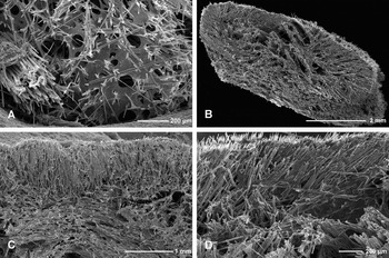
Protosuberites lacustris. (A) Morphology of the sponge in incident light; (B) semi-oblique view of the fractured sponge ectosome as seen by SEM, showing radial canals (left) and brushes of protruding spicules; (C) perpendicular view of the sponge surface at the growth margin as seen by SEM; note discrete bunches of spicules protruding in the form of bouquets; (D) disorganized spicule arrangement in the sponge's interior.
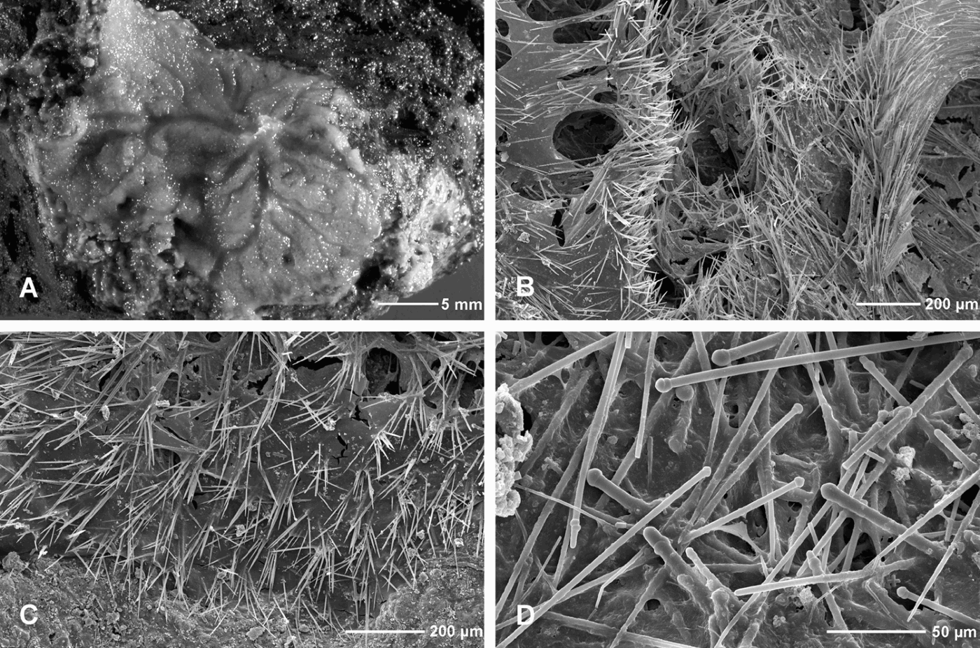
Thinly encrusting Suberitidae, with choanosomal tylostyles in bundles and/or single without orientation and ectosomal tylostyles in palisade arrangement or forming brushes at the sponge surface, points outward (cf. van Soest Reference van Soest, Hooper and van Soest2002: p. 234.)
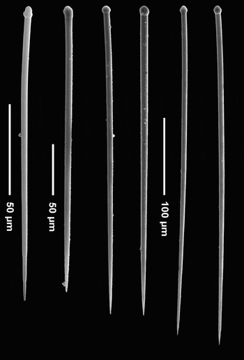
Protosuberites lacustris. Tylostyles of various shape and size.

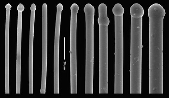
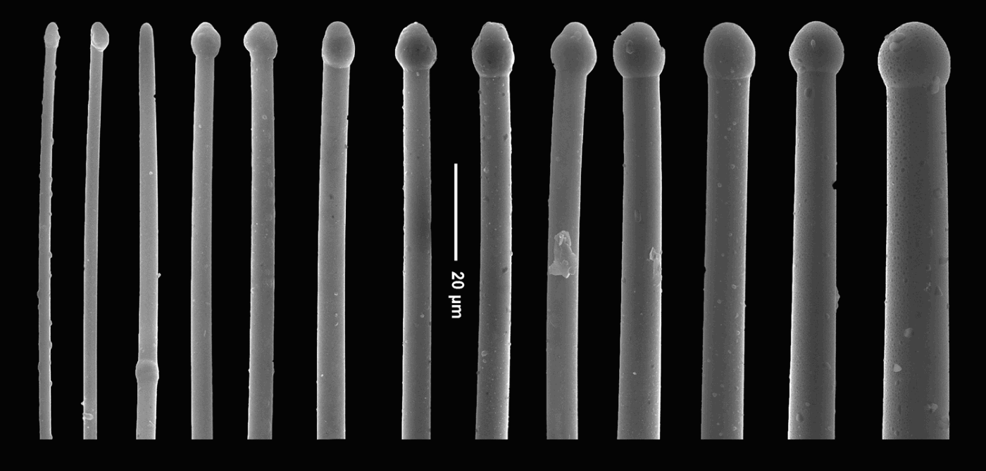
Protosuberites lacustris. Variation in tylostyle heads.

MATERIAL
S-216A; S-250 (A1); S-250 (B); S-Bioprobe 0-1 m (B1, B2); S-Bioprobe (C).
DESCRIPTION
Habit: thin crusts (0.5–2 mm), grey colour in ethanol (yellow alive), pronounced radial excurrent canals. Growing on and in the interstices of crustose coralline algal rock.
Skeleton: in the choanosome, spicules in strands alternating with others without orientation; in the ectosome, spicules single in perpendicular orientation, or fanning out from choanosomal fibres to form surface brushes. All spicule sizes occur intermixed throughout the body; there is no specialization of the smaller forms exclusive to the ectosome.
Spicules: large size-range of tylostyles, most of which are widest at the neck (just below the tyle). Many subtylote forms, some with tyles subterminal or missing (styles), a few oxeote modifications; none of these have a particular location within the skeleton.
Measurements (range of length × maximum width (head width)): large tylostyles, 400–540 × 10 µm (13–15 µm); medium tylostyles; 220–300 × 9 µm (11 µm); small tylostyles, 160–180 × 2.5–4 µm (5 µm); accessory oxeas/styles, 280 × 3 µm.
REMARKS
We transfer this species from Laxosuberites to the genus Protosuberites because Laxosuberites Topsent was found to be a junior synonym of Hymeniacidon Bowerbank (Halichondrida) (van Soest, Reference van Soest, Hooper and van Soest2002: p. 235). The investigated sponge has the same thinly encrusting habit, size-range and shape of tylostyles, and tolerance to a low salinity environment as the type series described from Chilka Lake (east coast of India). In the same paper, Annandale (Reference Annandale1915) redescribes a closely related species, Suberites aquaedulcioris Annandale, Reference Annandale1914 (as Laxosuberites), which has a similar skeleton structure but differs in having smaller tylostyles (330 × 6 µm maximum); it too is thinly encrusting and occurs in brackish water. From its skeletal properties, Suberites aquaedulcioris should also be transferred to Protosuberites. Protosuberites lacustris comb. nov. was referred to by Reitner et al. (Reference Reitner, Wörheide, Arp, Hooper and Kempe1999) as Laxosuberites.
Our material did not include gemmules, asexual reproductive bodies that help the sponges overcome periods of adverse environmental conditions; presumably the specimens were collected at a time (October 1986, i.e. end of dry season) that gemmules were not developed. However, Annandale (Reference Annandale1915) describes these structures as being formed in groups at the base of the sponge, encased in spongin and persistently bright yellow in colour.
Crustose to massive Suberitidae, with disorganized central choanosomal skeleton of large tylostyles, changing to dense, radial spicule strands toward periphery; with smaller category of ectosomal tylostyles oriented perpendicular to the surface, arranged in palisade or as brushes over the fibre terminals, points outward. Microscleres (centrotylote and microstrongyles) may be present in some species (cf. van Soest, Reference van Soest, Hooper and van Soest2002: p. 240).
Suberites sp., SEM images. (A) Sponge surface showing brushes of small, ectosomal tylostyles and a few large, perpendicular spicules, possibly expelled from the choanosome; (B) cross-section of the sponge body showing disorganized to radial skeleton arrangement and major canals; (C) details of spicule arrangement; note radial orientation (in palisade) of smaller tylostyles in the ectosome (top) and criss-cross organization in the choanosome (bottom); (D) detail of the ectosomal zone (top) in (C).

Suberites sp. Tylostyles of various shape and size; an oxeote modification to the far right.

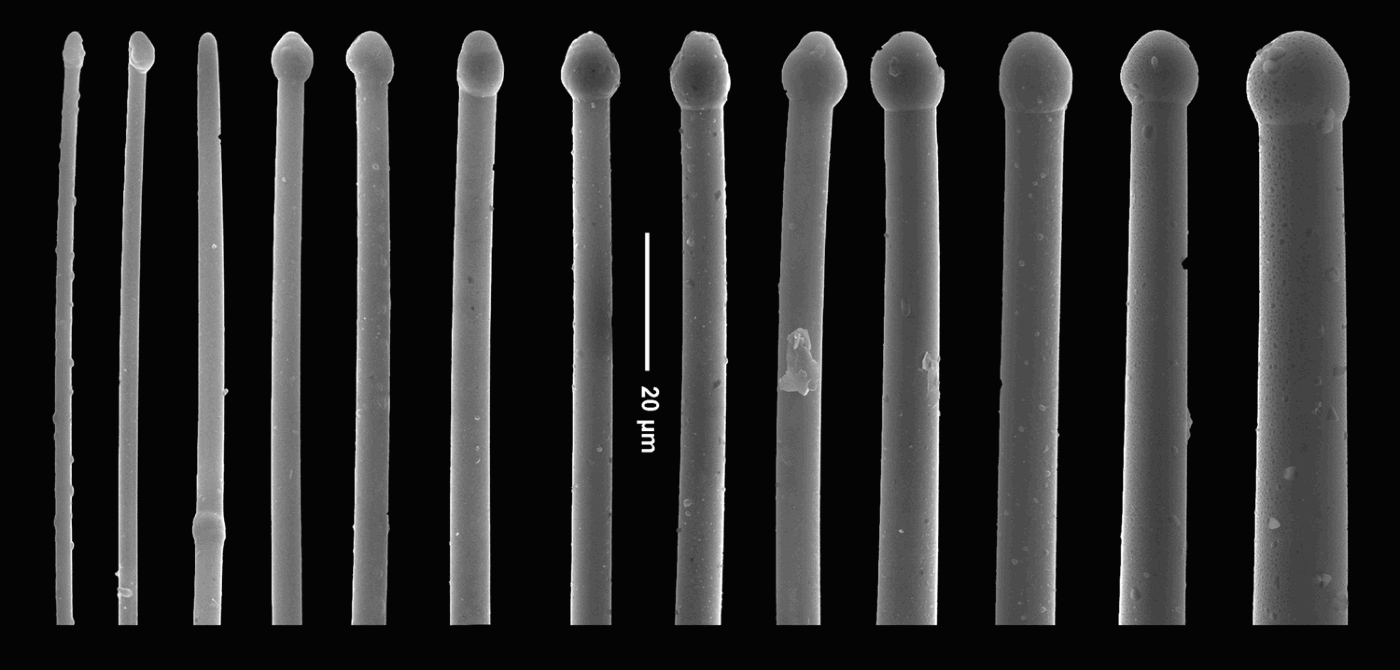
Suberites sp. Tylostyle head modifications.
MATERIAL
S-250 (A2); S-289 (A, B); S-295; S-Bioprobe 0–1 m (A); S-Bioprobe 0–1 m (B3).
DESCRIPTION
Habit: thin crust or cushion, tan colour (in ethanol); growing mainly on and incorporating filamentous red algae and (dead) bryozoan (?) stolons; also massive with branches (in deeper water) growing on rocky substrate, yellow to orange when alive.
Skeleton: criss-cross organization of spicules in the sponge interior, radial orientation toward the ectosome; small category of tylostyles forming surface palisade or bouquets.
Spicules: large size-range of tylostyles, most of which are widest near the upper third (toward the tyle) or in the middle of the shaft. Many subtylote forms, some tyles subterminal or missing (styles); small accessory oxea and centrotylote oxea (malformations), some extremely thin and delicate, similar ultra-thin tylostyles (resembling raphids; probably early growth stages) and siliceous debris made up primarily of diatom shells. No microscleres occur.
Measurements (range of length × maximum width (head width)): large, 520–750 × 7.5–16 µm (10–16 µm); medium, 500–520 × 10 µm (11 µm); small, 170–240 × 5–6 µm (5 µm); accessory ultrathin oxeas, styles, and tylostyles: 150–220 × 1–3 µm.
REMARKS
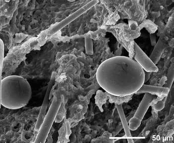
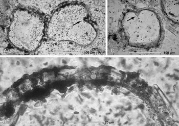
The species described here resembles Suberites sericeus Thiele, 1898, from Japan, which was also found by Annandale (Reference Annandale1915) in brackish Chilka Lake (India), in some places in nearly freshwater conditions, and described and illustrated in detail. Annandale emphasizes the stout shape of the small, ectosomal tylostyles, which are almost as thick as the larger, choanosomal spicules. This disparity with our material is significant enough to persuade us not to use that name and, in the absence of larger specimen series and suitably fixed samples, not to describe this sponge as a new species. Just as we observed, Annandale (Reference Annandale1915) describes S. sericeus as having a basal layer of gemmules and attaching to and incorporating algae and stolons of hydroids or bryozoans. The structures we identified as gemmules (Figure 10) are spherical and unattached, have a complex wall structure, show clear foramina and are enveloped by an 11-µm-thick layer of orange amorphous material (spongin) coat (Figure 10A, C). The gemmules in our specimens measure about 130–160 µm in diameter. Small (47–72 µm in diameter), smooth spherical bodies (Figure 9) were observed in the choanosome of this species, which we interpret as oocytes or embryos.
Suberites sp. SEM view of choanosome showing spicules and possible oocytes or embryos (rounded bodies).

Resting bodies (gemmules, probably of Suberites sp.) as seen in substratum thin section; note complex spongin wall structure (enlarged in B); arrows (A, C) point to foramina, structures uncommon in gemmules of marine sponges.

The morphology of this sponge corresponds to that of the specimens discussed by Reitner et al. (Reference Reitner, Wörheide, Arp, Hooper and Kempe1999) as Suberites.
DISCUSSION
The alkaline conditions and the high supersaturation of carbonates caused the marine molluscs in the Satonda crater lake to die out, except for one abundant gastropod species, Cerithium corallium (Kempe & Kaz'mierczak, Reference Kempe and Kaz'mierczak1993). Instead, 0.5- to 1.2-m-thick reefs of calcareous red algae and in situ calcifying cyanobacteria began to grow at rocky points, forming microbialitic fringe reefs along Satonda's perimeter, its most striking feature (Figure 1). These microbialites are unique among all other modern in situ calcifying cyanobacterial mats because of their association with marine organisms in a hydrochemically marine-derived environment (see above). The mats are formed by stenohaline organisms such as corallinacean (Lithoporella melobesioides) and squamariacean (Peyssonnelia squamaria) red algae and nubecullinid foraminifers. They are composed of thin layers of subglobular aggregates of coccoid cells (Pleurocapsa group; see Rippka et al., Reference Rippka, Waterbury, Herdman, Castenholz, Boon and Castenholz2001). Permineralized cyanobacterial mats intergrow with the arcuate thalli of red algae and, together with agglomerations of nubecullinid foraminifers and serpulids, generate massive calcareous mounds and irregularly shaped crusts along the lakeshore. Living calcifying cyanobacterial biofilms have been observed at the lake surface down to the O2–H2S interface. Three groups of cyanobacterial microbialites can be recognized in the lake (cf. Kaz'mierczak & Kempe, Reference Kaz'mierczak and Kempe1990, Reference Kaz'mierczak and Kempe1992; Kempe & Kaz'mierczak, Reference Kempe and Kaz'mierczak1993). The first group is represented by crusts composed of tightly adhering laminated columns, 3–7 mm high and ~1–3 mm in diameter, growing on tufted thalli of siphonocladalean green algae. These microstromatolites occur mainly in very shallow water down to 20–30 cm but can be found as tiny individual columns at depths almost reaching the chemocline. The second group, representing the bulk of Satonda microbialites, is a dense agglomeration of elongated and irregularly twisted microlaminated bodies. Their centres are occupied by one, rarely by two or three, cylindrical tubes, 90–220 µm in diameter, representing moulds of filaments of siphonocladalean green algae (Cladophoropsis sp.) around which the microbialites (microstromatolites) accreted. Because Cladophoropsis grows in depths of 10 m or less, these microbialites occur only in that range. The third group is laminated cystose and tubuloid microbialitic structures occurring in interstices and crevices between the foliaceous thalli of calcareous red algae and agglomerations of nubecullinid foraminifers. The surfaces of the microbialites are home to a sparse population of sponges. They occur from the water table (Figure 1D) all the way down to the chemocline, at ~20 m (Figure 1C).
In contrast to the diversified sponges (45 species, 26 genera and 2 families) known from four anchihaline lakes of Indonesia (de Voogd et al., Reference Voogd de, Weerdt de, van Soest, Custódio, Lôbo-Hajdu, Hajdu and Muricy2006), the sponge population of Satonda Lake is extremely sparse and consists of only two species from two related genera, Protosuberites and Suberites. Interestingly, species of the genus Suberites are among the most common in other Indonesian lakes (de Voogd et al., Reference Voogd de, Weerdt de, van Soest, Custódio, Lôbo-Hajdu, Hajdu and Muricy2006). In addition, the genus Suberites is reportedly common in some marine lakes of North Vietnam (Calcinai et al., Reference Calcinai, Azzini, Bavestrello, Cerrano, Pansini and Thung2006; Cerrano et al., Reference Cerrano, Azzini, Bavestrello, Calcinai, Pansini, Sarti and Thung2006; Azzini et al., Reference Azzini, Calcinai, Cerrano, Bavestrello, Pansini, Custódio, Lôbo-Hajdu, Hajdu and Muricy2007), notably in those that lack any visible connection with the sea, and thus whose temperatures and also water chemistry are quite different from that of normal seawater. Suberites species are also found in saline lakes in New South Wales, Australia (Barnes, Reference Barnes, Custódio, Lôbo-Hadju, Hadju and Muricy2006), as well as in the hypersaline parts of Shark Bay, Australia (Hentschel, Reference Hentschel, Michaelsen and Hartmeyer1909). All these settings are characterized also by temperatures often periodically higher than those in an open sea, a factor that may stress or even cause mass mortality of marine organisms (Cerrano et al., Reference Cerrano, Bavestrello, Bianchi, Cattaneo-Vietti, Bava, Morganti, Sara, Schiaparelli, Siccardi and Sponga2000). Such occurrences suggest that some suberitid species adapt better to the stressful conditions of changing water chemistry and temperature than most other sponges do. In his study of sponges in brackish Chilka Lake, Annandale (Reference Annandale1915) suggested that the low salinity may have led to morphological or genetic differentiation, as in the example of the species pair ‘Laxosuberites’ (=Protosuberites) aquaedulcioris and ‘L.’ lacustris.
The presence of gemmules in suberitids from Satonda Lake was first reported by Arp et al. (Reference Arp, Reimer and Reitner2004). We, too, found structures that we interpreted as resting bodies (Figure 10), but they differ from those designated gemmules by Arp et al. (Reference Arp, Reimer and Reitner2004, figure 5A, B), which are subspherical, are attached to the substrate, and have unilayered, structureless walls. The structures we identified as gemmules (Figure 10) are spherical and unattached, have a complex wall structure, show clear foramina (Figure 10A, C), and consist of orange amorphous material.
Resting bodies (gemmules) are rare in marine sponges but are known to occur in the families Suberitidae and Clionaidae (Hadromerida) and Haliclonidae (Haplosclerida) (Simpson & Fell, Reference Simpson and Fell1974; Connes et al., Reference Connes, Carriere and Paris1978; Simpson, Reference Simpson1984; Schönberg, Reference Schönberg2002). On the other hand, gemmules are typical of most freshwater sponges and help them survive unfavourable conditions. This adaptation clearly allowed sponges to colonize the extreme, unstable environments of Satonda crater lake and similar environments. More detailed studies of suberitid sponges from extreme environments and comparisons with populations in normal oceanic habitats are needed to determine taxonomic relationships and morphological or genetic adaptations to harsh environments, including gemmules development.
SUMMARY
Owing to its high alkalinity, carbonate supersaturation, and seasonal salinity changes, Satonda's quasi-marine crater lake (Sumbawa, Indonesia) is inhabited by only few marine macroscopic organisms, among them two sponge species, Protosuberites lacustris comb. nov. and Suberites sp. These sponges belong to a family (Suberitidae) that is known to tolerate low-salinity environments elsewhere, which suggests they are especially well adapted to stressful habitat conditions. This adaptation may be facilitated by the production of resting bodies (gemmules), rare in marine forms but common in freshwater sponges, allowing them to survive unfavourable conditions over long periods and thus colonize extreme, unstable environments.
ACKNOWLEDGEMENTS
We are indebted to G. Landmann, A. Lipp, A. Reimer, J. Reitner, T. Strecker and G. Wörheide for their assistance in the field and in the laboratory. Field research was funded by the Deutsche Forschungsgemeinschaft (DFG) (Grant Ke 287 9/1-4, Satonda). Financial support by the Polish Academy of Sciences and the Committee for Scientific Research (KBN) (Grant 6 6209 92 03) is gratefully acknowledged. SYNTHESYS grants (EU) to A. Pisera are also greatly appreciated.