The conformation of a folded protein is stabilised by a network of hydrophobic, electrostatic and hydrogen-bonding interactions involving residues distributed throughout the polypeptide chain (Ref. Reference Dobson1). During co- or post-translational protein folding, hydrophobic surfaces that are buried in the final native structure become transiently exposed and available for incorrect interactions with neighbouring molecules in the cell. These interactions promote protein aggregation, a process that competes with correct folding. Similarly, polypeptides that lack a stable folded structure even in their native soluble states (referred to as ‘natively unfolded’) expose hydrophobic surfaces and, therefore, have a high propensity to form aggregates at high concentrations. Under normal conditions, improper interactions leading to protein aggregation are inhibited by molecular chaperones – cellular factors that enhance the efficiency of protein folding by shielding exposed hydrophobic surfaces (Fig. 1). However, in the presence of cellular stresses that induce protein unfolding and aggregation, chaperones are no longer able to keep up with preventing incorrect protein interactions, and aggregates begin to accumulate. Examples of cellular stresses that trigger protein unfolding include heat shock, the introduction of amino acid substitutions as a result of gene mutations, and protein damage due to post-translational modifications (Fig. 1).
Model illustrating protein misfolding, aggregation and clearance inside the cell, along with opportunities for therapeutic intervention. (a) A folded protein is converted to a misfolded species (shown enclosed in a red circle) by various stresses, including oxidative damage, gene mutations and proteolysis. The red asterisk represents a site of modification on the misfolded protein as a result of one of these stresses. (b) Under normal conditions, misfolded proteins are degraded by the ubiquitin–proteasome pathway (shown in the figure) or by chaperone-mediated autophagy (not shown). (c) At high concentrations, a misfolded protein (or a natively unfolded polypeptide such as α-synuclein) can be induced to form prefibrillar, β-sheet-rich oligomers by various stresses, including phosphorylation, oxidative damage, gene mutations and proteolysis. (d) The oligomers are transported to perinuclear aggresomes, where they may form more mature, fibrillar aggregates such as those present in Lewy bodies. (e) Ultimately, protein aggregates at the aggresome are targeted for degradation via macroautophagy. (f) If β-sheet-rich oligomers accumulate to a point that exceeds the capacity of both the ubiquitin–proteasome pathway and the aggresome–macroautophagy pathway, then they may induce cell death via apoptosis. (g) Misfolding and aggregation of a normally folded protein can be suppressed by stabilising the native conformation (an example of Therapeutic Strategy I in the text). (h) Antioxidant compounds (e.g. curcumin) may inhibit protein misfolding and/or aggregation induced by oxidative stress (Therapeutic Strategy II). Curcumin also prevents the formation of toxic aggregates by directly interfering with β-sheet stacking (Therapeutic Strategy I). (i) Protein aggregation can also be blocked by upregulating molecular chaperones with an Hsp90 inhibitor, including the novobiocin analogue A4 (Therapeutic Strategy III). Inhibition of Hsp90 results in the release of previously bound, inactive HSF1, which undergoes trimerisation and phosphorylation coupled with nuclear transfer and induces the expression of heat-shock proteins including Hsp90, Hsp70, Hsp40 and Hsp27. These heat-shock proteins inhibit protein aggregation, and Hsp27 also suppresses oxidative stress. (j) Aggregate clearance can be upregulated via pharmacological inhibition of mTOR, a kinase that inhibits macroautophagy (Therapeutic Strategy III), for example by the classic mTOR inhibitor rapamycin. The chemical structures of curcumin, the novobiocin analogue A4 and rapamycin are shown below the schematic.

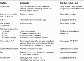
Several diseases are characterised by protein misfolding and aggregation. Although the formation of protein aggregates can trigger cell death by depleting proteins essential for cell survival, in many cases cytotoxicity results from harmful properties of the aggregates themselves (Ref. Reference Dobson1). Diseases in which protein aggregates cause cell death are said to occur via a toxic gain-of-function mechanism (examples discussed in this review are listed in Table 1). A hallmark feature of these disorders is the presence in diseased tissue of pathological lesions containing β-sheet-rich, fibrillar protein assemblies. Genetic evidence strongly suggests that protein aggregation plays a critical role in the pathogenesis of these diseases. For example, mutations in the gene encoding the amyloid precursor protein (APP) are thought to lower the age of onset of Alzheimer disease by increasing the production of a fibrillogenic variant of the amyloid-beta peptide, Aβ1–42, relative to the shorter, less fibrillogenic variant Aβ1–40. In addition, mutations that expand the polyglutamine repeat region of huntingtin (by increasing the number of consecutive glutamine residues) are thought to cause the onset of Huntington disease by increasing the propensity of huntingtin to form cytotoxic aggregates.
Proteins involved in misfolding diseases (examples discussed in the text)a

aFor more detailed information, see the following review articles: Refs Reference Rochet and Lansbury16, Reference Muchowski and Wacker155, Reference Kelly206, Reference Zoghbi and Orr207, Reference Taylor, Hardy and Fischbeck208, Reference Caughey and Lansbury209, Reference Ross and Poirier210, Reference Chaudhuri and Paul211.
Protein aggregation occurs via a complex self-assembly pathway involving the formation of small oligomers, larger complexes called ‘protofibrils’, and mature, β-sheet-rich fibrils. An intensely debated question is whether prefibrillar aggregates (oligomers, protofibrils) or fibrils are responsible for the underlying cytotoxicity in protein-misfolding diseases. Multiple lines of evidence suggest that oligomers or protofibrils play a more important role than fibrils in pathogenesis. Oligomeric forms of Aβ are more toxic than mature fibrils in neuronal cell culture (Ref. Reference Dahlgren2), and they suppress hippocampal long-term potentiation and elicit memory deficits in rats in vivo (Refs Reference Walsh3, Reference Lesne4). Evidence from ground-breaking work in transgenic Caenorhabditis elegans overexpressing Aβ1–42 suggests that Aβ toxicity is suppressed not only via inhibition of protein aggregation by molecular chaperones, but also via a secondary pathway involving conversion of toxic protofibrils into larger, less harmful aggregates (Ref. Reference Cohen5). Data from another study indicate that the formation of huntingtin inclusions correlates with enhanced neuronal survival in a cell-culture model of Huntington disease (Ref. Reference Arrasate6). In addition, oligomeric forms of the mammalian prion protein (PrP) have a greater propensity to transmit disease than larger fibrils (Ref. Reference Silveira7), and low molecular weight oligomers of transthyretin (TTR) exhibit greater toxicity than fibrillar species in neuronal cells (Ref. Reference Reixach8). The reason for the enhanced toxicity of oligomeric assemblies compared with fibrils is uncertain, although data from several studies suggest that oligomers or protofibrils might have a greater propensity to trigger harmful membrane permeabilisation (Refs Reference Volles9, Reference Lashuel10, Reference Kayed11).
The purpose of this review is to provide a comprehensive overview of novel therapeutic strategies for the treatment of protein-misfolding diseases. Three general approaches are considered here: (Reference Dobson1) directly targeting proteins that undergo misfolding and aggregation; (Reference Dahlgren2) interfering with post-translational modifications that promote protein misfolding and aggregation; and (Reference Walsh3) inhibiting a build-up of protein aggregates by upregulating molecular chaperones or stimulating aggregate clearance. Strategies that target cellular pathways modulated by protein aggregation (e.g. apoptosis, transcription) are not included owing to space constraints and because these approaches have been covered extensively in other reviews (for example, see Refs Reference Hashimoto12, Reference Mattson and Sherman13, Reference Rego and de Almeida14). The discussion in this review is focused primarily on research carried out during the past five years.
Therapeutic strategy I: direct targeting of proteins that undergo misfolding and aggregation
One approach for developing new therapies to treat protein-misfolding disorders has been to identify agents that bind an aggregating polypeptide and keep it from forming toxic assemblies. To target fibrillogenic proteins or peptides that lack a stable fold under native conditions, the main strategy has been to identify inhibitors that interfere directly with aggregation. These inhibitors consist of small molecules or peptides identified via structure-based design or high-throughput screening. To target proteins that are folded under native conditions, a powerful strategy has been to identify chemical entities that stabilise the native conformation and prevent unfolding leading to aggregation. Examples of these different approaches are discussed below.
Inhibition of fibril formation: use of structure-based approaches
Over the past five years there have been remarkable advances in our understanding of the structures of amyloid fibrils and protofibrils. In turn, these advances have stimulated the development of therapeutic approaches targeting structural motifs required for protein self-assembly.
Structure of amyloid fibrils
Many proteins that are susceptible to misfolding and aggregation form extracellular deposits termed ‘amyloid fibrils’ (Ref. Reference Nelson and Eisenberg15). Electron microscopy has shown that amyloid fibrils formed from different proteins are straight, rigid and unbranched, with diameters ranging from 5 to 13 nm. Each amyloid fibril consists of multiple fibrillar subunits, named ‘protofilaments’, that wind around each other in a helical fashion (typically, there are two to six protofilaments per fibril). Amyloid fibrils produce an X-ray diffraction pattern that suggests that the amyloid protofilament consists of a ‘cross-β’ structure [or ‘cross-β spine’ (Ref. Reference Nelson and Eisenberg15)], with β-strands orthogonal to and backbone hydrogen bonds parallel to the fibril axis. [Note that the terms ‘protofilament’ and ‘protofibril’ have different meanings: whereas the former is a structural unit of the amyloid fibril, the latter is a kinetic intermediate of fibrillisation (Ref. Reference Rochet and Lansbury16).]
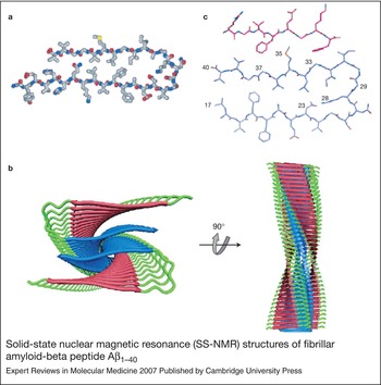
Because amyloid fibrils are insoluble and noncrystalline, it is generally not possible to solve their atomic structures using traditional methods such as X-ray crystallography or solution nuclear magnetic resonance (NMR) (Ref. Reference Petkova, Yau and Tycko17). However, recent solid-state NMR (SS-NMR) data have enabled investigators to characterise amyloid fibril structure with unprecedented detail. An advantage of SS-NMR is that it provides constraints for modelling fibril secondary structure (folding of the polypeptide chain into β-strand or non-β-strand segments), tertiary structure (organisation of β-strands into parallel or antiparallel β-sheets), and quaternary structure (arrangement of β-sheets relative to each other) (Ref. Reference Petkova, Yau and Tycko17). The most intensive SS-NMR studies have been carried out on fibrils formed from Aβ1–40. Each Aβ1–40 fibril consists of a bundle of two to five protofilaments (Refs Reference Petkova, Yau and Tycko17, Reference Petkova18). Two-dimensional SS-NMR data indicate that each Aβ1–40 molecule in a protofilament consists of a disordered N-terminal region spanning residues 1–9, followed by two β-strands encompassing residues 10–22 and 30–40 (Fig. 2a). Residues 23–29 form a structured loop enabling the two β-strands to fold over each other and interact via contacts between side-chains (Ref. Reference Petkova, Yau and Tycko17). An array of Aβ1–40 ‘strand–loop–strand’ motifs projects along the fibril axis to form a two-layered structure (Fig. 2b). Each layer in this structure consists of an extended β-sheet with individual β-strands aligned parallel and in-register (Ref. Reference Petkova, Yau and Tycko17). Mass per unit length data obtained via scanning transmission electron microscopy (STEM) indicate that each Aβ1–40 protofilament consists of four β-sheet layers, equivalent to two molecular layers (Fig. 2b) (Refs Reference Petkova, Yau and Tycko17, Reference Petkova18). The protofilament is stabilised by (Reference Dobson1) ‘internal’ quaternary interactions between two β-strands in the same molecular layer, and (Reference Dahlgren2) ‘external’ quaternary interactions between strands from different molecular layers (Fig. 2b) (Ref. Reference Petkova, Yau and Tycko17).
The structure of fibrillar Aβ1–40 described above is consistent with constraints determined using other methods (Refs Reference Torok19, Reference Williams20, Reference Whittemore21, Reference Kheterpal22, Reference Shivaprasad and Wetzel23) and similar to the structure of the Aβ1–42 fibril determined by hydrogen–deuterium exchange NMR (HX-NMR) and complementation mutagenesis (Ref. Reference Luhrs24). In addition to these studies of Aβ, biophysical analyses of other fibrillar assemblies have yielded remarkable structural insights. For example, α-synuclein, a protein that accumulates in brain disorders referred to as ‘synucleinopathies’ (e.g. Parkinson disease, dementia with Lewy bodies, and multiple system atrophy), forms fibrils with a multilayered structure. Each layer consists of a parallel, in-register β-sheet similar to the model described for fibrillar Aβ (Refs Reference Der-Sarkissian25, Reference Del Mar26, Reference Heise27). In addition, X-ray diffraction analysis of crystals formed by two amyloidogenic peptides revealed an extended cross-β spine structure in which the β-sheets are held together by highly stabilising ‘zipper-like’ contacts involving side-chains of amino acids (Refs Reference Nelson and Eisenberg15, Reference Makin28, Reference Sambashivan29). In principle, these advances in our understanding of fibrillar structure make it possible to develop structure-based therapeutics to inhibit protein fibrillisation. Examples of this type of ‘rational’ approach to designing novel inhibitors of Aβ fibril formation are outlined below.
Structure-based design of peptides that inhibit fibrillisation
In previous work, a peptide spanning residues 16–20 of Aβ with N-methyl groups at positions 17 and 19 was found to interfere with fibril formation by Aβ1–40 (Ref. Reference Sciarretta30). The rationale for the design of this peptide was that N-methyl groups positioned on the same side of a β-strand should prevent β-sheet formation by disrupting interstrand hydrogen bonding. Structural evidence that fibrillar Aβ1–40 consists of two β-sheet layers (see above) suggested that fibrillisation should be blocked more effectively by targeting both layers simultaneously rather than either layer alone. This hypothesis was addressed by testing the inhibitory effects of introducing a pair of alternating N-methyl amino acids in each β-strand of Aβ1–40 (i.e. at positions 17 and 19 in the N-terminal β-strand and at positions 37 and 39 of the C-terminal β-strand) (Ref. Reference Sciarretta30). The authors showed that disrupting the β-sheet structure in either layer was insufficient to completely prevent fibril formation. By contrast, the Aβ1–40 isoform with N-methyl groups in both β-strands failed to form fibrils altogether, instead remaining mostly soluble and monomeric. These results imply that a mixture of N- and C-terminal peptides targeting both β-sheet layers should be used to inhibit Aβ1–40 fibrillisation. N-methyl peptides are particularly effective inhibitors because: (Reference Dobson1) they are water soluble, and therefore do not form fibrils themselves; and (Reference Dahlgren2) they adopt a stable, extended conformation that enables them to ‘cap’ fibril growth via sterically favourable interactions with edge β-strands (Ref. Reference Sciarretta30). In a related study, an analogue of the islet amyloid polypeptide (IAPP) with two N-methyl groups was shown to be a soluble, noncytotoxic, highly potent inhibitor of IAPP fibrillisation (Ref. Reference Yan31). Given that peptides may have limited therapeutic potential because of their immunogenicity, susceptibility to proteolysis, and weak bioavailability, it is uncertain whether the N-methyl peptides described above can be used to treat Alzheimer disease or other protein-misfolding diseases (Ref. Reference Sciarretta, Gordon and Meredith32). Nevertheless, these peptides are powerful tools to investigate mechanisms of fibrillisation and to test new inhibitory strategies, and they may provide an entry point for developing agents with greater drug-like character (e.g. peptidomimetics).
Another way to inhibit Aβ fibrillisation involves targeting interactions between two β-sheet layers. SS-NMR data indicate that fibrils formed by Aβ1–40 or Aβ1–42 are held together by external quaternary contacts involving the peptide segment 33GLMVG37 from two stacked molecules (Fig. 2) (Refs Reference Petkova, Yau and Tycko17, Reference Sato33). Because the β-strands of Aβ are organised in parallel, in-register β-sheets, the glycine residues in the 33GLMVG37 peptide form grooves on the outside face of the C-terminal layer (Ref. Reference Sato33). Bulky residues on the surface of the opposite molecular layer form ridges that fit into the grooves and stabilise the fibrillar structure. Upon examination of these external quaternary contacts, it was hypothesised that a peptide with alternating small and bulky residues on the same face of a β-strand should interact in a complementary manner with grooves and ridges of fibrillar Aβ (Ref. Reference Sato33). In support of this idea, peptides with the sequence motif RxTzExKz, where x is glycine, alanine or serine and z is leucine, phenylalanine, tyrosine or tryptophan, were found to inhibit Aβ1–40 fibrillisation and cause depolymerisation of preformed Aβ fibrils. The structure of the complex formed by Aβ1–40 and the inhibitory peptide RGTFEGKF (determined by SS-NMR) confirmed that binding occurs via complementary interactions (Fig. 2c). These interactions are expected to block fibrillisation by interfering with β-sheet stacking. This strategy might be widely applicable, given that fibrils formed from other proteins such as α-synuclein presumably also contain surface grooves and ridges. Although inhibitory peptides may not be ideal drug candidates (see above), smaller, more therapeutically suitable molecules with appropriately spaced aromatic groups (e.g. the natural product curcumin) may also interfere with fibrillisation by disrupting β-sheet stacking (Fig. 1) (Ref. Reference Sato33).
Solid-state nuclear magnetic resonance (SS-NMR) structures of fibrillar amyloid-beta peptide Aβ1–40. (a) Structure of the strand–loop–strand motif spanning residues 9–40 (residues 1–8 are not shown because they are disordered). Side-chains pointing inside the motif participate in interactions at the internal interface, whereas side-chains pointing to the exterior from the C-terminal strand participate in contacts at the external interface. (b) Structure of an Aβ protofilament with the longitudinal fibril axis perpendicular or parallel to the page (left or right views, respectively). The mature amyloid fibril consists of a bundle of two to five protofilaments. Each Aβ molecule contains two β-strands, depicted in red and blue (N- and C-terminal strands, respectively). Strands from neighbouring molecules are organised in separate, parallel β-sheets with 4.8 Å displacements along the longitudinal axis and an arbitrarily imposed twist of 0.833°/Å. The internal interface is located between the red and blue β-sheets, whereas the external interface is located between two blue β-sheets. Internal quaternary interactions involve contacts among the side-chains of L17, F19, I32, L34 and V36 in a hydrophobic core (Ref. Reference Petkova, Yau and Tycko17). External quaternary interactions involve contacts between the side-chains of I31 and M35 from one molecular layer and ‘grooves’ formed by G33, G37 and G38 in another (Refs Reference Petkova, Yau and Tycko17, Reference Sato33). (c) Structure of a complex formed by Aβ1–40 and a peptide inhibitor. Gly6 from the inhibitory peptide is in contact with Met35 of Aβ, whereas Gly33 and Gly37 of Aβ pack against the two aromatic side-chains of the inhibitor (Phe4 and Phe8). Panel (a) was reprinted from Ref. Reference Petkova, Yau and Tycko17 [Petkova, A.T., Yau, W.M. and Tycko, R. (2006) Experimental constraints on quaternary structure in Alzheimer's beta-amyloid fibrils. Biochemistry 45, 498–512 (Published 2006 American Chemical Society)]; panel (b) was reprinted, with permission, from Ref. Reference Tycko212 [Tycko, R. (2006) Molecular structure of amyloid fibrils: insights from solid-state NMR. Q Rev Biophys 39, 1–55 (© 2006 Cambridge University Press)]; panel (c) was reprinted, with permission, from Ref. Reference Sato33 [Sato, T. et al. (2006) Inhibitors of amyloid toxicity based on beta-sheet packing of Abeta40 and Abeta42. Biochemistry 45, 5503–5516 (© 2006 American Chemical Society)].

Potential pitfalls of fibrillisation inhibitors
Although structure-based design of fibrillisation inhibitors may yield new therapies for treating protein-misfolding disorders, this approach has an important limitation: namely, one cannot be certain that fibrils produced from synthetic or recombinant polypeptides have the same underlying molecular structure as toxic aggregates involved in disease. One reason for this uncertainty is that fibril growth conditions may differ markedly between in vitro and in vivo conditions, such that assemblies with distinct structures are formed in the two contexts. This point is highlighted by the demonstration that synthetic Aβ1–40 forms fibrils with significantly different conformations depending on the incubation conditions (Ref. Reference Petkova18). Additional differences in fibrillar structure might result from the fact that fibrillogenic polypeptides undergo distinct post-translational modifications in vitro and in vivo (see below). Given that fibrils generated from synthetic or recombinant polypeptides may be structurally distinct from aggregates involved in disease, fibrillisation inhibitors designed on the basis of a single high-resolution structure might not have the desired mitigating effects on toxicity. One approach to address this issue may be to solve the structures of fibrils formed from peptides with different post-translational modifications under various growth conditions. In parallel, fibrils with different molecular structures should be characterised in terms of toxicity in cell-culture or animal models, as was demonstrated for different structural variants of fibrillar Aβ1–40 (Ref. Reference Petkova18). These analyses will make it possible to correlate differences in fibrillar structure with differences in toxicity, and therefore to identify structural features that are shared among all toxic assemblies. In turn, the development of fibrillisation inhibitors targeting these shared structural features would be a sound therapeutic strategy. An alternative approach might be to develop ‘cocktails’ of inhibitors that target multiple structural features identified across various toxic assemblies [for example, strand–strand inhibitors could be paired with molecules targeting β-sheet stacking interactions (Ref. Reference Sato33)].
Another limitation of inhibiting fibril formation is that this strategy may lead to a build-up of toxic oligomers or protofibrils. One way to address this problem is to confirm that soluble oligomers do not accumulate in the presence of a fibrillisation inhibitor (Refs Reference Conway34, Reference Rochet35). Data from gel filtration analyses indicated that Aβ1–40 with N-methyl groups in both β-strands remained monomeric even upon prolonged incubation (see above), suggesting that a mixture of N- and C-terminal peptides targeting both β-sheet layers may prevent a build-up of toxic oligomers (Ref. Reference Sciarretta30). It is also imperative to verify that fibrillisation inhibitors suppress toxicity in cellular or animal models. Two peptides with the sequence motif RxTzExKz suppress cell death induced by Aβ1–42, suggesting that they inhibit Aβ fibrillisation without causing a build-up of toxic oligomers (Ref. Reference Sato33).
A more challenging approach may be to develop therapies that target protofibril formation directly. This strategy requires a detailed understanding of how the structure of protofibrils differs from that of mature fibrils. A difficulty associated with characterising protofibrillar structure using high-resolution methods is that preparations of protofibrils tend to be polydisperse, consisting of mixtures of oligomeric species with potentially nonidentical structures. In a ground-breaking study, the structure of protofibrillar Aβ1–40 was characterised at the level of individual residues via hydrogen-exchange mass spectrometry (HX-MS) combined with proteolysis (Ref. Reference Kheterpal22). The protofibrils were shown to have a similar structure to Aβ1–40 fibrils, with a β-sheet-rich core comprising residues 20–34 flanked by relatively unstructured segments spanning residues 1–19 and 35–40. Importantly, however, the β-sheet-rich core was less ordered in the protofibrils than in the fibrils. These findings suggest that a therapeutic strategy aimed at inhibiting the formation of Aβ1–40 protofibrils as well as fibrils should target residues forming the most highly structured part of the protofibrillar core. The use of methods such as HX-MS to determine protofibrillar structure will be essential for the rational design of inhibitors that target protofibril formation in various protein-misfolding disorders. As in the case of fibrillisation inhibitors, molecules that block protofibril formation must be validated with respect to their therapeutic potential by showing that they abolish toxicity in cellular or animal models.
Inhibition of protein aggregation: use of high-throughput screening
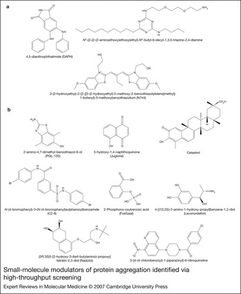
A complementary approach has been to identify small molecules that inhibit the formation of cytotoxic protein aggregates via high-throughput screening. In general, high-throughput screening is carried out using cell-free or cell-based assays. An advantage of cell-based assays is that they enable the identification of small molecules that inhibit protein aggregation indirectly (e.g. by acting on cellular regulatory factors), in addition to compounds that bind the aggregating protein and interfere with self-assembly directly. Recent high-throughput screening initiatives have led to the discovery of compounds that inhibit the aggregation of Aβ, tau, expanded polyglutamine proteins, or PrP. Importantly, the chemical diversity of ‘hit’ molecules identified via high-throughput screening greatly exceeds the diversity that would normally be achieved using structure-based methods (this point is well illustrated by the diversity of compounds in Fig. 3). As in the case of structure-based drug design, it is critical to ensure that fibrillisation inhibitors identified via high-throughput screening do not in fact stabilise toxic prefibrillar intermediates.
Small-molecule modulators of protein aggregation identified via high-throughput screening. Selected examples from the text are shown. (a) Molecules that inhibit the fibrillisation of amyloid-beta peptide Aβ or tau. (b) Molecules that modulate the aggregation of proteins with expanded polyglutamine repeats. C2-8, fosfosal, levonordefrin and nadolol inhibit aggregation, whereas 5-[4-(4-chlorobenzoyl)-1-piperazinyl]-8-nitroquinoline promotes the formation of large, relatively nontoxic aggregates. The compounds were identified by screening chemical libraries in cell-free systems, Escherichia coli, yeast or mammalian cell lines (see text for details). The pronounced structural variation among compounds with the same molecular target (e.g. Aβ, mutant huntingtin fragment) illustrates the chemical diversity that can be accessed via high-throughput screening.

Aβ and tau
A library of 3780 biologically active compounds was screened using a fluorescence-based assay of Aβ fibrillisation in a cell-free system (Ref. Reference Blanchard36). These efforts led to the identification of 4,5-dianilinophthalimide (DAPH; Fig. 3a) as a novel inhibitor of Aβ fibrillisation that also disrupts preformed fibrils. Coincubation of Aβ with DAPH interfered with the conversion of the peptide to a form that elicits toxicity in a neuronal cell line (IC50 = ~0.7 µm), suggesting that DAPH inhibits Aβ self-assembly without stabilising a toxic oligomer. In a second study, a library of ~1000 compounds was screened using an assay that involves monitoring a decrease in the fluorescence of a fusion of Aβ1–42 and green fluorescent protein (GFP) as a readout of Aβ aggregation in an Escherichia coli expression system (Ref. Reference Kim37). This screen led to the discovery of a triazine derivative (Fig. 3a) that suppressed Aβ fibrillisation with an IC50 of ~30 µm in a secondary cell-free assay. An advantage of the E. coli screen is that it is designed to identify compounds that inhibit early oligomer formation, whereas the same is not necessarily true of cell-free assays of Aβ fibrillisation. Specifically, prefibrillar oligomers formed by the Aβ1–42–GFP fusion in E. coli are expected to be nonfluorescent, so that compounds must disrupt these oligomers to be detected by the E. coli screen (Ref. Reference Kim37). In a third study, a small-molecule library was screened using a fluorescence-based cell-free assay monitoring the self-assembly of tau, a protein that forms aggregates called ‘neurofibrillary tangles’ (NFTs) in diseases referred to as ‘tauopathies’ (Table 1) (Ref. Reference Chirita, Necula and Kuret38). This screen resulted in the identification of a planar aromatic compound (N744; Fig. 3a) that inhibited tau polymerisation with high potency (IC50 = ~300 nm) and caused disaggregation of preformed filaments.
Expanded polyglutamine proteins
In a pioneering study, an automated filter-trap assay of huntingtin aggregation was used to screen a library of ~184 000 compounds (Ref. Reference Heiser39). In this assay, fibrils formed by an N-terminal fragment of polyglutamine-expanded (‘mutant’) huntingtin were trapped on a filter membrane in 384-well format and quantified via immunoblotting. The screen led to the identification of 300 huntingtin-aggregation inhibitors, 25 of which were benzothiazole derivatives (IC50 = ~1–10 µm). Although one of these compounds (PGL-135; Fig. 3b) inhibited huntingtin aggregation in a mammalian cell line and in hippocampal slice cultures (IC50 = ~40 µm), it was found to be metabolically unstable and therefore unsuitable for testing in vivo (Ref. Reference Hockly40). A filter-trap assay was also used to screen the NINDS Custom Collection of 1040 drugs and bioactive compounds approved by the US Food and Drug Administration (FDA) (Ref. Reference Wang41). This screen yielded ten hits that suppressed aggregation with an IC50 < 15 µm. Two of these compounds, the natural products juglone and celastrol (Fig. 3b), prevented the abnormal relocalisation of full-length mutant huntingtin in a striatal cell line. In another study, a library of 16 000 compounds was screened using a yeast-based assay of huntingtin aggregation and toxicity (Ref. Reference Zhang42). This screen resulted in the discovery of a compound (C2-8; Fig. 3b) that suppressed aggregation by an N-terminal fragment of mutant huntingtin in a neuronal cell line (IC50 = 0.05 µm) and in cultured hippocampal slices from a transgenic mouse model of Huntington disease. The compound also rescued photoreceptor neurodegeneration in huntingtin transgenic flies, suggesting that it may protect against huntingtin aggregation and toxicity in vivo. A cell-based screen was also used to identify molecules that inhibit the aggregation of two expanded polyglutamine proteins (N-terminal fragments of the androgen receptor and huntingtin) monitored via a sensitive fluorescence resonance energy transfer (FRET) assay (Ref. Reference Desai43). Of ten hits identified by this screen, five partially suppressed photoreceptor neurodegeneration in huntingtin transgenic flies, and three of these (fosfosal, levonordefrin and nadolol; Fig. 3b) are FDA-approved drugs that could potentially be rapidly translated into the clinic. Finally, a library of 37 000 compounds was screened using a cell-based assay that monitors the fluorescence of a huntingtin–GFP fusion as a readout of aggregation (Ref. Reference Bodner44). This screen led to the identification of a compound (5-[4-(4-chlorobenzoyl)-1-piperazinyl]-8-nitroquinoline; Fig. 3b) that enhanced huntingtin aggregation and suppressed huntingtin-induced proteasome dysfunction in mammalian cells. This compound also increased α-synuclein inclusion formation and reduced α-synuclein toxicity in a neuroglioma cell line (Ref. Reference Bodner44). Collectively, these findings suggest that novel therapeutics in the treatment of protein-misfolding diseases may prevent cell death by inhibiting the formation of toxic oligomers or by promoting the conversion of misfolded proteins to large, relatively benign inclusions.
PrP
The mammalian prion protein PrP exists in two states: a soluble, nonpathogenic form (PrPc) and an aggregated form (PrPsc) that is thought to be responsible for prion diseases in the brain. A library of 10 000 drug-like compounds was screened using an innovative cell-free assay referred to as SIFT (‘scanning for intensely fluorescent targets’) to monitor the association of PrPc with PrPsc (Ref. Reference Bertsch45). This screen led to the identification of six compounds (four with a common N'-benzylidene-benzohydrazide core structure) that suppressed PrPsc formation in a neuronal cell-culture model (IC50= ~2–6 µm). In another study, a library of 2000 drugs and natural products was screened using a high-throughput immunoblotting assay (Ref. Reference Kocisko46). This screen resulted in the discovery of 15 novel, potent inhibitors of PrPsc formation, including polyphenols, antimalarials, antihistamines, phenothiazine analogues, and statins (IC50= ~0.1–1 µm). Unfortunately, the most efficacious inhibitor in the cell-free assay (mefloquine, an antimalarial compound) was shown to be inactive in vivo (Ref. Reference Kocisko and Caughey47). This finding emphasises the importance of testing hits from high-throughput screens in preclinical animal models.
Inhibition of protein misfolding and aggregation via native-state stabilisation
Proteins with a compact, globular structure must undergo unfolding (or, in the case of oligomeric proteins, subunit dissociation followed by unfolding) prior to forming fibrils (Ref. Reference Colon and Kelly48). A strategy to prevent the fibrillisation of these proteins is to suppress subunit dissociation and/or unfolding using small molecules that stabilise the native state (Fig. 1). This approach has two important advantages over interfering with downstream self-assembly. First, inhibition of early misfolding precludes a build-up of toxic oligomers or protofibrils. Second, compounds that stabilise the native state typically bind in a deep pocket or cavity of a folded protein, and, therefore, they avoid pitfalls faced by molecules that disrupt protein–protein interactions (e.g. weak affinity, poor selectivity) (Refs Reference Johnson49, Reference Fry50).
Native-state stabilisation of transthyretin
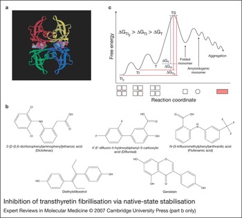
The strategy of inhibiting fibrillisation via native-state stabilisation has been explored extensively in a series of elegant papers on TTR (reviewed in Ref. Reference Johnson49). TTR is a 55 kDa homotetrameric protein with two binding sites for the hormone thyroxine (T4) (Fig. 4a). The amyloid diseases senile systemic amyloidosis and familial amyloid polyneuropathy are characterised by the fibrillisation of wild-type and mutant TTR, respectively. Amino acid substitutions in patients with familial amyloid polyneuropathy destabilise the native tetramer (Ref. Reference Johnson49). Biophysical data indicate that TTR fibrillisation occurs via rate-limiting tetramer dissociation, followed by partial unfolding and aggregation of the dissociated monomer. Previous work showed that T4 suppresses subunit dissociation by wild-type or mutant TTR (Ref. Reference Johnson49). Although T4 would not be a useful therapeutic inhibitor because of its hormonal activity, it was hypothesised that the binding of other small molecules to the hormone-binding pocket might stabilise tetrameric TTR and suppress pathogenesis (Ref. Reference Johnson49). Targeting the T4-binding pockets was a reasonable therapeutic strategy because TTR is only a minor carrier of T4 in the periphery, with <1% of the T4-binding sites occupied. Therefore, a high proportion of sites are available for inhibitor binding (Ref. Reference Adamski-Werner51). A number of biaryl compounds with structures similar to T4, including the nonsteroidal anti-inflammatory drugs (NSAIDs) diclofenac, diflunisal and flufenamic acid (Fig. 4b), were found to suppress TTR fibrillisation by binding to the native tetramer and inhibiting subunit dissociation under partially denaturing conditions (Refs Reference Johnson49, Reference Hammarstrom52). Isothermal titration calorimetry data indicated that the binding of inhibitor to one or both T4-binding pockets increases the activation energy for tetramer dissociation by stabilising the ground state relative to the transition state (Fig. 4c) (Refs Reference Johnson49, Reference Hammarstrom52).
Inhibition of transthyretin fibrillisation via native-state stabilisation. (a) The X-ray structure of the complex formed by transthyretin (TTR) and thyroxine (T4) (both hormone-binding sites are occupied). Each monomer in the TTR tetramer is depicted in a different colour. (b) Small-molecule inhibitors of TTR fibrillisation. (c) Free-energy diagram illustrating how native-state stabilisation inhibits TTR fibrillisation. When an inhibitor binds selectively to one or both T4-binding sites (yielding TI or TI2, respectively), the activation energy for tetramer dissociation (ΔGT) is increased due to stabilisation of the ground state relative to the transition state (TS) for subunit dissociation (ΔGTI22 > ΔGTI > ΔGT). As a result, drug binding leads to a decrease in the level of folded monomer with a high propensity to misfold and aggregate. The geometric shapes at the bottom of the graph represent different forms of TTR along the reaction coordinate (square, folded subunit; circle, unfolded subunit; tinted rectangle, aggregate; red dash, drug bound to tetrameric TTR). Panel (a) was reprinted, with permission, from Ref. Reference Adamski-Werner51 [Adamski-Werner, S.L. et al. (2004) Diflunisal analogues stabilize the native state of transthyretin. Potent inhibition of amyloidogenesis. J Med Chem 47, 355–374 (© 2004 American Chemical Society)]; panel (c) was adapted, with permission, from Ref. Reference Johnson49 [Johnson, S.M. et al. (2005) Native state kinetic stabilization as a strategy to ameliorate protein misfolding diseases: a focus on the transthyretin amyloidoses. Acc Chem Res 38, 911–921 (© 2005 American Chemical Society)].

Because of its inhibitory activity against TTR fibrillisation and its high oral bioavailability, diflunisal is currently being tested in a clinical trial for familial amyloid polyneuropathy (Ref. Reference Johnson49). Nevertheless, diflunisal and other NSAIDs may not provide a long-term therapeutic solution because chronic treatments with these compounds are likely to produce gastrointestinal side effects. Accordingly, recent efforts have focused on identifying inhibitors of TTR dissociation that lack NSAID activity. In one study, a library of 55 diflunisal analogues with various patterns of substitutions by halogen, hydroxyl or carboxyl groups was screened for inhibitory activity against TTR fibrillisation (Ref. Reference Adamski-Werner51). The screen yielded three inhibitors that bound the T4-binding pocket, and two of these analogues bound TTR selectively over other proteins in human plasma. In other studies, compounds structurally unrelated to diflunisal, including diethylstilbestrol (Fig. 4b) (Ref. Reference Morais-de-Sa53), bisaryloxime ethers (Ref. Reference Johnson54) and dibenzofurans (Ref. Reference Petrassi55), were found to inhibit TTR subunit dissociation and fibrillisation with high binding selectivity. The identification of TTR fibrillisation inhibitors from various structural classes represents an important advance in the development of therapeutics that lack NSAID activity. Although most inhibitors occupy both T4-binding sites at sufficiently high concentrations, recent data indicate that binding to a single site is sufficient for kinetic stabilisation (Ref. Reference Wiseman, Green and Kelly56). In turn, this finding suggests that one can minimise toxic side effects in vivo by administering a dose of inhibitor no greater than that required to occupy a single T4-binding site. Finally, genistein (Fig. 4b), the primary flavone in soy, has been shown to inhibit TTR subunit dissociation and fibrillisation, suggesting that this natural product may be used therapeutically with little risk of toxic side effects (Ref. Reference Green, Foss and Kelly57).
Native-state stabilisation of SOD1
Superoxide dismutase 1 (SOD1), like TTR, undergoes subunit dissociation, partial unfolding, and aggregation as a result of disease-linked mutations. SOD1 is a homodimer whose quaternary structure is destabilised by mutations linked to familial amyotrophic lateral sclerosis (Ref. Reference Hough58). Small molecules that bind to the dimer interface of SOD1 were identified by in silico screening (Ref. Reference Ray59). These compounds were found to inhibit the aggregation of mutant SOD1, suggesting that they may be a new class of drugs to treat familial amyotrophic lateral sclerosis (Ref. Reference Ray59).
A related strategy for the treatment of various protein-misfolding disorders is to rescue folding defects with ‘pharmacological chaperones’. A pharmacological chaperone is a small molecule (e.g. a ligand or substrate) that binds the folded, functional state of a protein and shifts the equilibrium away from partially unfolded conformers, thereby enabling folding and trafficking to the correct subcellular compartment (reviewed in Ref. Reference Loo and Clarke60).
Therapeutic strategy II: targeting post-translational modifications that promote protein misfolding and aggregation
Proteins undergo various types of post-translational modifications in cells under normal or stressed conditions. In some cases, the modified protein has reduced conformational stability and therefore an increased propensity to misfold and aggregate. Three post-translational modifications that promote the aggregation of some proteins are considered below: proteolysis, phosphorylation and oxidation (Fig. 1). Inhibiting these modifications may be a useful therapeutic strategy in the treatment of some protein-misfolding disorders.
Inhibition of proteolysis
A number of fibrillogenic polypeptides are released from larger precursor proteins by proteolysis, and the proteases involved in catalysing these processing events are potential therapeutic targets. The most intensively studied example of a fibrillogenic polypeptide generated by proteolysis is Aβ. Other examples discussed below include expanded polyglutamine proteins, α-synuclein, tau and gelsolin.
Targeting Aβ formation via inhibition of β- and γ-secretase
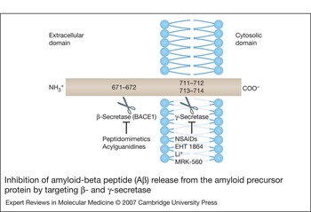
Aβ is released from the transmembrane amyloid precursor protein (APP) via successive cleavages by two aspartyl proteases: β- and γ-secretase (Fig. 5). β-Secretase, also referred to as beta-site APP-cleaving enzyme 1 (BACE1) or memapsin 2, is a transmembrane enzyme that cleaves APP to yield a membrane-bound C-terminal fragment (CTFβ or C99) (Ref. Reference De Jonghe61). γ-Secretase is a transmembrane multiprotein complex that cleaves CTFβ within the membrane, resulting in the secretion of Aβ1–40 or Aβ1–42 (Refs Reference De Jonghe61, Reference Wolfe62, Reference Hamaguchi, Ono and Yamada63). Several lines of evidence suggest that presenilin (presenilin 1 or 2), a component of the multiprotein complex, acts as the catalytic subunit of γ-secretase (Ref. Reference Wolfe62).
Inhibition of amyloid-beta peptide (Aβ) release from the amyloid precursor protein by targeting β- and γ-secretase. The amyloid precursor protein (APP) is depicted as a transmembrane protein in the phospholipid bilayer (phospholipid headgroups are shown as spheres; acyl chains are drawn as ‘tails’ associated with each headgroup). The β- and γ-secretase cleavage sites are indicated in the schematic representation of APP: β-secretase (also referred to as beta-site APP-cleaving enzyme 1, BACE1) cleaves between residues 671 and 672; and γ-secretase cleaves between residues 711 and 712, yielding Aβ1–40, or 713 and 714, yielding Aβ1–42. β- and γ-Secretase inhibitors discussed in the text are listed below the corresponding cleavage sites. The use of a combination of β- and γ-secretase inhibitors may be the best strategy to lower Aβ levels while minimising toxic side effects (see text for details). EHT 1864, inhibitor of the small GTP-binding protein Rac1; Li+, lithium ion; MRK-560, N-[cis-4-[(4-chlorophenyl)sulfonyl]-4-(2,5-difluorophenyl)cyclohexyl]-1,1,1-trifluoromethanesulfonamide (selective γ-secretase inhibitor from Merck Sharp and Dohme); NSAIDs, nonsteroidal anti-inflammatory drugs.

In addition to its role in APP proteolysis, γ-secretase is responsible for the intramembranous cleavage of Notch, a signalling molecule involved in specifying cell fate during embryogenesis (Ref. Reference Wolfe62). Presenilin knock-out mice have an embryonic lethal phenotype similar to that produced by deleting the Notch gene, and a number of γ-secretase inhibitors have undesired consequences that result from disruption of Notch processing (Refs Reference Wolfe62, Reference Hamaguchi, Ono and Yamada63). Accordingly, major efforts have focused on identifying compounds that block APP cleavage without perturbing Notch signalling (Fig. 5). An important breakthrough came from the discovery that NSAIDs selectively reduce Aβ1–42 levels in cell culture and in vivo without targeting Notch, an effect that is independent of cyclooxygenase inhibition (Ref. Reference Weggen64). Evidence suggests that NSAIDs inhibit γ-secretase activity by binding to an allosteric site remote from the active site (Ref. Reference Beher65). In turn, this binding could trigger a conformational change that specifically disrupts the enzyme's cleavage of APP but leaves Notch processing unaffected. NSAIDs may also block the formation of Aβ1–42 by preventing activation of the small GTP-binding protein Rho, apparently by interfering with Rho prenylation (Ref. Reference Zhou66). Another compound, EHT 1864, prevents activation of the small GTP-binding protein Rac1 and suppresses Aβ formation (in neuroblastoma cells and in vivo) without affecting Notch cleavage (Ref. Reference Desire67). These findings implicate Rho and Rac1 in the selective control of APP processing and suggest that targeting these small GTP-binding proteins is a valid strategy to prevent the accumulation of Aβ in Alzheimer disease. Consistent with this idea, Rho and Rac1 are involved in modulating the assembly or clustering of lipid rafts, and these membrane microdomains are a site for γ-secretase cleavage of APP but not of Notch (Refs Reference Desire67, Reference Manes68). Lithium, used in the clinic to treat bipolar disorder, is another agent that selectively targets γ-secretase cleavage of APP (Ref. Reference Phiel69). Various kinase inhibitors (including a metabolite of Gleevec) inhibit Aβ production without affecting Notch processing, apparently by interacting with an ATP-binding site on presenilin 1 (Refs Reference Netzer70, Reference Fraering71). The binding of a kinase inhibitor to this site may induce a conformational change in a γ-secretase domain involved in cleaving APP but not Notch (Ref. Reference Fraering71). Although several tyrosine kinase inhibitors are known to be safe for use in humans (Ref. Reference Netzer70), the suitability of these compounds to attenuate amyloid plaque formation may be limited by their weak penetrance of the blood–brain barrier. Finally, recent data indicate that the potent γ-secretase inhibitor MRK-560 suppresses amyloid plaque formation and Aβ generation in APP transgenic mice without perturbing the Notch signalling pathway (Ref. Reference Best72). Because MRK-560 is orally bioavailable and penetrates the blood–brain barrier, this compound holds promise as a new, selective inhibitor of amyloid formation in the treatment of Alzheimer disease.
As an alternative to inhibiting γ-secretase, several groups have focused on the strategy of lowering Aβ levels with small molecules that target β-secretase (BACE1) (Fig. 5). The ideal BACE1 inhibitor should be both highly potent and selective for BACE1 over other aspartyl proteases not involved in APP processing, including BACE2 (memapsin 1), cathepsin D, and pepsin. An advantage of targeting BACE1 rather than γ-secretase is that a cocrystal structure of the BACE1 protease domain complexed with a transition-state analogue has been solved (Ref. Reference Hong73). In turn, this information has facilitated structure-based design of BACE1 inhibitors with optimised potency and selectivity (Refs Reference Cole74, Reference Freskos75, Reference Ghosh76). A series of transition-state inhibitors containing a nonhydrolysable Leu–Ala dipeptide isostere was described in a recent report (Ref. Reference Ghosh76). One of these compounds was highly potent in an assay of recombinant BACE1 activity (K i = 0.12 nm) and exhibited high selectivity for BACE1 over BACE2 and cathepsin D. Other peptidomimetics (Refs Reference Freskos75, Reference Hanessian77) and a series of acylguanidines (Ref. Reference Cole74) were also reported to inhibit BACE1 in cell-free and cellular assays. A potential limitation of some of these compounds is that they may exhibit weak penetration of the blood–brain barrier because of their ‘peptidic character’ (Ref. Reference Stachel78). Various strategies have been reported to improve the blood–brain barrier permeability and Aβ-lowering ability of BACE1 inhibitors in vivo, including covalent attachment of a carrier peptide (Ref. Reference Chang79) and restriction of conformational flexibility via macrocyclisation (Ref. Reference Stachel78).
The intense interest in developing BACE1 inhibitors has been motivated largely by evidence from early studies that BACE1 null mice are apparently normal (Refs Reference Wolfe62, Reference Hamaguchi, Ono and Yamada63). However, this notion has been challenged by data from more recent knock-out studies suggesting that BACE1 is involved in modulating synaptic plasticity (Ref. Reference Laird80) and nerve myelination (Refs Reference Hu81, Reference Willem82). These findings suggest that careful dosing of BACE1 inhibitors will be critical to minimise toxic side effects. One strategy to minimise the doses of BACE1 and γ-secretase inhibitors while maintaining therapeutic efficacy might be to treat patients with both types of drugs simultaneously. The rationale for this type of approach is that the degree of γ-secretase inhibition required to maintain Aβ levels below a toxic threshold may be reduced if the steady-state levels of CTFβ substrate are decreased by BACE1 inhibition. Conversely, the extent to which the generation of CTFβ must be suppressed by BACE1 inhibition may be lower if the downstream conversion of CTFβ to Aβ is largely abrogated by γ-secretase inhibition. A combination therapy involving BACE1 and γ-secretase inhibitors would also alleviate the accumulation of potentially toxic fragments resulting from a single cleavage of APP. For example, inhibition of γ-secretase alone might lead to a build-up of C-terminal fragments of APP, a phenomenon linked to memory deficits in presenilin knock-out mice (Ref. Reference Saura83). Dual inhibition of BACE1 and γ-secretase would be expected to reduce this toxic effect. The results of a recent study indicate that NSAIDs downregulate expression of BACE1 by activating the peroxisome proliferator-activated receptor γ (PPARγ), a repressor of BACE1 gene transcription (Ref. Reference Sastre84). Therefore, NSAIDs may block APP cleavage by reducing BACE1 and γ-secretase activity simultaneously. Another report showed that various APP-binding molecules inhibit cleavage by both BACE1 and γ-secretase (Ref. Reference Espeseth85). Because NSAIDs and the APP-binding agents interfere with processing by both enzymes, they are excellent candidates for anti-amyloid therapies in the treatment of Alzheimer disease.
In addition to targeting BACE1 and γ-secretase, another strategy to reduce the amyloid levels in the brain may be to enhance the destruction of Aβ via proteolysis (Refs Reference Leissring86, Reference Choi87). Interestingly, the activity of one of the major Aβ-processing enzymes, neprilysin, is upregulated in cortical neurons by the neuropeptide somatostatin (Ref. Reference Saito88). These data suggest that the activation of somatostatin receptors might be another therapeutic strategy in the treatment of Alzheimer disease.
Role of proteolysis in the aggregation of polypeptides other than Aβ
Evidence suggests that fibrillogenic polypeptides other than Aβ are generated from larger precursor proteins via proteolysis. Intense research activity has focused on the role of proteolysis in the polyglutamine diseases. Recent data indicate that blocking calpain cleavage reduces huntingtin aggregation and toxicity in a cell-culture model (Ref. Reference Gafni89), and caspase-6 processing is necessary for huntingtin-induced neurodegeneration in mice (Ref. Reference Graham90). In addition, the aggregation of polyglutamine-expanded ataxin-3 is triggered by the removal of an N-terminal segment, possibly via caspase-mediated cleavage (Refs Reference Berke91, Reference Haacke92). The cleavage of huntingtin at some sites on the polypeptide chain may be associated with the normal function of the protein (Ref. Reference Cattaneo, Zuccato and Tartari93), implying that not all forms of huntingtin proteolysis are appropriate targets for therapy. Proteolysis has also been implicated in modulating the aggregation and toxicity of the following proteins: (Reference Dobson1) α-synuclein, cleaved C-terminally by the 20S proteasome to yield a truncated, fibrillogenic species that seeds aggregation of the full-length protein (Refs Reference Li94, Reference Liu95); (Reference Dahlgren2) tau, processed at its C-terminus by multiple caspases to yield a truncated species with a high propensity to form fibrils (Refs Reference Gamblin96, Reference Yin and Kuret97); and (Reference Walsh3) gelsolin, cleaved successively by furin and MT1-matrix metalloproteinase to yield 5 kDa and 8 kDa amyloidogenic peptides (Ref. Reference Page98). A reasonable therapeutic strategy may be to suppress the aggregation of these proteins using compounds that inhibit their corresponding proteases. However, as in the case of small molecules targeting β- and γ-secretase, a challenge will be to develop potent, selective inhibitors with minimal toxic side effects.
Inhibition of phosphorylation
The aggregation behaviour of some proteins is influenced by phosphorylation, and kinases and phosphatases that promote the formation of toxic aggregates are potential therapeutic targets. The prototypical protein that undergoes aggregation modulated by phosphorylation is tau. A second example discussed below is α-synuclein.
Role of phosphorylation in the aggregation of tau
A characteristic feature of tau from NFTs in human tauopathies is the hyperphosphorylation of the protein at multiple serine and threonine residues. A total of 38 serine/threonine phosphorylation sites have been identified in filamentous tau, compared with only 15 sites in tau from control adult brain (Ref. Reference Chen99). Hyperphosphorylated tau has a weakened affinity for microtubules and readily forms filaments, implying that phosphorylation promotes NFT formation by releasing a pool of unbound, fibrillogenic tau in the cytosol (Refs Reference Chen99, Reference Alonso100, Reference Duff and Planel101). However, evidence from in vivo studies suggests that tau hyperphosphorylation correlates with neurotoxicity but not with NFT assembly per se. Expression of the human tau gene in Drosophila induced age-dependent neurodegeneration and the accumulation of phosphorylated tau without NFT formation (Ref. Reference Wittmann102). Although phosphorylated tau and NFTs were detected in transgenic mice expressing human tau, the accumulation of filamentous tau was inversely correlated with the presence of apoptotic nuclei in individual neurons (Ref. Reference Andorfer103). In transgenic mice expressing a repressible human tau construct, downregulation of transgene expression after the onset of NFT formation led to increased neuronal counts and improved memory function without affecting the progression of tau pathology (Ref. Reference Santacruz104). Inhibition of tau hyperphosphorylation with a kinase inhibitor suppressed motor dysfunction without reducing NFT levels in transgenic mice expressing P301L human tau, a mutant form of the protein involved in frontotemporal dementia with parkinsonism-17 (Ref. Reference Le Corre105). These findings suggest that tau hyperphosphorylation triggers neurodegeneration by a mechanism unrelated to NFT formation. Consistent with this idea, hyperphosphorylated tau was found to disrupt microtubule assembly by a dominant-negative mechanism that involved trapping unmodified tau in an inactive complex, whereas the polymerisation of hyperphosphorylated tau eliminated this potentially neurotoxic inhibitory effect (Ref. Reference Alonso Adel106).
Regardless of whether NFTs are toxic, benign or protective, tau hyperphosphorylation generally correlates with neurodegeneration in animal models or humans (Refs Reference Duff and Planel101, Reference Le Corre105, Reference Shulman and Feany107). Therefore, the inhibition of tau kinases may be a reasonable therapeutic strategy in the treatment of tauopathies. Multiple kinases have been implicated in tau phosphorylation (Ref. Reference Avila108). Notable examples include glycogen synthase kinase 3β (GSK3β) (Refs Reference Lucas109, Reference Jackson110), cyclin-dependent kinase 5 (CDK5) (Refs Reference Cruz111, Reference Noble112), protein kinase A (PKA) (Ref. Reference Liu113) and tau-tubulin kinase 1 (Ref. Reference Sato114). Conditional overexpression of GSK3β induces tau hyperphosphorylation and neurotoxicity in a transgenic mouse model (Ref. Reference Lucas109), and Drosophila co-expressing tau and GSK3β exhibit a more severe neurodegenerative phenotype than flies expressing tau alone (Ref. Reference Jackson110). Oligomeric Aβ activates GSK3β via downregulation of the Akt pathway (Refs Reference Avila108, Reference Hoshi115, Reference Takashima116). Accordingly, GSK3β plays a critical role in the pathogenesis of Alzheimer disease, linking Aβ aggregation with tau hyperphosphorylation. Inhibition of GSK3 with lithium or the small molecule AR-A014418 results in decreased tau phosphorylation and aggregation and reduced neurodegeneration in transgenic mice expressing the P301L mutant (Ref. Reference Noble117). The inhibitor AR-A014418 is a promising lead compound for treating Alzheimer disease and other tauopathies because it is potent (IC50 = 104 ± 27 nm) and highly selective for GSK3 over other kinases (Ref. Reference Bhat118).
Another kinase considered to be a potential therapeutic target in tauopathies is CDK5. Two activator proteins, p35 and its more stable truncated form, p25, modulate CDK5 activity in neurons (Ref. Reference Patrick119). Levels of p25 are increased in Alzheimer disease brain, resulting in deregulated CDK5 activity. Overexpression of p25 elicits tau hyperphosphorylation, tau aggregation, and neurodegeneration in transgenic mice (Refs Reference Cruz111, Reference Noble112). Similarly, co-expression of CDK5 and its activator, p35, enhances tau toxicity in a Drosophila model (Ref. Reference Shulman and Feany107). A lentivirally encoded CDK5 inhibitory peptide derived from p35 was found to suppress tau hyperphosphorylation and apoptosis in cortical neurons overexpressing CDK5/p25 or treated with Aβ1–42 (Ref. Reference Zheng120). These results imply that CDK5/p25 may be a useful therapeutic target in Alzheimer disease and other tauopathies. By contrast, data from another study indicate that p25-mediated CDK5 activation results in the inhibition of GSK3, in turn suggesting that a decrease in CDK5/p25 activity could lead to enhanced tau hyperphosphorylation (Ref. Reference Plattner, Angelo and Giese121). Given these contradictory findings, it would seem that GSK3 is a more favourable target than CDK5 to suppress tau hyperphosphorylation and neurotoxicity.
Role of phosphorylation in the aggregation of α-synuclein
The most abundant modified form of α-synuclein in Lewy bodies (characteristic inclusions in the brains of synucleinopathy patients) is a species phosphorylated at Ser129 (Refs Reference Fujiwara122, Reference Anderson123). Although this phosphorylated species is also detected in the soluble portion of normal and diseased brain tissue, it is highly enriched in Lewy bodies. These observations suggest that the phosphorylation of α-synuclein at Ser129 occurs under normal physiological conditions, and this modification converts the protein to a form with a high propensity to aggregate. In support of this idea, phosphorylation at Ser129 is necessary for the formation of α-synuclein-containing inclusions in a neuroblastoma cell line (Ref. Reference Smith124). By contrast, evidence from studies of α-synuclein transgenic Drosophila suggests that phosphorylation at Ser129 prevents the formation of detergent-insoluble aggregates but increases neurotoxicity, perhaps by eliciting a build-up of soluble α-synuclein oligomers (Ref. Reference Chen and Feany125). Although it is still unclear how phosphorylation at Ser129 affects α-synuclein aggregation, the fact that this modification is involved in neurotoxicity suggests that targeting serine/threonine kinases may be a reasonable therapeutic strategy in the treatment of Parkinson disease. Kinases proposed to play a role in phosphorylating α-synuclein at Ser129 include casein kinases 1 and 2 and G-protein-coupled receptor kinase 2 (Refs Reference Smith124, Reference Chen and Feany125, Reference Kim126). DYRK1A, a member of the dual-specificity tyrosine-regulated kinase family, has been shown to promote α-synuclein aggregation in a hippocampal cell line and protofibril formation in a cell-free system (Ref. Reference Kim126). Interestingly, DYRK1A selectively phosphorylates α-synuclein at Ser87, suggesting that phosphorylation at sites other than Ser129 may play a role in α-synuclein aggregation and/or toxicity. Because serine/threonine kinases involved in phosphorylating α-synuclein have many different substrates, the feasibility of suppressing α-synuclein toxicity by targeting these kinases may be limited by harmful off-pathway effects.
Inhibition of oxidation
Protein aggregation and oxidative stress are coupled phenomena: not only does protein oxidation generally promote aggregation, but protein aggregates also tend to elicit a build-up of reactive oxygen species (ROS), thereby creating a vicious cycle (Ref. Reference Tabner127). These findings raise the possibility that antioxidants may be beneficial in the treatment of protein-misfolding diseases. Two proteins that exhibit aggregation behaviour and/or toxicity modulated by oxidative stess are Aβ and α-synuclein. Other examples discussed below are huntingtin, tau and SOD1.
Role of oxidation in the aggregation and toxicity of Aβ
A large proportion of Aβ in the brains of Alzheimer disease patients is oxidised at Met35 to the corresponding sulphoxide (Refs Reference Barnham128, Reference Butterfield and Boyd-Kimball129). Evidence from several groups indicates that oxidation at this position inhibits the conversion of Aβ1–40 or Aβ1–42 to oligomers or fibrils (Refs Reference Hou130, Reference Palmblad, Westlind-Danielsson and Bergquist131, Reference Bitan132). This inhibitory effect can be rationalised by examining the SS-NMR structure of fibrillar Aβ1–40: methionine sulphoxide is substantially more polar than methionine, and an increase in polarity at position 35 would be predicted to disrupt external quaternary interactions necessary for self-assembly (Fig. 2a) (Ref. Reference Petkova, Yau and Tycko17). Although the presence of methionine sulphoxide at position 35 interferes with Aβ aggregation, redox reactions involving Met35 are thought to contribute to Aβ-induced oxidative stress and neurotoxicity. As one possibility, one-electron oxidation of Met35 in oligomeric membrane-bound Aβ may result in the formation of a sulphuranyl radical, which in turn can induce peroxidation of neighbouring lipids and a build-up of ROS (Ref. Reference Butterfield and Boyd-Kimball129). Alternatively, the oxidation of Met35 may be coupled with the reduction of metal ions (e.g. Cu2+, Fe3+) bound to Aβ (Ref. Reference Barnham128). The reduced forms of these metal ions may then react with O2 to generate H2O2 (Refs Reference Tabner127, Reference Barnham128).
Recent data indicate that various oxidised metabolites stimulate Aβ aggregation (Ref. Reference Zhang133). A ketoaldehyde derivative of cholesterol and its aldol product accelerated the conversion of Aβ1–40 to spherical aggregates and mature fibrils in a cell-free system by lowering the critical concentration for self-assembly. This effect was attributed to the reaction of Aβ1–40 with the oxidised cholesterol metabolites, resulting in the formation of covalent adducts. These findings may account for the enhanced deposition of wild-type Aβ in patients with sporadic Alzheimer disease compared with healthy individuals – namely, Alzheimer disease patients may have increased levels of oxidised metabolites that covalently modify wild-type Aβ, converting it to a form with a high propensity to aggregate. Oxidised cholesterol metabolites are formed via inflammatory ozonolysis reactions associated with atherosclerosis, providing a rationale for observed links among hypercholesterolaemia, inflammation, atherosclerosis and sporadic Alzheimer disease (Ref. Reference Zhang133).
Given the role of oxidative stress in Aβ aggregation and toxicity, antioxidant therapy may be beneficial in the treatment of Alzheimer disease. Consistent with this idea, a number of polyphenols with the ability to scavenge ROS inhibit Aβ fibrillisation and destabilise preformed Aβ fibrils in cell-free systems at concentrations between 0.1 and 1 µm (Refs Reference Ono134, Reference Masuda135). The polyphenol curcumin (Fig. 1) prevents the conversion of Aβ to potentially toxic oligomers in addition to fibrils (Ref. Reference Yang136). Curcumin is a promising therapeutic candidate because it crosses the blood–brain barrier and reduces amyloid formation in aged APP transgenic mice (Ref. Reference Yang136). The pineal hormone melatonin is another promising candidate because it penetrates the blood–brain barrier, attenuates oxidative damage, and prevents Aβ accumulation in the brains of APP transgenic mice, and it has been used safely in the clinic to treat sleep disorders (Refs Reference Matsubara137, Reference Feng138). Antioxidant molecules such as curcumin and melatonin may inhibit Aβ aggregation by scavenging ROS and/or interfering with intermolecular interactions that drive Aβ self-assembly (Refs Reference Yang136, Reference Matsubara137). Another antioxidant compound, acetyl-l-carnitine, suppresses Aβ1–42-induced neurotoxicity and oxidative damage by increasing cellular levels of the endogenous antioxidant glutathione (Ref. Reference Abdul139). Finally, the metal chelator clioquinol reduces Aβ neurotoxicity in cell culture, presumably by disrupting interactions between Aβ and metal ions (Ref. Reference Barnham128). The results of a pilot Phase II clinical trial imply that clioquinol may slow cognitive decline in severely affected Alzheimer disease patients (Ref. Reference Ritchie140).
Role of oxidation in the aggregation and toxicity of α-synuclein
Various covalent oxidative modifications affect the conversion of α-synuclein to high molecular weight species in cell-free systems. α-Synuclein modified by nitration (Refs Reference Norris141, Reference Yamin, Uversky and Fink142), metal-catalysed oxidation (Ref. Reference Cole143) or dopamine quinone (Refs Reference Conway34, Reference Rochet35) does not fibrillise, but instead accumulates as soluble, potentially toxic oligomers. Oligomers also accumulate in mixtures of methionine-oxidised and unoxidised α-synuclein (Refs Reference Conway34, Reference Glaser144). α-Synuclein species containing nitrotyrosine, methionine sulphoxide, and methionine sulphone were detected in a cellular model of Parkinson disease (Ref. Reference Mirzaei145), implying that these modifications play a role in Parkinson disease pathogenesis. Oxidised cholesterol metabolites were shown to accelerate the formation of α-synuclein protofibrils and fibrils in a cell-free system, apparently via a noncovalent mechanism (Ref. Reference Bosco146). These metabolites are elevated in the brains of synucleinopathy patients, suggesting that they promote α-synuclein aggregation and toxicity associated with Lewy body disorders (Ref. Reference Bosco146). Several compounds with antioxidant activity inhibit α-synuclein fibrillisation and destabilise preformed fibrils. Examples include the polyphenols curcumin, myricetin, nordihydroguaiaretic acid, rosmarinic acid and tannic acid, and the flavonoid baicalein (Refs Reference Masuda135, Reference Zhu147, Reference Ono and Yamada148). A potential limitation of these findings is that the compounds may block fibrillisation without suppressing the formation of toxic protofibrils. For example, the inhibition of α-synuclein fibrillisation by baicalein results in a build-up of soluble oligomers (Ref. Reference Zhu147). Accordingly, it will be critical to determine whether antioxidants suppress α-synuclein toxicity in cellular and animal models of Parkinson disease and other synucleinopathies.
Role of oxidation in the aggregation and toxicity of huntingtin, tau and SOD1
Various reports indicate that oxidative stress plays a central role in the aggregation and toxicity of other proteins involved in protein-misfolding disorders, in addition to Aβ and α-synuclein. For example, the toxicity of a mutant huntingtin fragment was shown to be suppressed in yeast lacking the gene encoding kynurenine 3-monooxygenase (KMO), an enzyme involved in the degradation of tryptophan (Ref. Reference Giorgini149). Two kynurenine pathway metabolites – 3-hydroxykynurenine and quinolinic acid – are known to induce the formation of ROS. Accordingly, it was hypothesised that mutant huntingtin causes cell death by triggering oxidative stress via upregulation of the kynurenine pathway (Ref. Reference Giorgini149). In support of this hypothesis, the expression of mutant huntingtin led to a build-up of ROS in Saccharomyces cerevisiae, and this effect was abolished by genetic or pharmacological disruption of KMO activity. Data from studies in mammalian cell culture suggest that a mutant huntingtin fragment causes a build-up of ROS via protein aggregation and iron-dependent redox chemistry, and this oxidative stress is suppressed by antioxidants and heat-shock proteins (Refs Reference Wyttenbach150, Reference Firdaus151, Reference Firdaus152). In a Drosophila model of human tauopathies, tau-induced neurodegeneration was enhanced or suppressed in flies with reduced or increased expression of antioxidant enzymes, respectively (Ref. Reference Dias-Santagata153). Importantly, neurodegeneration was also suppressed in flies treated with the antioxidant compound α-tocopherol (vitamin E). Finally, the formation of disulphide-linked (i.e. oxidised) SOD1 dimers and oligomers was found to be a critical early step in amyotrophic lateral sclerosis pathogenesis (Ref. Reference Deng154). Together, these findings underscore the important role of oxidative stress in various protein-misfolding disorders, and they suggest that antioxidants are reasonable therapeutic candidates in the treatment of these diseases.
Therapeutic strategy III: upregulating molecular chaperones or aggregate clearance
Cells have evolved various quality-control mechanisms to prevent a build-up of toxic protein aggregates (Ref. Reference Muchowski and Wacker155). An initial line of defence is provided by molecular chaperones – cellular factors that inhibit protein misfolding and/or interfere with non-native interactions leading to aggregate formation (Fig. 1). In some cases, chaperones promote the correct folding of their misfolded protein substrates. Alternatively, chaperones target misfolded polypeptides for degradation by the ubiquitin–proteasome pathway or by chaperone-mediated autophagy, a clearance mechanism in which a target protein is translocated into the lysosome with the assistance of the cytosolic chaperone Hsc70 (Ref. Reference Muchowski and Wacker155). Aggregates formed under conditions where normal homeostasis mechanisms are overwhelmed by a high burden of protein misfolding are eliminated by ‘macroautophagy’, a lysosomal clearance pathway in which cellular material is engulfed in a vesicular body termed an ‘autophagosome’ (Fig. 1) (Ref. Reference Cuervo156). Enhancing chaperone function and stimulating aggregate clearance might be useful therapeutic strategies for the treatment of protein-misfolding disorders.
Enhancing chaperone function
Several molecular chaperones are referred to as heat-shock proteins because they are upregulated in response to heat and other forms of stress that cause a build-up of misfolded polypeptides (Ref. Reference Muchowski and Wacker155). Examples of molecular chaperones associated with the heat-shock response include Hsp27, Hsp40, Hsp70 and Hsp90 (Fig. 1). Evidence suggests that the upregulation of heat-shock proteins suppresses the aggregation of Aβ, α-synuclein and polyglutamine proteins.
Effect of chaperones on the aggregation and toxicity of Aβ
Data from studies in a cell-free system indicate that fibril formation by Aβ1–42 is inhibited by Hsp90 or by Hsp70 in combination with its co-chaperone, Hsp40 (Ref. Reference Evans, Wisen and Gestwicki157). The Hsp70–Hsp40 inhibitory effect is dependent on ATP hydrolysis, suggesting that these two chaperones expend energy to convert Aβ to a form with a weak propensity to aggregate. A combination of Hsp90, Hsp70 and Hsp40 causes dissociation of preformed oligomers but not fibrils, implying that the chaperones target oligomeric intermediates on the Aβ aggregation pathway. Other studies have shown that the small heat-shock proteins Hsp20, Hsp27 and αB-crystallin bind Aβ1–40, and this binding correlates with inhibition of Aβ aggregation and attenuation of Aβ toxicity (Ref. Reference Wilhelmus158). Down-regulation of heat-shock factor 1 (HSF1), a transcription factor that induces the expression of heat-shock proteins, results in accelerated paralysis and increased protein aggregation in transgenic worms overexpressing Aβ1–42 in the body-wall muscles (Ref. Reference Cohen5). These findings suggest that molecular chaperones associated with the heat-shock response play a critical role in preventing Aβ aggregation and toxicity in vivo. Given that they are located primarily in intracellular compartments, molecular chaperones are likely to protect against Aβ toxicity by binding oligomeric species inside the cell, as opposed to extracellular oligomers or plaques (Ref. Reference Evans, Wisen and Gestwicki157).
Effect of chaperones on the aggregation and toxicity of α-synuclein
The overexpression of Hsp70 causes a decrease in α-synuclein toxicity in cell culture and a reduction in high molecular weight and detergent-insoluble forms of α-synuclein in the brains of α-synuclein transgenic mice (Ref. Reference Klucken159). Hsp70 has also been found to prevent the accumulation of high molecular weight α-synuclein species in dopaminergic cells treated with the complex I inhibitor rotenone (Ref. Reference Zhou160). Although Hsp70 inhibits dopaminergic cell death in α-synuclein transgenic flies, the chaperone has no effect on the number, size or morphology of Lewy-like inclusions in this model (Ref. Reference Auluck161). Hsp70 has been shown to bind protofibrils but not fibrils formed from recombinant α-synuclein (Refs Reference Dedmon162, Reference Huang163), a result that may explain why Hsp70 has no discernible effect on fibrillar inclusions in α-synuclein transgenic flies. In other studies, Hsp27 has been shown to interfere with α-synuclein aggregation and toxicity in cell-culture models (Refs Reference Zourlidou, Payne Smith and Latchman164, Reference Outeiro165). Together, these findings suggest that Hsp70 and Hsp27 may delay neurodegeneration associated with synucleinopathies by inhibiting the formation of toxic α-synuclein aggregates in the brain.
Effect of chaperones on the aggregation and toxicity of polyglutamine proteins
Atomic force microscopy data indicate that Hsp70 and Hsp40 block the conversion of a mutant huntingtin fragment to spherical and ring-like oligomers by binding and sequestering the monomeric protein (Ref. Reference Wacker166). Evidence from several groups suggests that expanded polyglutamine proteins bound to Hsp70 are targeted for degradation via a pathway mediated by CHIP (‘C-terminus of Hsp70-interacting protein’), an Hsp70 co-chaperone with E3 ubiquitin ligase activity (Refs Reference Miller167, Reference Jana168, Reference Al-Ramahi169). Other studies have shown that Hsp70 acts in cooperation with the cytosolic chaperonin TRiC (CCT) to inhibit the conversion of expanded polyglutamine proteins to toxic oligomers, perhaps by promoting the formation of benign, off-pathway assemblies (Refs Reference Behrends170, Reference Kitamura171, Reference Tam172). Collectively, these data suggest that Hsp70, Hsp40, CHIP and TRiC interfere with protein aggregation and neurotoxicity associated with polyglutamine disorders.
Pharmacological upregulation of chaperone function
The observations outlined above suggest that enhancing the function of molecular chaperones might be a useful strategy to inhibit toxic aggregate formation in various protein-misfolding diseases. This type of approach may have fewer pitfalls compared with directly targeting aggregation with small-molecule inhibitors: whereas chaperones are designed to bind exposed hydrophobic regions of misfolded polypeptides, small molecules occupy an insufficient volume to efficiently disrupt aberrant protein–protein interactions involving these exposed surfaces (Ref. Reference Gestwicki, Crabtree and Graef173).
Emerging data suggest that it may be possible to upregulate molecular chaperones using one of several pharmacological strategies. Notably, the Hsp90 inhibitor geldanamycin (GA) and its derivatives 17-AAG (17-allylamino-demethoxygeldanamycin) and 17-DMAG [17-(dimethylaminoethylamino)-17-demethoxygeldanamycin] induce the expression of Hsp70 and Hsp27 in cancer models (Refs Reference Drysdale174, Reference McCollum175), raising the possibility that these agents may be useful for treating protein-misfolding diseases (Fig. 1). Consistent with this idea, GA was shown to inhibit α-synuclein toxicity in a cell-culture model (Ref. Reference McLean176) and in transgenic Drosophila (Ref. Reference Auluck, Meulener and Bonini177). The protective effect of GA occurred in parallel with a decrease in α-synuclein aggregation in cell culture (Ref. Reference McLean176), whereas it correlated with an increase in detergent-resistant, presumably nontoxic α-synuclein species in the brains of transgenic flies (Ref. Reference Auluck, Meulener and Bonini177). GA and another Hsp90 inhibitor, radicicol, suppressed huntingtin toxicity and aggregation in organotypic slice cultures derived from huntingtin transgenic mice (Ref. Reference Hay178). The data from these studies support a model in which GA inhibits the formation of toxic protein aggregates by upregulating Hsp70 (Refs Reference McLean176, Reference Auluck, Meulener and Bonini177, Reference Hay178). In turn, Hsp70 upregulation is thought to involve GA-mediated release of HSF1 from its inactive complex with Hsp90, followed by HSF1 trimerisation, phosphorylation and nuclear localisation (Fig. 1) (Refs Reference Shen179, Reference Ansar180). 17-AAG and 17-DMAG are already being tested in Phase I and II clinical trials as potential anticancer agents (Ref. Reference McCollum175), suggesting that they may be suitable for administration to patients with protein-misfolding disorders. A disadvantage of these molecules is that they provide a narrow therapeutic window because the concentrations at which they induce Hsp70 upregulation are similar to the concentrations at which they elicit toxicity as a result of degradation of Hsp90 client proteins (Ref. Reference Ansar180). This problem was addressed by developing a series of novobiocin analogues that target a different domain of Hsp90 (the C-terminal ATP-binding domain, in contrast to the N-terminal ATP-binding pocket targeted by GA and its derivatives) (Ref. Reference Ansar180). One analogue (A4; Fig. 1) was found to upregulate Hsp70 and suppress Aβ-induced neurodegeneration in a cellular model of Alzheimer disease without causing toxicity, suggesting that this compound may provide a relatively broad therapeutic window.
In addition to Hsp90 inhibitors, other small molecules have been shown to induce the heat-shock response. Examples include acetyl-l-carnitine, a compound that is naturally present in mitochondrial membranes and upregulates Hsp72 (Hsp70) (Ref. Reference Abdul139), and celastrol, a component of Chinese herbal medicines that activates HSF1 (Ref. Reference Westerheide181). These small molecules may also be useful in the treatment of protein-misfolding diseases.
Enhancing aggregate clearance
In cells with excess amounts of misfolded proteins, the capacity of chaperones and the ubiquitin–proteasome pathway to prevent protein aggregation is exceeded (Ref. Reference Muchowski and Wacker155). Under these conditions, misfolded proteins form aggregates that are transported along microtubules to the perinuclear region, where they are recruited into inclusion bodies termed ‘aggresomes’ (Fig. 1) (Ref. Reference Johnston, Ward and Kopito182). At the aggresome, it is hypothesised that protein aggregates are targeted for destruction via macroautophagy (Refs Reference Iwata183, Reference Ravikumar184). Examples of aggregated proteins that may be processed via aggresome formation and macroautophagy are α-synuclein and proteins with expanded polyglutamine repeats.
Clearance of aggregated α-synuclein
The overexpression of α-synuclein in mammalian cells leads to the formation of spherical, prefibrillar oligomers, followed by the appearance of aggresome-like inclusions enriched with fibrillar α-synuclein (Ref. Reference Lee and Lee185). Prefibrillar α-synuclein oligomers, but not the mature fibrils, are eliminated from neuronal cells via lysosomal degradation, and inhibition of this process augments α-synuclein-induced cell death (Ref. Reference Lee186). Data from another study suggest that macroautophagy plays a role in the clearance of α-synuclein from neuronal cells (Ref. Reference Webb187). Together, these data support a model in which toxic α-synuclein oligomers are transported to the aggresome as part of a protective mechanism. Once at the aggresome, the oligomers are either converted into relatively benign fibrils or cleared from the cell via macroautophagy. Consistent with the idea that aggresome formation is cytoprotective, the presence of aggresomes was found to correlate poorly with the presence of apoptotic markers in a mammalian cell line overexpressing α-synuclein and its binding partner, synphilin-1 (Ref. Reference Tanaka188). In addition to clearance via the aggresome–macroautophagy pathway, evidence suggests that α-synuclein is eliminated from neuronal cells via chaperone-mediated autophagy (Ref. Reference Cuervo156).
Clearance of expanded polyglutamine proteins
Aggresome-like inclusion bodies are formed in mammalian cells expressing expanded polyglutamine proteins, including N-terminal huntingtin fragments, ataxin-3, and the androgen receptor (Refs Reference Waelter189, Reference Taylor190, Reference Iwata191). Disruption of macroautophagy by RNA interference (RNAi) or with small-molecule inhibitors causes a reduction in the clearance of cytosolic polyglutamine aggregates (Refs Reference Iwata191, Reference Ravikumar, Duden and Rubinsztein192). The accumulation of autophagic proteins and lysosomes near the aggresome and the elimination of polyglutamine aggregates require intact microtubule-dependent transport (Refs Reference Iwata183, Reference Ravikumar184). Collectively, these findings suggest that recruitment of the autophagic machinery to the aggresome is necessary for the efficient removal of protein aggregates from the cytosol (Refs Reference Iwata183, Reference Taylor190, Reference Iwata191).
Pharmacological upregulation of aggregate clearance
Advances in understanding aggregate-clearance mechanisms have motivated efforts to activate these pathways as a strategy to treat protein-misfolding disorders (Ref. Reference Iwata191). Multiple lines of evidence suggest that the accumulation of cytosolic protein aggregates can be suppressed dramatically via pharmacological upregulation of macroautophagy. This approach may have greater therapeutic potential than the enhancement of proteasome activity, given that upregulation of the ubiquitin–proteasome pathway (in contrast to autophagy) is likely to increase the risk of cancer by causing depletion of short-lived proteins with anti-tumourigenic activity (e.g. p53) (Ref. Reference Rubinsztein193). The macrolide antibiotic rapamycin (also known as ‘sirolimus’) activates macroautophagy by blocking the activity of mTOR (‘mammalian target of rapamycin’), a kinase that inhibits autophagosome formation (Fig. 1). Rapamycin enhances autophagic clearance and reduces the toxicity of α-synuclein, tau and polyglutamine proteins in cellular and in vivo models (Refs Reference Webb187, Reference Ravikumar194, Reference Berger195). Rapamycin derivatives with improved water solubility (CCI-779, RAD001 and AP23576) are already under clinical development as possible anticancer drugs, and, therefore, they may also be suitable for use in humans to treat protein-misfolding diseases (Refs Reference Rubinsztein193, Reference Vignot196). At the same time, the potential for toxic side effects associated with long-term administration of rapamycin derivatives has motivated a search for alternative pharmacological approaches to stimulate macroautophagy. Recent findings suggest that macroautophagic clearance of protein aggregates is stimulated in an mTOR-independent manner by lithium (Ref. Reference Sarkar197) and the disaccharide trehalose (Ref. Reference Sarkar198).
Research in progress and outstanding research questions
There has been tremendous progress over the past five years in the development of novel therapeutic approaches targeting protein-misfolding diseases. Advances in our understanding of fibrillar structure have enabled the rational design of peptides that inhibit fibrillisation. Chemically diverse compounds that block aggregation or, alternatively, that promote the formation of large, nontoxic assemblies have been identified using powerful high-throughput screening technologies. In the case of proteins that normally have a compact, globular structure, misfolding and aggregation have been inhibited successfully by compounds that stabilise the native conformation. A large body of evidence suggests that the formation of toxic protein aggregates can be suppressed by interfering with post-translational modifications (e.g. proteolysis, phosphorylation and oxidation), increasing chaperone function, or upregulating aggregate-clearance mechanisms such as macroautophagy. Several of these highly promising therapeutic strategies are highlighted in the model shown in Figure 1.
What new developments can be expected during the years ahead? One reasonable prediction is that the structures of protofibrillar and fibrillar assemblies will continue to be solved using methods such as SS-NMR, and this information will enable the structure-based design of many more protein-aggregation inhibitors. In parallel, continued high-throughput screening efforts will lead to the identification of additional compounds that interfere with protein self-assembly. A high priority will be to assess whether drug candidates prevent the formation of oligomers, protofibrils or fibrils, and whether they reduce toxicity in animal models of misfolding diseases. Future developments will also likely include the implementation of new therapeutic strategies that have not been discussed in this review. For example, emerging data suggest that the depletion of aggregating proteins using RNAi methods in vivo may be a powerful approach to prevent misfolding diseases (Refs Reference Miller199, Reference Saito200, Reference Sapru201).
There are a number of outstanding research questions that must be addressed by future research. Which aggregated species (oligomers, protofibrils or fibrils) are responsible for cell death in protein-misfolding diseases? Why is the pathology of protein-aggregation disorders generally limited to specific types of cells or tissues? Which cytotoxic pathways are triggered by protein aggregation? Studies carried out in genetically manipulable model organisms including yeast, Drosophila and C. elegans will provide a powerful approach to answer the third question by driving the discovery of new pathogenic mechanisms involved in protein-misfolding diseases (see Ref. Reference Cooper202 for a recent example). The use of proteomic technologies (Refs Reference Zhou160, Reference Poon203) and genome-wide RNAi (Ref. Reference Nollen204) will also provide important clues about disease mechanisms. In turn, these insights will make it possible to devise therapeutic strategies that modulate cellular pathways disrupted by protein misfolding and aggregation, including apoptosis (Ref. Reference Hashimoto12), signal transduction (Refs Reference Mattson and Sherman13, Reference Morfini205) and transcription (Ref. Reference Rego and de Almeida14). Ultimately, drug combinations targeting protein aggregation as well as the resulting perturbation of cellular pathways will provide the most effective treatment for patients suffering from protein-misfolding diseases.
Acknowledgements and funding
Current funding is provided by the National Institute of Aging, National Institute of Neurological Disorders and Stroke, American Parkinson Disease Association, Parkinson's Disease Foundation, and National Parkinson Foundation. I am grateful to the peer reviewers for their insightful comments and to the members of my research group for stimulating discussions.