INTRODUCTION
Fasciola hepatica is the causative agent of fasciolosis, a food-borne zoonotic disease that affects grazing animals and humans of great importance for livestock and public health (Mas-Coma et al. Reference Mas-Coma, Bargues and Valero2005). According to Piedrafita et al. (Reference Piedrafita, Spithill, Smith and Raadsma2010), fasciolosis constitutes a veterinary problem, since affected animals show reduced meat and milk production, poor feed conversion, and decreased work capacity, causing economic losses to farmers. Fasciolosis is considered an important clinical and epidemiological health problem, mostly due to the occurrence of snails, which are intermediate hosts of F. hepatica, associated with the existence of a large variety of reservoir hosts, contributing to maintain the infection worldwide, with up to 2·4 million people infected and approximately 180 million at risk (Mas-Coma et al. Reference Mas-Coma, Bargues and Valero2014).
Oxidative stress is considered a disturbance in the antioxidant/oxidant status due to excessive production of free radicals or their lower removal (Winterbourne, Reference Winterbourne2015). This process leads to biomolecular damage caused by free radicals or other reactive oxygen species (ROS), causing alterations in molecular structure and biological properties, which may be detrimental to the cell when antioxidant mechanisms are compromised or ROS scavenging is ineffective (Fujita, Reference Fujita2002). Accumulation of ROS and oxidative tissue damage have been implicated in a variety of diseases (Sena and Chandel, Reference Sena and Chandel2012), such as those caused by Schistosoma mansoni, another important trematode parasite (De Oliveira et al. Reference De Oliveira, Senger, Vasques, Gasparotto, Dos Santos, Pasqualli, Moreira, Silva and Gelain2013), as well as F. hepatica (Baldissera et al. Reference Baldissera, Mendes, Doleski, Bottari, Casali, Cardoso, Henker, Christ, Stedille, Stefani and Da Silva2016; Bottari et al. Reference Bottari, Mendes, Baldissera, Bochi, Moresco, Schetinger, Christ, Gheller, Marques and Da Silva2016; Doleski et al. Reference Doleski, Mendes, Leal, Piva, Da Silva, Gabriel, Lucca, Schwertz, Giacomim, Morsch, Schetinger, Baldissera and Da Silva2016). During the migration of juvenile forms of F. hepatica into the host liver, an intense inflammatory reaction is observed followed by fibrosis and cirrhosis, associated with chemical alterations in the cell, such as lipid peroxidation and changes in the antioxidant/oxidant status, which lead to an oxidative stress process mediated by ROS (Kolodziejczyk et al. Reference Kolodziejczyk, Siemieniuk and Skrzydlewska2005; Bottari et al. Reference Bottari, Mendes, Lucca, Schwertz, Henker, Olsson, Piva, Sangoi, Campos, Moresco, Jaques and Da Silva2015). Recently, a study conducted by Kamel et al. (Reference Kamel, Sarhan and Saad2015) demonstrated that the antioxidant/oxidant status in liver tissue is altered in patients with chronic fasciolosis, and this contributes directly to the evolution of the disease, as well as the relationship between stage of disease and the activities of antioxidant enzymes, such as catalase (CAT) and superoxide dismutase (SOD).
Adenosine deaminase (ADA) is an important enzyme in the purine metabolism that catalyses the deamination of 2-deoxyadenosine to inosine. Adenosine is an endogenous purinenucleoside that acts as a homeostatic network regulator in all living systems (Fredholm et al. Reference Fredholm, Johansson and Wang2011). ADA also acts as an endogenous regulator of the innate immunity, protecting the host tissue from the damage caused by strong inflammation and oxidative stress (Mills et al. Reference Mills, Kim, Krenz, Chen and Boyne2012), which are observed during F. hepatica infection. Recently, a study conducted by Baldissera et al. (Reference Baldissera, Bottari, Mendes, Schwertz, Lucca, Dalenogare, Bochi, Moresco, Morsch, Schetinger, Rech, Jaques and Da Silva2015) demonstrated alterations on seric and hepatic ADA activities of rats experimentally infected by F. hepatica, that contributed directly to the pathophysiology of fasciolosis.
ROS levels are maintained due to the balance between production and removal through enzymatic antioxidants, such as CAT and SOD. However, excessive ROS production is associated with oxidative stress, leading to hepatic lesions in rats experimentally infected by F. hepatica, contributing to the disease evolution (Bottari et al. Reference Bottari, Mendes, Lucca, Schwertz, Henker, Olsson, Piva, Sangoi, Campos, Moresco, Jaques and Da Silva2015). Taking this into consideration, the aim of this study was to evaluate the occurrence of oxidative stress in serum and liver tissue and ADA activity in cattle experimentally infected by F. hepatica.
MATERIALS AND METHODS
Animals and experimental design
Ten Friesian male cattle (25–30 months old) were used in this study. These animals went through an adaption period of 20 days. Hemogram (erythrocyte and leucocyte counts) and biochemical analyses (alanine aminotransferase, gamma glutamyltransferase, creatinine, total protein, albumin, globulins and urea) were performed 5 days before infection. All results indicated that the animals were healthy (data not shown), that is, the variables studied were in accordance with reference values (Thrall et al. Reference Thrall, Weiser, Allison and Campbell2012). During the adaptation period (day 15 days pre-infection) the presence of antibodies against F. hepatica was assessed by ELISA (capture enzyme-linked immunosorbent assay) according to Salimi-Bejestani et al. (Reference Salimi-Bejestani, McGarry, Felstead, Ortiz, Akca and Williams2005), and all animals were negatives. Also, antibodies against F. hepatica were not detected during and in the end of the experiment [days 20 and 80 post-infection (PI)] in the negative control group. Cattle were kept in paddocks without the presence of the intermediate host, fed with silage, hay and concentrate (12% protein), as well as water ad libitum. The animals were free of ectoparasites, and helminths as revealed by parasitological examinations (fluctuation and sedimentation techniques according to Ueno and Gonçalves, Reference Ueno and Gonçalves1994), conducted every 10 days since the beginning of the experiment, as well as on day 0 of the experiment.
Animals were divided into two groups: the group A consisted of five healthy animals (uninfected), used as a control group; the group B consisted of five animals that were orally infected by F. hepatica. Clinical signs, mainly apathy, anorexia and weight loss were evaluated once a week.
In this study, the Weybridge strain (F. hepatica) was used. First, one sheep was infected in order to obtain parasite eggs, its feces were collected directly from the rectum once a week and washed through a sequence of 100-mesh (150 µm), 170-mesh (90 µm) and 200-mesh (75 µm) sieves. Eggs were separated and maintained at 24 °C for 10 days, and mass hatching was induced by exposure to light. Snails of Pseudosuccinea columella species with shell lengths ranging from 5 to 6 mm were infected with three miracidia in 3 mL of water in a well of a 24-well plate (Corning, New York, USA). Metacercariae were collected 40 days PI and stored at 4 °C for 60 days. Prior to use, metacercariae viability was established to 98%. The inoculum (infective doses) was composed of 200 viable metacercariae, and was kept at room temperature for 24 h prior to oral infection.
Blood collection and euthanasia
Animals were restrained for blood sampling by jugular venipuncture on days 20, 40, 60 and 80 PIto evaluate variables of the oxidative stress. Total blood was collected (4 mL) and allocated into tubes without anticoagulant to separate the serum.
On day 100 PI, animals were slaughtered in a commercial abattoir under state inspection. After evisceration, five liver samples were collected from each animal and fixed in formalin 10% for histopathology, and another liver sample was frozen for biochemical analyses. Also, duodenum was ligated proximally and distally to the ductus choledochus (8–10 cm), and the liver was removed. Flukes were carefully removed from gallbladders and bile ducts. Finally, the liver was cut into small pieces of 1 cm, and washed in hot water to collect the remaining parasites.
ADA activity assay
Seric ADA activity was determined as previously described by Giusti and Galanti, (Reference Giusti and Galanti1984), and results were expressed as units per liter (U µL−1). A liver fragment was homogenized in sodium phosphate buffer (50 mm, pH 7·0), and each homogenate was centrifuged at 14 000 g for 30 min at 4 °C, and the supernatant was collected for analysis. Hepatic ADA activity was quantified by Giusti and Gakis (Reference Giusti and Gakis1971) method, adapted by Baldissera et al. (Reference Baldissera, Bottari, Mendes, Schwertz, Lucca, Dalenogare, Bochi, Moresco, Morsch, Schetinger, Rech, Jaques and Da Silva2015). The results were expressed as U mg−1 of protein.
Oxidative stress markers in serum and liver
A fragment of liver was removed, weighed, and homogenized in Tris–HCl (50 mm, pH 7·4 with 1 mm EDTA) at a dilution of 1:10 (w/v). The homogenate was centrifuged at 2200 g during 10 min, and the supernatant was collected and frozen at −20 °C until analyses. Blood was allocated into tubes without anticoagulant, serum was obtained after centrifugation at 3500 g for 10 min at 4 °C, and stored at −20 °C. Serum was used to determine the thiobarbituric acid reactive substances (TBARS) and ROS levels.
Lipid peroxidation
Lipid peroxidation was determined by TBARS levels, using the methods described by Ohkawa et al. (Reference Ohkawa, Ohishi and Yagi1979) for liver samples and by Jentzsch et al. (Reference Jentzsch, Bachmann, Furts and Biesalski1996) for sera samples. Hepatic TBARS results were expressed as nm malondialdehyde (MDA) mg−1 of protein, and as nm of MDA mL−1 for sera TBARS results.
ROS measurement
The oxidation of 2′−7′-dichlorofluorescein levels were determined in serum and liver as an index of peroxide produced by cellular components according to the literature by the modified method of Colpo et al. (Reference Colpo, de Bem, Pieniz, Schettert, dos Santos, Farias, Bertoncello, Moreira, Barbosa, Moretto and Rocha2008), aiming to determine the levels of ROS (Halliwell and Gutteridge, Reference Halliwell and Gutteridge2007; Murphy et al. Reference Murphy, Holmgren, Larsson, Halliwell, Chang, Kalyanaraman, Rhee, Thornalley, Partridge, Gems, Nyström, Belousov, Schumacker and Winterbourn2011). For the assay, S1 (50 µL) of liver and serum (10 µL) were incubated with DCF (12 µL) 1 mm at 37 °C during 1 h in the dark. Fluorescence was determined using 488 nm for excitation and 520 nm for emission. Fluorescence measurements were normalized to time 0 values and rates of increased fluorescence (reflecting ROS levels). The results were expressed U DCF mL−1 (serum) and U DCFA mg−1 protein (liver).
Antioxidants enzymatic activities in the liver
CAT and SOD
Hepatic CAT activity was measured according to Nelson and Kiesow (Reference Nelson and Kiesow1972), modified by Aebi (Reference Aebi1984), and results were expressed in nm mg−1 of protein. SOD activity was measured from the liver supernatants by spectrophotometry according to Misra and Fridovich (Reference Misra and Fridovich1972), and results were expressed in U mg−1 protein.
Glutathione peroxidase (GPx), glutathione reductase (GR) and glutathione S-transferase (GST)
GPx activity was determined using GR and NADPH in liver (Paglia and Valentine, Reference Paglia and Valentine1967), and results were expressed as nm min−1 mg−1 of protein. GR activity was measured as described by Carlberg and Mannervik in liver (Reference Carlberg and Mannervik1985), and results expressed as nm min−1 mg−1 of protein. GST activity was measured as described by Habig et al. (Reference Habig, Pabst and Jakoby1974) in the liver, and results were expressed as nm GS-DNB min−1 mg−1 of protein.
Protein determination
Protein concentration of hepatic samples was determined by the Coomassie blue method (Bradford, Reference Bradford1976), using bovine serum albumin as the standard.
Liver histopathology
Tissue samples were collected from the left (four) and right (one) hepatic lobes, fixed in 10% buffered formalin and embedded in paraffin wax. Tissue sections were stained with haematoxylin and eosin (HE) for histopathological examination. Liver sections were evaluated by a pathologist on a blind manner, and tissue lesions were described and scored as followed: absent, mild, moderate, severe and very severe.
Statistical analysis
The data from all parameter measured were first analysed by descriptive statistics for contingency of the information and for further assumptions. The data presented as descriptive are the mean and s.d. Seric ADA, ROS, TBARS, CAT, SOD, GPx, GR and GST were tested for normality of variance by Shapiro–Wilk test, skewness, kurtosis and homogeneity by Levene's test (Box, Reference Box1953), for further parametric analysis. If the data matched the above criteria, then a Student's t-test was performed. The comparison between groups was done comparing mean for each given time of the study (days 20, 40, 60 and 80). The analyses were performed using SAS statistical package (SAS Institute, Cary, NC, USA), and P value < 0·05 was considered significant. All data were expressed as mean ± standard deviation (s.d.).
RESULTS
Clinical signs and histopathology
No clinical signs of fasciolosis were observed. No parasites were found in animals from the group A. The average of F. hepatica found in the liver of animals from the group B was 20 per animal. All infected animals showed histopathological changes consisted of moderate fibrous perihepatitis, chronic tracts composed of an abundant infiltrate of macrophages containing brown pigment, lymphocytes, plasma cells and fibrosis. Bile ducts showed marked hyperplasia associated with fibrosis, and inflammatory infiltration of eosinophils, lymphocytes and plasma cells was observed in portal spaces.
Oxidative stress markers
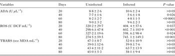
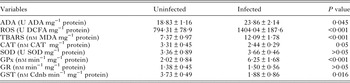
ROS levels in serum and liver homogenate are shown in Tables 1 and 2, respectively. Infected animals showed increased levels (P < 0·05) of ROS on days 20, 40, 60 and 80 PI in sera samples, as well as in liver samples on day 100 PI compared with uninfected control animals. Regarding TBARS, infected animals showed higher (P < 0·05) hepatic levels on day 100 PI compared with uninfected control animals. No differences were observed (P > 0·05) between groups regarding TBARS levels in serum on days 20, 40, 60 and 80 PI.
Adenosine deaminase (ADA) activity, reactive oxygen species (ROS), and thiobarbituric acid reactive species (TBARS) levels in serum of cattle experimentally infected by Fasciola hepatica on days 20, 40, 60 and 80 post-infection (PI)

Note: data represented by means ± s.e.
Adenosine deaminase (ADA) activity, reactive oxygen species (ROS) levels, thiobarbituric acid reactive substances (TBARS), catalase (CAT), superoxide dismutase (SOD), glutathione peroxidase (GPx), glutathione reductase (GR) and glutathione S-transferase (GST) activities in liver of cattle experimentally infected by Fasciola hepatica on day 100 post-infection (PI)

Note: all data represented as means ± s.e.
Infected animals showed lower (P < 0·05) CAT and GST activities, while hepatic GPx activity was higher (P < 0·05) on day 100 PI compared with uninfected control animals. No differences (P > 0·05) were observed between groups regarding hepatic SOD and GR activities on day 100 PI.
ADA activity in serum and hepatic tissue
Results of seric ADA activity are shown in Table 1. Infected animals showed lower ADA activity (P < 0·05) only on days 40 and 60 PI compared with the uninfected control (the group A). ADA results from hepatic samples are shown in Table 2. Infected animals showed the highest (P < 0·05) ADA activity on day 100 PI compared with the uninfected control.
DISCUSSION
The results of this study indicate that experimental fasciolosis in cattle is accompanied by changes in hepatic antioxidant systems, followed by increases on oxidative variables, as observed by Bottari et al. (Reference Bottari, Mendes, Lucca, Schwertz, Henker, Olsson, Piva, Sangoi, Campos, Moresco, Jaques and Da Silva2015), while studying rats experimentally infected by F. hepatica on days 20 and 150 PI. These authors demonstrated that oxidative stress is a significant feature of fasciolosis in rats, evidenced by increased levels of MDA, indicating lipid peroxidation. Likewise, Kolodziejczyk et al. (Reference Kolodziejczyk, Siemieniuk and Skrzydlewska2006) showed that in the liver of mice experimentally infected by F. hepatica, lipid peroxidation processes were evidenced by increased levels of MDA, followed by alterations in CAT and SOD activities. According to Fiss et al. (Reference Fiss, Adrien, Marcolongo-Pereira, Assis-Brasil, Sallis, Riet-Correa, Ruas and Schild2013), oxidative cell injury may occur during fasciolosis as a consequence of tissue destruction caused by toxic secretions produced by the larvae, as observed in the histopathological examinations of liver samples in this work that showed bile duct multifocal hyperplasia associated with moderate inflammatory infiltrate of eosinophils.
ROS levels have a great impact on the normal function of biomolecules such as proteins and cell membrane phospholipids. However, excessive production and/or inadequate removal of ROS results in destructive irreversible damage to the host cells (Lopaczyski and Zeisel, Reference Lopaczyski and Zeisel2001), along with imbalanced oxidant/antioxidant system (Khadija et al. Reference Khadija, Mohammed, Saad and Mohamed2009), as observed in the present study. Seric and hepatic ROS levels were increased in infected animals, as reported by Sarin et al. (Reference Sarin, Kumar, Prakash and Sharma1993). According to these authors, the presence of flukes in hepatic ducts causes chemical alterations in the hepatocytes, such as ROS production, which may react and damage biological membranes, contributing directly to the pathogenesis of parasitic infections (Kilic et al. Reference Kilic, Yazar, Saraymen and Ozbilge2003). Lipid peroxidation was identified by increased seric and hepatic MDA levels of infected animals, which reflected the severity of cellular damage. Lipid peroxidation in cattle infected by F. hepatica enhanced the oxidation of polyunsaturated fatty acids and caused lipid peroxidation, as reported by Kamel et al. (Reference Kamel, Sarhan and Saad2015) in humans. The increase of MDA and ROS levels occurred at similar periods in this experiment. This may occur because F. hepatica triggers the generation of ROS in cattle at the beginning of the infection, causing lipid peroxidation. Additionally, antioxidant enzymes decreased CAT and GST, and increased GPx during fasciolosis, contributing to oxidative damage and clinical signs of the disease.
SOD is considered the first line of defence against ROS, and is active on catalysing detoxification of superoxide radical, producing hydrogen peroxide (H202) (Shao et al. Reference Shao, Zhu, Dong, Wang, Wang, Xie, Zhang, Du and Zhu2012). In the present study, no difference was observed for SOD activity between groups, which could have contributed to the increase on seric and hepatic ROS levels in animals infected by F. hepatica. Although, a study conducted by Kamel et al. (Reference Kamel, Sarhan and Saad2015) demonstrated a different result, that SOD activity increased in patients with chronic fasciolosis in attempt to reduce increased levels of seric ROS. In the present study, the ROS levels significantly increased with concomitantly depletion of CAT activity. This can be explained since the antioxidant enzyme was unable to remove the excess of ROS in a timely manner, resulting on decreased cellular antioxidant activity beyond regulating levels, which finally influenced the synthesis of CAT or even caused enzymatic activity restrain (Lushchak, Reference Lushchak2011). Recently, a study conducted by Bottari et al. (Reference Bottari, Mendes, Lucca, Schwertz, Henker, Olsson, Piva, Sangoi, Campos, Moresco, Jaques and Da Silva2015) demonstrated that rats experimentally infected by F. hepatica showed lower total antioxidants levels in serum and liver compared with uninfected animals. This finding could have contributed to decrease CAT activity in infected cattle.
According to Halliwell and Gutteridge (Reference Halliwell and Gutteridge1999), the CAT and GPx represent a major cell pathway to metabolize H2O2. Therefore, its suppression leads to H2O2 accumulation up to toxic levels, as observed by Kamel et al. (Reference Kamel, Sarhan and Saad2015) in hepatic tissue of patients with chronic fasciolosis. However, in this study increased hepatic GPx activity was observed in infected animals by F. hepatica, probably due to compensatory mechanisms for reduced CAT activity. Depletion of CAT activity leads to H2O2 hepatic accumulation, which may have been converted into H2O and O2 through increased GPx activity. Thus, the increase in GPx activity is an attempt to reduce the accumulation of H2O2 and consequently, prevent or reduce hepatic damage. GST plays a key role in the detoxification system and can inhibit lipid peroxidation through ROS inactivation (Choi et al. Reference Choi, An and An2008). Decreased hepatic GST activity in animals infected by F. hepatica on day 100 PI may be due to GSH, as a substrate, since it was largely consumed, and many intermediate metabolites were produced in the process of detoxification, which could reduce GST activity or competitively inhibit GST substrates. Study conducted by Kolodziejczyk et al. (Reference Kolodziejczyk, Laszczynska, Masiuk, Grabowska and Skrzydlewska2015) demonstrated that rats experimentally infected by F. hepatica showed reduced GSH, a substrate for GST enzyme, corroborating to the hypothesis of decreased GST activity.
The reduction on seric ADA activity observed on days 40 and 60 PI could contribute to restrict the inflammatory response, and the subsequent cellular damage (Abbracchio and Ceruti, Reference Abbracchio and Ceruti2007), since it would increase serum adenosine levels, an anti-inflammatory molecule, a fact also observed by Baldissera et al. (Reference Baldissera, Bottari, Mendes, Schwertz, Lucca, Dalenogare, Bochi, Moresco, Morsch, Schetinger, Rech, Jaques and Da Silva2015) in rats experimentally infected by F. hepatica on days 20 and 150 PI. However, hepatic ADA activity increased at day 100 PI on infected cattle that contributed to the intense inflammatory process and oxidative damage due to the depletion of adenosine levels, an anti-inflammatory molecule, which was corroborated to the histopathological analyses of liver with intense inflammatory infiltrate.
Some minor limitations may have affected this study. Firstly, we have to mention that these animals were used in another study. Because of the above reason, we did not present a day zero as the starting point, what is reasonable if we take into account that all animals were clinical and haematological healthy at the beginning of the experiment.
In summary, we conclude that oxidative stress occurred in cattle experimentally infected by F. hepatica is mainly due to excessive ROS production in the course of fasciolosis. Increased ROS levels lead to lipid peroxidation that decreased CAT and GST activities, while GPx activity increased, contributing to the oxidative stress process in serum and liver, as well as hepatic damage, since increased ADA activity in the liver may contribute to the inflammatory process.
ETHICS COMMITTEE
The procedure was approved by the Animal Welfare Committee of the Instituto Federal Catarinense (IFC), under protocol number 001/2014.
ACKNOWLEDGEMENTS
Work supported by grant from the National Council for Scientific and Technological Development (CNPq), No. 487996/2013-5.
FINANCIAL SUPPORT
This research did not receive any specific grant from funding agencies.