The incidence rate of juvenile idiopathic arthritis ranges from 1.6–23 cases per 100,000 children annually, and systemic juvenile idiopathic arthritis accounts for 5%–15% of its subtypes.Reference Thierry, Fautrel, Lemelle and Guillemin1 Macrophage activation syndrome is a rare but life-threatening complication that can occur in systemic inflammatory disorders, including juvenile idiopathic arthritis.Reference Ravelli, Minoia and Davì2 We report the case of a 12-year-old girl with systemic juvenile idiopathic arthritis who had an intracardiac mass in the mitral valve, which was diagnosed as valvulitis associated with systemic rheumatic disease. Although she suffered from macrophage activation syndrome, she successfully underwent mass excision and mitral valve replacement after immunosuppressant therapy.
Case report
A 12-year-old girl visited another hospital due to chest discomfort and palpitation lasting 5 days without fever. Six years ago, she was diagnosed with systemic juvenile idiopathic arthritis and treated with steroid pulse therapy for associated macrophage activation syndrome. Four years ago, she underwent pericardiotomy for pericarditis with cardiac tamponade. She had recurrence of mild pericarditis and was treated with steroid pulse therapy 1 year prior. Over the past year, she continued to take prednisolone, azathioprine, naproxen, and aspirin. Considering her history of recurrent pericarditis, echocardiography was performed when she arrived at the hospital. Echocardiography revealed a mass-like lesion in the mitral valve and severe mitral regurgitation. Vancomycin and gentamicin were started as infective endocarditis was suspected. After 4 days of treatment, there was no change in mass size. She was transferred to our hospital for consideration of early surgical treatment.
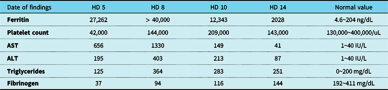
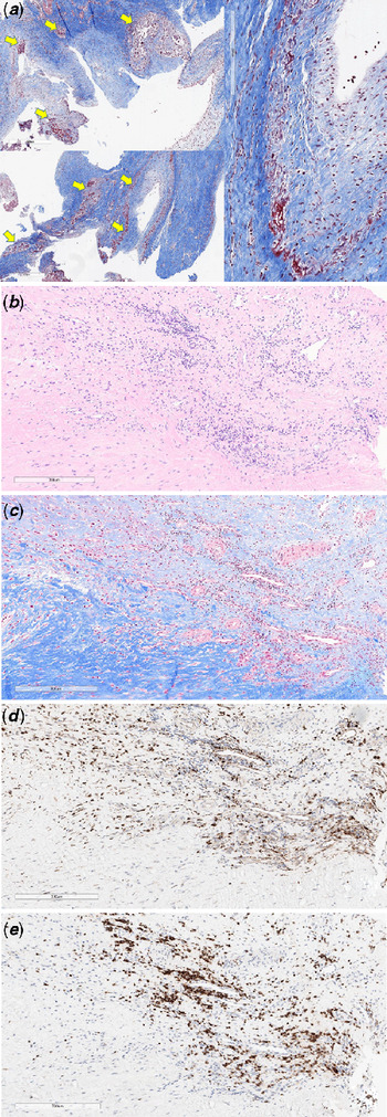
On admission to our hospital, her blood pressure, pulse rate, and body temperature were 102/66 mmHg, 126 beats per minute, and 36.5°C, respectively. A grade 2 to 3/6 systolic murmur was auscultated on the left lower sternal border with clear lung sounds. Laboratory tests revealed mild leukocytosis (11,150/uL) and normal C-reactive protein (0.98 mg/dL). Echocardiography showed an irregular mass (13.8 × 12.3 mm) on the anterior leaflet of the mitral valve and severe mitral regurgitation through the mass (Fig. 1A-B). We suspected infective endocarditis, continued to administer the same antibiotics, and planned urgent surgery for removal of the mass. On the 2nd day of admission, she developed fever and hypotension. We started continuous infusions of norepinephrine, epinephrine, dopamine, and vasopressin, but hypotension persisted. Intravenous immunoglobulin and meropenem were administered for possible septic shock. We could not rule out adrenal insufficiency, considering that she was on long-term steroid treatment; therefore, we decided to administer stress doses of hydrocortisone. However, on the 4th day of hospitalisation, fever and hypotension persisted. Microorganisms were not detected in the initial blood cultures performed in the previous hospital or in subsequent blood cultures performed in our hospital. Based on the results obtained on the 5th day after admission (Table 1), we suspected reactivated macrophage activation syndrome and started methylprednisolone pulse therapy, following which, the laboratory findings worsened (Table 1). Chest X-ray showed bilateral pleural effusion (Fig. 1C). Brain and abdominal CT revealed no evidence of embolic infarction. We discussed whether to delay surgery after the stabilisation of systemic inflammatory reaction, or to perform early surgical removal of the mass due to a risk of embolism. On the 9th day of hospitalisation, we decided to administer tocilizumab, a monoclonal antibody against the interleukin-6 receptor, to treat the cytokine storm before performing the surgery. After tocilizumab infusion, the laboratory findings improved (Table 1), and all inotropes were discontinued. She underwent delayed open-heart surgery on the 14th day of hospitalisation. Operative findings revealed that the mass involved almost the entire anterior leaflet of the mitral valve; therefore, the anterior leaflet was removed, and mitral valve replacement with On-X® 23 mm (On-X Life Technologies, Inc, Austin, Texas) was performed. Histopathology of the resected mitral valve showed granulomatous and lymphocytic inflammation, degeneration and calcification of collagen fibres, and vascular proliferation (Fig. 2). These findings resulted in the final diagnosis of valvulitis caused by systemic rheumatic disease. All antibiotics were discontinued on the 19th day of admission, because tissue culture revealed no microorganism isolation. After the surgery, she was administered warfarin and tocilizumab infusion every 2 weeks. She was discharged on the 33rd day of hospitalisation. At 6-month follow-up, chest radiograph revealed improved cardiomegaly and pleural effusion (Fig. 1D), and echocardiography showed good mechanical valve motion without significant paravalvular leakage or stenosis.
(A). Parasternal long-axis view reveals an irregular, multi-lobulated mass (arrow) on the anterior leaflet of the mitral valve. (B). Severe mitral valve regurgitation seen using apical four-chamber view. (C). The amount of pericardial effusion increases gradually on chest radiograph taken on the 6th day of hospitalisation. (D). Six months later, improved cardiomegaly and pleural effusion are seen on chest radiographs obtained at the outpatient clinic. AAo = ascending aorta; LA = left atrium; LV = left ventricle; RA = right atrium; RV = right ventricle.

Laboratory findings on days 5, 8, 10, and 14 after admission

AST = aspartate aminotransferase; ALT = alanine aminotransferase; HD = hospital day
HD 8 – the day after steroid pulse therapy
HD 10 – the day after tocilizumab infusion
HD 14 – the day of surgery
(A). Multifocal collagen necrosis and dystrophic calcification (yellow arrows). Note collagen degeneration begins at the margin of the fibrous core and continues to the loose connective tissue zone at the atrial side (atrialis). (B–E). Heavy inflammation at the atrialis part of the valve. Thickening of the leaflet is due to the fibrosis, vascular proliferation, and inflammatory infiltrates. Inflammatory cells are mostly T-cells, but histiocytes are also scattered. B, haematoxylin and eosin stain; C, Massonʼs trichrome stain; D, CD68 antibody stain, E, CD3 antibody stain.

Discussion
Juvenile idiopathic arthritis could involve multiple organs, including the heart, and valvular diseases may be associated with high-risk valve surgery and poor prognosis.Reference Koca, Sahin, Adrovic, Barut and Kasapcopur3 Systemic juvenile idiopathic arthritis may be accompanied by macrophage activation syndrome. One-third of patients with macrophage activation syndrome required treatment in the ICU, and the mortality rate was 8%.Reference Minoia, Davì and Horne4 Early treatment of macrophage activation syndrome is important to control the disease activity. However, reactivated autoimmune diseases may present with similar symptoms and signs to those seen in an acute infectious disease. Therefore, timely differential diagnosis and determination of treatment direction are challenging.Reference Lin, Yu and Lee5, Reference Parodi, Davì and Pringe6 Owing to the high risk of infection in patients treated with tocilizumab, the use of such biological agents in patients with autoimmune diseases without excluding severe infection requires careful differential diagnoses. The clinical features of macrophage activation syndrome include uncontrolled high fever, hepatomegaly, splenomegaly, lymphadenopathy, and haemorrhage, that could contribute to multiple-organ failures. It might be difficult to reach initial diagnoses based on clinical features only.Reference Ravelli, Magni-Manzoni and Pistorio7 Consequently, Ravelli et al. proposed diagnostic criteria for an early accurate diagnosis of macrophage activation syndrome in patients with systemic juvenile idiopathic arthritis.Reference Ravelli, Minoia and Davì2 These diagnostic criteria include ferritin >684 ng/ml and any two of the following: platelet count ≤181 × 109/L, aspartate aminotransferase >48 U/L, triglycerides >156 mg/dL, or fibrinogen ≤36 mg/dL.
An accurate differential diagnosis of an intracardiac mass, including vegetation, tumour, and thrombus detected by echocardiography alone is challenging.Reference Mazzeffi, Reich, Adams and Fischer8 In the case of a vegetation mass in patients with infective endocarditis, the echocardiographic findings show irregularly shaped mass with grey-scale echogenicity when compared to the normal myocardium. This vegetation mass is typically attached to the low-pressure side of the cardiac valves. In addition, it may also be found on the upstream side of the mitral and tricuspid valves; outflow tract side of the aortic and pulmonary valves; and along the leaflet coaptation zone.Reference Sedgwick and Scalia9 Other echocardiographic findings for the diagnosis of infective endocarditis include abscess or pseudoaneurysm and a new prosthetic cardiac valve dehiscence.Reference Habib, Lancellotti and Antunes10 Another example of an intracardiac mass is myxoma, which is one of the most common cardiac tumours. It often occurs in the left atrium, but also shows atypical features that provide potential limitations for differential diagnosis based on echocardiography alone. Cardiac rhabdomyomas, which are seen in children (< 4 years old) with tuberous sclerosis, often spontaneously regress and are detected by their bright echogenicity when compared to the surrounding myocardium.Reference Bruce11 Therefore, other imaging techniques, such as MRI and CT, may be required to make a differential diagnosis.Reference Caruso, Ventura, Viliani and Della Sala12 Cardiac MRI effectively distinguishes benign from malignant lesions, and the pathological findings are reported as a gold standard for the differential diagnosis of an intracardiac mass.Reference Mousavi, Cheezum and Aghayev13
Although the mass can sometimes be distinguished by its shape and location, the treatment direction should be determined based on additional evidence, including clinical symptoms, laboratory and pathology findings, and multimodality imaging studies.
Conclusion
Non-infective valvulitis mimicking infective endocarditis can occur in patients with systemic rheumatic diseases. Differential diagnosis of an intracardiac mass based on its detection on echocardiography is difficult but is important in determining a strategy for treatment. The patient’s medical history is vitally important and all efforts must be extended to obtain an accurate diagnosis through a multifaceted approach.
Acknowledgements
The authors wish to acknowledge Professor Kwang Nam Kim, Department of Pediatrics, Myongji Hospital, Kwandong University College of Medicine, Goyang, Korea. The authors appreciate Dr. Kwang Nam Kimʼs advice regarding the immunologic diagnosis and treatment of this patient.
Financial support
This research received no specific grant from any funding agency, or from commercial, or not-for-profit sectors.
Conflicts of interest
None.
Ethical Standards
The authors assert that all procedures contributing to this work comply with the ethical standards of the relevant national guidelines on human medical regulations and with the Helsinki Declaration of 1975, as revised in 2008, and has been approved by the Institutional Review Board (IRB) of Seoul National University Hospital (date of approval: September 26th, 2019, IRB number: H-1909-105-1066).