Highlights
-
• Vertigo in cerebellar infarction (CI) is predominantly associated with the involvement of the PICA or AICA territories.
-
• Cerebellar lesions can present with a spectrum of nystagmus and ocular motor dysfunction.
-
• Differentiating CI from acute unilateral vestibulopathy (AUVP) based solely on nystagmus can be challenging, so additional ocular motor tests can help.
Introduction
Cerebellar stroke is a common cause of vascular vestibular syndrome. Reference Lee1 Cerebellar stroke accounts for approximately 1.5% of all stroke etiologies. Reference Lee and Kim2 Cerebellar stroke typically results in severe ataxia, visual impairments, headache and vertigo. Reference Volgger and Gürkov3 The misdiagnosis rate of cerebellar stroke in brain CT scans ranges from 60% to 90%. Reference Lee and Kim2 Early MRI examinations (performed within <48 h of symptom onset) yield false-negative results in approximately 12% of cases. Reference Kattah, Talkad, Wang, Hsieh and Newman-Toker4 The National Institute of Health Stroke Scale is not as sensitive for detecting vertebrobasilar stroke as it is for strokes in the anterior circulation. Reference Elhfnawy, Abd El-Raouf, Volkmann, Fluri and Elsalamawy5 While vertigo attributed to cerebellar stroke is typically accompanied by other neurological symptoms or signs, approximately 10% of cerebellar stroke patients exhibit only dizziness and nausea, which may mimic acute peripheral vestibulopathy (pseudo-APV). Reference Lee1,Reference Lee, Sohn and Cho6,Reference Nelson and Viirre7 The infarction sites are often located in the medial branches of the posterior inferior cerebellar artery (mPICA), which supplies the nodulus and uvula of the cerebellum – crucial structures with strong connections to the ipsilateral vestibular nuclei. Reference Ogawa, Suzuki, Oishi, Kamei, Shigihara and Nomura8 These patients are frequently misdiagnosed, and even mild symptoms of ataxia may go unnoticed during the initial onset of the condition. Reference Ogawa, Suzuki, Oishi, Kamei, Shigihara and Nomura8 Cerebellar infarction (CI) in the territory of the anterior inferior cerebellar artery (AICA) can produce a broad spectrum of audiovestibular dysfunctions. Vertigo is usually associated with other neurological symptoms or signs, such as hearing loss, facial weakness, facial sensory loss, crossed sensory loss, Horner syndrome, gait ataxia and limb ataxia. Reference Amarenco, Rosengart, DeWitt, Pessin and Caplan9–Reference Lee, Kim and Chung11 A stroke involving the territory of the superior cerebellar artery (SCA) has a low incidence of vertigo, which may be a clinical clue to differentiate it from PICA or AICA CI with acute vertigo and limb ataxia. In clinical examinations, patients with cerebellar damage may also exhibit abnormalities in eye movements, including spontaneous nystagmus (SN), gaze-evoked nystagmus, saccadic hypometria or hypermetria, smooth pursuit (SP) abnormalities, abnormalities in ocular tilt reaction, impaired angular vestibulo-ocular reflex (aVOR) function and central positional nystagmus (CPN). Reference Shemesh and Zee12
This study aims to analyze the clinical manifestations of lesions in different vascular supply regions of the cerebellum, comparing the vestibular and oculomotor features of patients with CI and acute peripheral vestibulopathy (AUVP).
Methods
Patient selection
We retrospectively enrolled 23 patients (mean age ± SD = 59.9 ± 11.9, 17 men) with CI and 32 patients (mean age ± SD = 46.2 ± 12.3, 20 men) with acute unilateral vestibulopathy (AUVP) who visited either the emergency department or the outpatient clinic of the Department of Neurology in Aerospace Center Hospital and Xiangyang Central Hospital from March 2021 to October 2022. The inclusion criteria were (1) acute dizziness or vertigo, (2) symptom duration ≥10 min and (3) age ≥ 18 years old. The exclusion criteria were (1) Individuals with cognitive or mental impairments unable to complete the informed consent form; 2) patients with conditions such as encephalitis, cerebral hemorrhage, head trauma, intracranial masses, etc.; (3) internal medical conditions such as orthostatic hypotension, drug intoxication, arrhythmias, etc.; (4) benign paroxysmal positional vertigo (BPPV); (5) not cooperative in completing the brain MRI; and (6) incomplete data. Dizziness refers to a sensation of disturbed or impaired spatial orientation without a false or distorted sense of motion. It does not include vertiginous sensations and should be distinguished from a feeling of impending faintness (presyncope), confusion or detachment from reality. Vertigo refers to the sensation of self-motion when no actual motion is occurring or the sensation of distorted self-motion during normal head movement or the false sensation that the visual surround is spinning or flowing. Vertigo encompasses both false spinning sensations and other false sensations, such as swaying, tilting or bouncing. Reference Bisdorff, Von Brevern, Lempert and Newman-Toker13
The diagnosis of CI was confirmed by diffusion-weighted MRIs with an onset time of <7 days. We excluded patients with brainstem infarction. MRI axial sections confirmed the location of the infarct. Thirty-one consecutive patients were identified in this period. Eight patients had to be excluded because of a lack of essential laboratory tests (n = 6) or not pure CI (n = 2). We defined isolated acute vestibular syndrome (AVS) from CI as follows: (1) acute onset of vertigo or imbalance without accompanying neurological symptoms; (2) absence of general neurological signs, including altered mental status, facial palsy, limb weakness, sensory deficits, visual field abnormalities, limb ataxia/dysmetria, dysarthria, dysphonia, dysphagia, other cranial nerve abnormalities or distinct ocular motor signs such as ocular motor nerve palsies, internuclear ophthalmoplegia and gaze palsy; (3) confirmation of infarcts within the CI as shown by MRI imaging. Reference Choi, Kim and Choi14
The diagnosis of AUVP was made based on the criteria proposed by the Barany Society Reference Strupp, Bisdorff and Furman15 and onset time of spontaneous vertigo or dizziness <7 days. Forty-eight consecutive patients were identified in this period. Fourteen patients were excluded because of a lack of essential medical records or laboratory tests (n = 11) and with a prior history of vestibulopathy with Meniere’s disease (n = 2) or vestibular migraine (n = 1).
The study conformed to the tenets of the Declaration of Helsinki and was approved by the Aerospace Center Hospital (IRB no. 2022 (023)).
Clinical data collection
The medical records of patients diagnosed with CI or AUVP were thoroughly reviewed, including disease course, onset form, duration, symptoms, signs, risk factors for stroke history, ocular motor examination, caloric test and video head impulse test (vHIT, Interacoustics, Middelfart, Denmark). Subjects were not taking vestibular suppressants or had stopped taking them at least 48 h before undergoing vestibular and ocular motor tests.
Eye movements were binocularly recorded using video-oculography (VOG) equipment (Interacoustics, Middelfart, Denmark). SN was recorded both with and without visual fixation while the patients were in a seated position. Additionally, gaze-evoked nystagmus, horizontal SP, horizontal saccades, optokinetic response, positional test and horizontal head-shaking nystagmus were assessed. Head-shaking nystagmus: the intensity of head-shaking nystagmus was determined by deducting the slow-phase velocity of SN from the maximal slow-phase velocity of the induced head-shaking nystagmus. Head-shaking nystagmus was considered present when it exceeded the values: horizontal ≥ 3°/s, vertical ≥ 2°/s and torsional ≥ 2°/s, and when the nystagmus lasted for ≥5 s. Reference Huh and Kim16
The subjective visual vertical (SVV) test was assessed using VertiSVV (ZT-SVV-I, Shanghai ZEHNIT Medical Technology Co., Ltd., Shanghai, China). The head-upright SVV (HU-SVV) outside the range of −2.5° to + 2.5° was considered abnormal. Reference Sakagami, Wada, Shiozaki, Ota and Kitahara17 CPN was diagnosed only when a discernible nystagmus was induced by certain position changes. We categorized the CPN into three types: (1) vertical nystagmus on straight-head hanging, lying down or Dix-Hallpike maneuver to either side, (2) vertical nystagmus on up-righting from straight-head hanging and (3) geotropic or apogeotropic nystagmus during head turn to either side while supine. Reference Lee, Kim, Lee, Choi, Choi and Kim18
The vHIT was employed to evaluate the functionality of all six semicircular canals by measuring the gain of the vestibulo-ocular reflex (VOR). According to the manufacturer’s recommendations, normal gain values are anticipated to fall within the range of 0.80–1.20 for horizontal canals and 0.70–1.20 for vertical canals. Reference Li, Liu and Si19
Statistical analyses
All statistical analyses were conducted using SPSS software (version 20.0, IBM SPSS Statistics, N.Y., USA). Continuous variables were presented as mean ± SD for parametric values and median (range) for nonparametric ones. Categorical variables were expressed as percentages. The normality of the data was assessed using the Shapiro–Wilk test. The chi-square test was employed to compare the proportions of gender, lesion side, hypertension, diabetes mellitus, hyperlipidemia, coronary heart disease, atrial fibrillation, smoking and history of cerebral infarction between patients with CI and AUVP. A significance level of p < 0.05 was considered statistically significant.
Results
Demographic and clinical characteristics
The detailed clinical profiles of the patients are presented in Table 1. Patients with CI were older than those with AUVP (p < 0.001, see Table 1). CI patients exhibited a higher prevalence of hypertension, diabetes mellitus, hyperlipidemia and coronary heart disease, as well as more than three vascular risk factors compared to AUVP patients (all p < 0.05, see Table 1). However, there were no differences in gender distribution, disease duration, lesion side, history of smoking, history of atrial fibrillation and history of cerebral stroke (all p > 0.05, see Table 1).
Clinical characteristics of patients with acute unilateral vestibulopathy and cerebellar stroke

AUVP = acute unilateral vestibulopathy; CI = cerebellar infarction.
Characteristics of vestibular symptoms by vascular territory
Among CI cases, 56.5% (13/23) occurred in the PICA territory, 17.4% (4/23) in the SCA territory and 8.7% (2/23) in the AICA territory. Additionally, 8.7% (2/23) involved both SCA and PICA territories, 4.3% (1/23) involved both SCA and AICA territories and 4.3% (1/23) involved both PICA and AICA territories. Among CI patients, 43.5% (10/23) presented with isolated AVS. Among these cases, 80% (8/10) involved the PICA territory, 10.0% (1/10) involved the SCA territory and 10.0% (1/10) involved both PICA and SCA territories (Figure 1). Out of the 23 patients, 52.2% (12/23) experienced vertigo, while 47.8% (11/23) experienced dizziness. Among those with vertigo, seven cases occurred in the PICA territory, two in the AICA territory, one in both SCA and PICA territories, one in both SCA and AICA territories and one in both PICA and AICA territories. Regarding dizziness, six cases were in the PICA territory, four in the SCA territory and one in both SCA and PICA territories (Figure 2).
The ratio of isolated and non-isolated acute vestibular syndrome (AVS) resulting from damage in various cerebellar vascular territories. AICA = anterior inferior cerebellar artery; PICA = posterior inferior cerebellar artery; SCA = superior cerebellar artery.

The ratio of vertigo and dizziness resulting from damage in various cerebellar vascular territories. AICA = anterior inferior cerebellar artery; PICA = posterior inferior cerebellar artery; SCA = superior cerebellar artery.

Spontaneous nystagmus and saccadic intrusion and oscillations
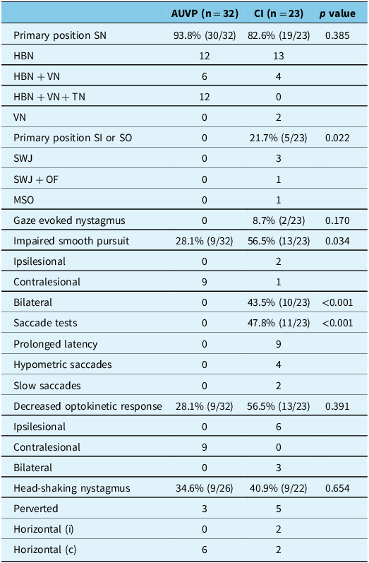
Of the 13 patients with unilateral PICA infarction, 11 (84.6%) exhibited SN. A horizontal component was observed in nine patients, primarily directed toward the side of the lesion (7/9, 77.8%). An upward vertical component was noted in one patient, while a downward component was observed in three patients. Among the two patients with unilateral AICA infarction, both showed SN. A horizontal component was observed in two patients, with half directed toward the lesion side (1/2, 50.0%). An upward vertical component was observed in one patient. Of the four patients with SCA infarction (three with unilateral SCA infarctions), three (3/4, 75.0%) exhibited SN. Among the three patients with unilateral SCA infarctions, two showed a horizontal component directed toward the lesion side (2/3, 66.7%). The patient with the remaining bilateral SCA infarction displayed spontaneous horizontal nystagmus. Of the two patients with combined PICA and SCA infarctions, one with right PICA and right SCA infarction showed SN beating to the right side. One patient with combined left PICA and left AICA infarctions displayed SN beating to the right side. One patient with combined left AICA and left SCA infarctions exhibited SN beating to the left side. Overall, 19 patients (82.6%) with CI exhibited SN: pure horizontal in 13 (ipsilateral horizontal in 9 and contralateral horizontal in 4), mixed horizontal-vertical in 4 (mixed ipsilateral horizontal-upbeat in 2, mixed ipsilateral horizontal-downbeat in 1, contralateral horizontal-downbeat in 1) and pure vertical in 2 (upbeat in 1 and downbeat in 1). Five patients with CI showed saccadic intrusion and oscillation (frequent square wave jerks in two, macrosaccadic oscillations in one and mixed frequent square wave jerks and ocular flutter in two). Thirty patients (93.8%) with AUVP exhibited SN: pure horizontal in 12 (ipsilateral horizontal in 1 and contralateral horizontal in 11), mixed contralateral horizontal-upbeat-contralateral torsional in 12 and mixed contralateral horizontal-upbeat in 6.
Positional nystagmus and head-shaking nystagmus
Ten patients with CI showed CPN: five of them showed vertical nystagmus while lying down or during the Dix-Hallpike maneuver to either side, and five of them exhibited apogeotropic nystagmus during head turns to either side while supine. Head-shaking nystagmus was noted in nine (9/22, 40.9%) CI subjects, ipsilesionally beating in two, contralesionally beating in two and down-beating in five. Head-shaking nystagmus was noted in nine (9/26, 34.6%) AUVP subjects, ipsilesionally beating in six, up-beating in two and down-beating in one (Table 2).
Characteristics of ocular motor tests in patients with acute unilateral vestibulopathy and cerebellar infarction

AUVP = acute unilateral vestibulopathy; c = contralesional; CI = cerebellar infarction; HBN = horizontal beating nystagmus; i = ipsilesional; MSO = macrosaccadic oscillations; OF = ocular flutter; SI = saccadic intrusion; SN = spontaneous nystagmus; SO = saccadic oscillation; SWJ = square wave jerks; TN = torsional nystagmus; VN = vertical nystagmus.
Gaze, saccade, smooth pursuit and optokinetic tests
Two patients (6.5%) with CI had horizontal gaze-evoked nystagmus (GEN). Horizontal saccades were abnormal in 11 patients (47.8%) with CI, with prolonged latency in 9, hypometria in 4 (contralesional hypometric in 3 and bilateral hypometric in 1) and slowness in 2. Vertical saccades were abnormal in nine patients (39.1%) with CI, with prolonged latency in nine, hypometria in three and slowness in two. All AUVP patients had normal horizontal and vertical saccades. In 13 patients (56.5%) with CI, horizontal SP was impaired, showing decreased gain in both directions in 10 cases and in one direction in 3 cases (ipsilesional side in 2 and contralesional side in 1). In nine patients (28.1%) with AUVP, horizontal SP was impaired, with decreased gain on the contralesional side in all cases (Figure 3). Primary saccadic intrusion and oscillation, abnormal SP and abnormal saccades were more prevalent in the CI group than in the AUVP group (all p < 0.05). No statistically significant differences were found between the groups regarding reduced optokinetic responses and head-shaking nystagmus (Table 2).
Comparison of smooth pursuit gain in patients with cerebellar infarction (CI) and acute unilateral vestibulopathy (AUVP).

Subjective visual vertical test
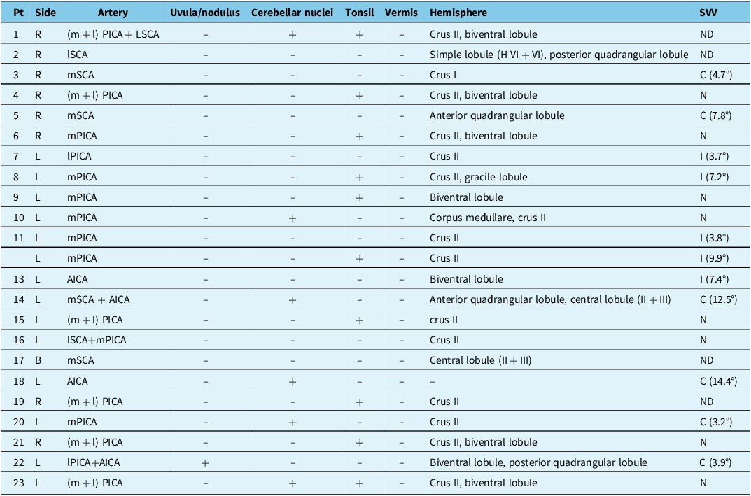
SVV tests were conducted on 19 patients with CI, resulting in an SVV range of −0.1° (−14.4°–9.9°). Among them, 26.3% (5/19) exhibited deviation toward the affected side, 31.6% (6/19) toward the unaffected side and 42.1% (8/19) fell within the normal range. In 30 patients with AUVP, the SVV range was 8.5° (−2.0°–27.7°), with 70.0% (21/30) showing deviation toward the affected side and 30.0% (9/30) falling within the normal range. The difference in SVV values between the two groups was statistically significant (Mann–Whitney U test, p < 0.001). The detailed relationship between lesion location and SVV deviation is shown in Table 3.
The detailed relationship between lesion location and SVV deviation

AICA = anterior inferior cerebellar artery; B = bilateral; C = contralesional; Crus I = superior semilunar lobule; Crus II = inferior semilunar lobule; I = ipsilesional; l = lateral; m = medial; ND = not done; N = normal; PICA = posterior inferior cerebellar artery; SCA = superior cerebellar artery; SVV = subjective visual vertical.
Caloric and head impulse tests
Seventeen patients with CI completed the caloric test, with one exhibiting contralesional horizontal canal paresis. Seventeen patients with CI underwent vHIT testing, indicating three patients showed unilateral vestibular dysfunction, involving the ipsilesional posterior canal in one, the contralesional posterior canal in one and both the contralesional horizontal and anterior canal in the remaining one case. Canal paresis on the caloric test was noted in all 32 patients with AUVP. In AVUP patients, impaired responses on vHIT toward the side of the lesion were noted in 25 out of 31 patients, involving the horizontal canal in 13 cases, both the horizontal and anterior canals in eight cases, the posterior canal in two cases, both the anterior and posterior canals in one case and all three canals in one case.
Discussion
Dizziness and vertigo are primary symptoms of posterior circulation stroke, especially when it involves the cerebellum, and it is typically the sole symptom. Reference Williams and Wilson20 In cases of acute dizziness and vertigo presentations, cerebellar lesions constitute roughly two-thirds of central lesions, comprising approximately 2.5%–6.5% of all acute cases. Reference Newman-Toker, Hsieh, Camargo, Pelletier, Butchy and Edlow21,Reference Kim, Kim and Kim22 The blood supply of the cerebellum comprises the PICA, derived from branches of the vertebral artery, and the AICA and the SCA, which originate from the basilar artery. Reference Wright, Huang, Strbian and Sundararajan23 PICA supplies the lateral medulla, the inferior cerebellum and the inferior cerebellar peduncle. The PICA provides blood supply to the lateral medulla, the inferior aspects of the cerebellar hemispheres, the choroid plexus and tela choroidea of the fourth ventricle, the tonsils, the inferior vermis and the inferior cerebellar peduncle. Reference Miao, Zhang, Wang, Jiao and Jiao24 In this study, the prevalence of infarctions in the PICA region is the highest. Specifically, the isolated AVS manifests predominantly in CIs within the PICA territory. These findings closely corroborate with previous research results. Reference Mehler25,Reference Kase, Norrving and Levine26 A study suggested that 3.7% of patients with isolated dizziness or vertigo had acute cerebellar strokes, with the lesions predominantly confined to the medial PICA vascular territory. Reference Perloff, Patel, Kase, Oza, Voetsch and Romero27 AICA supplies the inner ear, lower lateral pons, middle cerebellar peduncle and anteroinferior surface of the cerebellum, including the flocculus. Reference Fogwe, Sandhu, Goyal and Mesfin28 The labyrinthine artery, responsible for supplying the labyrinth and cochlea of the inner ear, along with the facial and vestibulocochlear nerves, typically originates as a branch of the AICA. Less frequently, it directly arises from the basilar artery. Reference Li, Zhang and Zhao29–Reference Ogawa, Suzuki, Takahashi, Akimoto, Kamei and Soma31 Therefore, patients with CI in the territory supplied by the AICA may present with symptoms resembling those of acute audiovestibulopathy. Reference Nam, Yoo and Lee32,Reference Kim33 In our study, vertigo is predominantly observed in patients with infarctions affecting PICA or AICA. The SCA supplies the pons, the superior half of the cerebellar hemispheres, the dentate nucleus, the vermis and the superior cerebellar peduncles. Reference Sarikaya, Steinlin, Manto and Huisman34,Reference Malicki, Szmyd and Bobeff35 Typical symptoms associated with infarction in the SCA vascular territory encompass dizziness, nausea, vomiting, tinnitus, headache, ataxia and dysarthria, with a relatively low incidence of vertigo. Reference Terao, Sobue, Izumi, Miura, Takeda and Mitsuma36 In our study, only dizziness was observed in four patients with lesions involving the SCA. This aligns with findings from a previous study, which documented that only 2 of the 14 patients with isolated CI in the medial branch territory of the SCA exhibited vertigo. Reference Ogawa, Suzuki, Oishi, Kamei, Shigihara and Nomura8 However, another study showed that nearly half of the patients complained of vertigo early during the SCA infarction. Reference Lee and Kim37
While SN usually beats toward the contralateral side in unilateral peripheral vestibulopathy, the patterns of SN can vary in central vestibular disorders. Reference Huh and Kim16 In our patients with a unilateral PICA or SCA infarction, the horizontal component of SN was predominantly directed toward the ipsilesional side. This aligns with findings from a previous study, which demonstrated ipsilesional SN in 11 out of 14 patients with a unilateral PICA infarction. Reference Huh and Kim16 One study has documented that all patients with an SCA infarction exhibit purely ipsilesional SN. Reference Lee and Kim37 Another study reported that roughly half of individuals with an SCA infarction exhibited predominantly ipsilesional SN. Reference Kase, Norrving and Levine26 The occurrence of ipsilesional SN in patients with unilateral CI can be attributed to damage to the vestibulocerebellum or its outflow tracts leading to the vestibular nuclei. The vestibulocerebellum receives a direct projection from the labyrinth and maintains robust connections with the vestibular nuclei. Reference Huh and Kim16 As the Purkinje fibers from the vestibulocerebellum typically exert an inhibitory effect on the ipsilateral vestibular nuclei, injury to this region or its outflow tracts is likely to elevate the tonic activity of the ipsilateral vestibular nuclei, leading to the manifestation of ipsilesional nystagmus. Reference Huh and Kim16
Among patients with CI, two patients exhibited frequent square wave jerks, one patient showed macrosaccadic oscillations and two patients with CI displayed a combination of frequent square wave jerks and ocular flutter. Square wave jerks are a common finding in healthy subjects, probably more so in the elderly. Reference Herishanu and Sharpe38–Reference Leigh and Zee40 Herishsanu and Sharpe documented a mean frequency of 4.7/min among young individuals and 27/min among the elderly. Reference Herishanu and Sharpe38 While square wave jerks can be observed in normal individuals, their frequent occurrence can serve as a significant indicator of central nervous system impairment. Reference Zachou, Armenis, Stamelos, Stratigakou-Polychronaki, Athanasopoulos and Anagnostou41 The physiopathologic mechanisms underlying square wave jerks are currently uncertain. A dysfunction in the brainstem oculomotor network is postulated, where the inhibitory effect of inhibitory burst neurons (IBNs) on omnipause neurons (OPNs) prevails over the usual inhibition of OPNs on IBNs. Following this, a brief burst of activity emerges in the excitatory burst neurons, generating a minor saccade. Subsequently, this gives rise to a slight retinal error detected in the superior colliculus, resulting in a second saccade in the opposite direction, thereby completing the square wave jerks. Reference Zachou, Armenis, Stamelos, Stratigakou-Polychronaki, Athanasopoulos and Anagnostou41,Reference Otero-Millan, Macknik, Serra, Leigh and Martinez-Conde42 The cerebellum, superior colliculus, basal ganglia and cortical regions are interconnected directly and indirectly in this network, thus influencing its functionality. Reference Anagnostou, Karavasilis and Potiri43 Furthermore, macrosaccadic oscillations occur in the presence of pontine or cerebellar lesions that affect either the OPNs or input from the fastigial nucleus to premotor burst neurons. Reference Choi, Choi, Choi and Choi44
In our study, two patients (6.5%) had horizontal GEN. One patient showed infarction involving the inferior cerebellar peduncle and dentate nucleus, while the other one showed infarctions involving the quadrangular lobule, central lobule (II + III), culmen and cerebellar nuclei. Structures of the cerebellar vermis, including the pyramid, uvula and tonsil, as well as portions of the biventer lobule and the inferior semilunar lobule, were indicated in previous studies to be implicated in the horizontal gaze-holding system. Reference Baier and Dieterich45 Additionally, there are few reported cases where the occurrence of GEN is associated with lesions in the rostral anterior cerebellum, including the ala of the central lobule and a portion of the quadrangular lobule, situated around the upper pons. Reference Wu, Ling and Zhao46 Thirteen patients with CI showed impaired horizontal SP in both directions in 10, to the lesion side in 2 and to the intact side in 1. Prior studies have indicated that SP may be impaired bilaterally in the presence of unilateral cerebellar lesions. Reference Lee, Kim, Lee, Choi, Choi and Kim18,Reference Sharpe47–Reference Lee, Park, Kim, Kim, Yunusov and Zee49 This could be clarified by an asymmetry in the tonic activity of the vestibular nuclei resulting in the decompensation of the entire SP system. Reference Baier, Stoeter and Dieterich48 However, in nine patients with AUVP, horizontal SP was impaired, all to the intact side. Therefore, bilateral SP impairment can serve as a characteristic for distinguishing between CI and AUVP.
In this study, 10 patients with CI had CPN: 5 of them showed vertical nystagmus while lying down or during the Dix-Hallpike maneuver to either side, and 5 of them exhibited apogeotropic nystagmus during head turns to either side while supine. CPN was more prevalent in lesions involving the cerebellum, followed by the fourth ventricle, isolated brainstem lesions and diffuse or nonspecific lesions. Reference Macdonald, Kaski, Saman, Al-Shaikh Sulaiman, Anwer and Bamiou50 In CPN, the lesions mostly overlapped in the vestibulocerebellum (nodulus, uvula and tonsil). According to the temporal profile of the nystagmus, CPN were divided into paroxysmal and persistent. Paroxysmal CPN was attributed to disinhibition and enhanced responses of the secondary neurons during positioning due to lesions involving the nodulus and uvula. Reference Choi, Kim, Kim, Glasauer and Kim51,Reference Ling, Kim, Lee, Choi, Yang and Kim52 Persistent CPN has been explained by erroneous neural processing within the velocity-storage circuit that functions in estimating the angular head velocity, gravity direction and inertia. Reference Choi, Glasauer, Kim, Zee and Kim53 Patients with AUVP presenting with SN with a vertical component may exhibit varying degrees of vertical component change during positional testing. This makes it challenging to definitively diagnose positional nystagmus as CPN. The differentiation of CPN becomes more conclusive when accompanied by additional neurological signs or positional nystagmus induced in multiple planes. CPN is often ruled out if the vHIT indicates damage to the vertical semicircular canal without other neurological deficits. Furthermore, it is crucial to differentiate between CPN and horizontal semicircular canal BPPV. CPN is typically distinguished if repeated manual maneuvers for BPPV are ineffective or if it is associated with other central signs. Reference Choi, Kim, Kim, Glasauer and Kim51,Reference Choi, Glasauer, Kim, Zee and Kim53
Studies have suggested that the deviation of the SVV varies depending on the cerebellar structures involved. Reference Feng, Zhao and Wu54 Lesions involving the nodulus/uvula and dentate nucleus may result in contraversive OTR/SVV tilt due to a loss of inhibition over the ipsilesional vestibular nuclei. This loss of inhibition can cause an increase in tonic resting activity in the ipsilesional VN. This could thus induce contraversive OTR/SVV tilt. Reference Baier, Bense and Dieterich55,Reference Choi, Lee, Kim and Kim56 However, ipsiversive OTR/SVV tilt deviations were observed when lesions involved the biventral lobule and inferior semilunar lobule. Reference Feng, Zhao and Wu54 This may be due to the disruption of inhibitory GABAergic efferents from the cerebellar cortex, leading to enhanced activity in the intact dentate nucleus and, consequently, a decrease in tonic resting activity in the vestibular nucleus. Reference Baier, Bense and Dieterich55 In our study, among the six patients with cerebellar infarcts displaying contraversive SVV, one had lesions in the unilateral nodulus, and three had lesions in the dentate nucleus. This supports the hypothesis that lesions affecting the dentate nucleus or nodulus may disinhibit the ipsilesional vestibular nuclear complex, resulting in contraversive OTR/SVV tilt. Reference Baier, Bense and Dieterich55,Reference Choi, Lee, Kim and Kim56 The remaining two patients with contraversive SVV showed infarctions involving the medial SCA territory, one in the superior semilunar lobule and the other in the anterior quadrangular lobule. In the case of isolated superior semilunar lobule or anterior quadrangular lobule damage causing OTR/SVV tilt abnormalities, there are relatively few reports, and the underlying mechanisms leading to abnormal SVV tilt need further investigation. Of the five patients with CI showing ipsiversive SVV tilt in our study, four had lesions in the PICA, mostly involving the tonsils, biventer lobule and inferior semilunar lobule, and one had a lesion of the biventer lobule involving the AICA territory. The ipsiversive SVV tilt may be due to the disruption of the inhibitory efferents from the cerebellar cortex, which may lead to an enhanced activity of the intact dentate nucleus. Reference Choi, Lee, Kim and Kim56
In clinical practice, the HINTS examination (head impulse, nystagmus, test of skew) and the HINTS-Plus assessment, which incorporates hearing loss evaluation, are reliable bedside tools for differentiating central lesions from AUVP. Reference Kattah, Talkad, Wang, Hsieh and Newman-Toker4,Reference Newman-Toker, Kerber and Hsieh57 However, the diagnostic accuracy of these methods depends significantly on the operator’s expertise and skill. False negatives can occur during the head impulse test when VOR impairment is subtle. Reference Jorns-Häderli, Straumann and Palla58–Reference Machner, Erber, Choi, Trillenberg, Sprenger and Helmchen60 To address these limitations, VOG-assisted HINTS has been demonstrated to outperform the bedside HINTS examination, offering improved diagnostic precision. Reference Korda, Wimmer and Zamaro61 This study indicates various ocular motor signs that can assist in differentiating CI from AUVP, including saccadic intrusions and oscillations, abnormal saccades and impaired bilateral SP. The reliability of these ocular motor evaluations, however, is also dependent on the clinician’s experience and proficiency. Utilizing video recordings to document and analyze these abnormalities can improve the consistency and standardization of assessments. The development and increased use of portable bedside video-oculography in clinical practice would greatly enhance the ability to distinguish between CI and AUVP, thereby improving diagnostic accuracy and reliability.
In conclusion, our study demonstrated that vertigo is mainly seen in PICA or AICA infarctions. SCA lesions mostly cause dizziness. There is significant overlap in the eye movement abnormalities observed in both AUVP and CI, with saccadic intrusion and oscillation, abnormal bilateral SP and abnormal saccades contributing to the distinction between the two conditions. Moreover, SVV deviation varies depending on the cerebellar structures involved.
Author contributions
Ling X and Chang LY acquired and analyzed the data and wrote the manuscript. Feng YF, Wu YX, Xu Y, Wang JR, Zhao GP, Kim JS, Yang X and Wang ZX analyzed and interpreted the data and revised the manuscript. Wang ZX and Yang X designed and conceptualized the study, interpreted the data and revised the manuscript.
Funding statement
This study was supported by the Peking University First Hospital (no. 2023SF14).
Competing interests
None.
Disclosures
Drs. X. Ling, L.Y. Chang, Y.F. Feng, Y.X. Wu, Y. Xu, J.R. Wang, G.P. Zhao, X. Yang and Z.X. Wang report no disclosures relevant to the manuscript. Dr J.S. Kim serves as an associate editor of Frontiers in Neuro-otology and on the editorial boards of the Journal of Clinical Neurology, Frontiers in Neuro-ophthalmology, Journal of Neuro-ophthalmology, Journal of Vestibular Research, Journal of Neurology and Medicine.