
Introduction
Folic acid (FA) is an important water-soluble vitamin B and is responsible for several functions in the human body. Since it cannot be synthesized by the human body, it is generally provided through the diet. Vitamin B occurs naturally as folate in vegetables, fruits, and meat, for examples, spinach, broccoli, bananas, lemons, beef liver, and kidney.[Reference Moll and Davis1,Reference Green, Allen, Bjørke-Monsen, Brito, Guéant, Miller, Molloy, Nexo, Stabler, Toh, Ueland and Yajnik2] The federal law has made FA mandatory to be included in cereals, bread, and flour.[Reference Liu, Hu and Su3] FA promotes the formation of red blood cells from bone marrow and lack of FA could possibly result in anemia. Pregnant women require an adequate quantity of FA to avoid abnormal growth of the baby's nerves. Furthermore, low levels of FA are also linked with cancer.[Reference Duthie4] This biomolecule also plays an important role in physiological functions such as synthesis and repair of nucleotides (DNA and RNA) and prevention of cerebral folate deficiency.[Reference Chakravarty, Dutta, Kalita and Sarma5] This rare disease occurs due to lack of FA in the brain, which can lead to lesions in the cranial nerve resulting in compromised metabolism and possibly death. Therefore, it is important to develop accurate analytical techniques for the detection of FA. Analytical techniques, such as high-performance liquid chromatography, electrochemical method, and chemiluminescence,[Reference Duthie4,Reference Póo-Prieto, Haytowitz, Holden, Rogers, Choumenkovitch, Jacques and Selhub6,Reference Chekin, Teodorescu, Coffinier, Pan, Barras, Boukherroub and Szunerits7] have been utilized for the detection of FA. However, some of them lack sensitivity at low concentrations and involved tedious sample preparation processes. Since most cases require the detection of FA at very low concentrations, the demand for detection techniques offering high selectivity at low concentrations has increased. Fluorometric methods have emerged as alternatives for selective estimation of such biomolecules as it offers a high sensitivity over very low concentration ranges, excellent selectivity, simplicity of operation, and fast detection.[Reference Manzoori, Jouyban, Amjadi and Soleymani8,Reference Yu, Cui, Chen, Gao, Wei and Ding9]
Quantum dots (QDs) have become an attractive fluorescent probe over the past decades due to their size-tunable emission properties, narrow emission spectra, and high-photoluminescence quantum yields (PLQY). They have been explored for the detection of various biomolecules including FA with high sensitivity and selectivity. The sensing mechanism generally involves the quenching of fluorescence of the QDs by the analyte. For instance, Geszke-Moritz et al.[Reference Geszke-Moritz, Clavier, Lulek and Schneider10] hydrothermally synthesized Cu–ZnS/ZnS and Mn–ZnS/ZnS core/shell QDs for the detection of FA. The addition of FA quenched the fluorescence of the QDs linearly in 10–100 µM. Li et al.[Reference Li and Chen11] reported a fluorescent magnetic nanocomposite consisting of Mn–ZnS QDs and Fe3O4 nanoparticles wrapped in SiO2. FA was reported to quench the fluorescence of the nanocomposite due to the electron transfer from the composite to the FA. The linear plot of fluorescence quenching against the FA concentration was obtained in the range of 0.23–1.13 µM with the limit of detection (LOD) of 21.7 nM. In attempts to improve selectivity, Ensafi et al.[Reference Ensafi, Nasr-Esfahani and Rezaei12] modified thioglycolic acid-capped CdTe QDs by capping them with molecularly imprinted polymers (MIPs) such as 3-aminopropyltriethoxysilane using tetraethoxysilane as a crosslinker. FA was found to quench the QD-MIPs fluorescence linearly between 0.5 and 20 µM concentrations with the LOD of 42 nM. The fluorescence quenching was attributed to the possible energy transfer from QD-MIPs to the analyte. Chakravarty et al.[Reference Chakravarty, Dutta, Kalita and Sarma5] fabricated a polyvinyl alcohol-based CdTe hybrid sensor for the detection of FA with the LOD of 1.3 pM. The fluorescence quenching of the hybrid sensor after FA addition was attributed to intramolecular charge transfer, which accounts for photoluminescence (PL) quenching.
Indeed, the studies so far show that the interaction between the QDs fluorescent probes and FA has led to the detection of FA through PL quenching. However, fluorescence intensity is easily affected by instrument conditions and environment, and thus, it is unstable. In this study, we demonstrated for the first time, the detection of FA based on the shift in the emission wavelength of the AgInS2 (AIS) QDs probe after the addition of FA. Since wavelength variations are rarely affected by interferences such as background noise, changes in the PL peak positions should exhibit better accuracy compared to changes in PL intensity. The synthesis of AIS QDs was based on our previous publication,[Reference May, Parani and Oluwafemi13] which involved the use of water-soluble precursors, low temperatures, and short reaction times. These ternary chalcopyrite QDs are much safer as they contain nontoxic elements and exhibit good biocompatibility compared to Cd-based QDs.[Reference Kang, Yang, Huang, Tao, Wang and Pan14] FA detection based on fluorescence quenching by utilizing mercaptopropionic acid (MPA)-capped ternary CuInS2 QD–ascorbic acid (AA) complex was previously reported by Liu et al.[Reference Liu, Hu and Su3] However, the synthesis involved high temperatures, longer reaction times, and longer detection times. Here, we report direct detection of FA via a hypsochromic (blue) shift of the emission peak of the QDs within a short period of time (30 s). The QDs were found to be sensitive and selective towards FA compared to other biomolecules. In addition, the FA was also detected in the presence of a mixture of many biomolecules.
Experimental
Materials
Silver nitrate (AgNO3, ≥99.0%), indium chloride (InCl3, 99.9%), sodium citrate dihydrate (C6H3Na3O7·2H2O, A.R.), thioglycolic (99%), sodium sulfide (Na2S, 98%), and sodium hydroxide (NaOH, 96%) were purchased from Sigma-Aldrich, South Africa. All the chemicals were used as received without further purification. Deionized water was used throughout the synthesis and analyses.
Synthesis of AgInS2 QDs
The synthesis was carried out according to our previous communication.[Reference May, Parani and Oluwafemi13] In a typical reaction, 0.0106 g (0.0625 mmol) of AgNO3, 0.055 g (0.25 mmol) of InCl3, and 0.2941 g (1 mmol) of sodium citrate were dissolved in a sequence in 88.4 mL of deionized water under stirring. Then, 22 µL (0.3 mmol) of thioglycolic acid was added to the solution, followed by the addition of 11.6 mL (0.406 mmol) of the Na2S solution. The mixture was refluxed at 95 °C for 3 h under stirring. The QDs obtained were purified by precipitation using ethanol followed by centrifugation.
Characterization
An Agilent Cary 50 ultraviolet–visible spectrophotometer was used for absorption measurement between a wavelength range of 200–900 nm. The measurements were carried out using a quartz cuvette (1 cm path length) at room temperature. The emission spectra were recorded at the excitation wavelength (λ exc) of 475 nm, using Shimadzu spectrophotometer with a Xenon lamp at room temperature and a 10 mm path length quartz cuvette. The surface chemistry of the as-synthesized QDs was investigated using a Spectrum Two Perkin Elmer Fourier transform infrared (FTIR) instrument at room temperature and fitted with an Attenuated total reflection sample holder. A high-resolution transmission electron microscope (HRTEM) image was recorded with a JEOL electron microscope (JEM-2100) at an accelerating voltage of 200 kV. The sample was prepared by placing a drop of the sample dilute solution on a copper grid and allowed it to dry completely at room temperature before being loaded into the microscope. The x-ray diffraction (XRD) measurements were performed on a D8 Focus diffractometer (Bruker) with Cu Kα radiation (λ = 0.15405 nm).
Detection of FA and interference studies
Fluorescence sensitivity of the QDs towards FA was carried out on a PL spectrophotometer. 1 mL of different concentrations of FA solution was added to 2 mL of the AIS QDs solution and analyzed after 30 s. The LOD was determined using the formula LOD = (3σ)/k, where σ is the standard deviation of the blank (QDs) and k is the slope. Fluorescence sensitivity of the QDs towards other biologically important molecules such as AA, citric acid, l-cysteine, glucose, and potassium sodium tartrate was also carried out at the same concentration (16.6 µM) in a similar manner for the selectivity studies. Interference studies were carried out by obtaining a mixture of these molecules with AIS QDs.
Results and discussion
Optical and structural properties
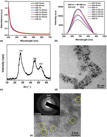
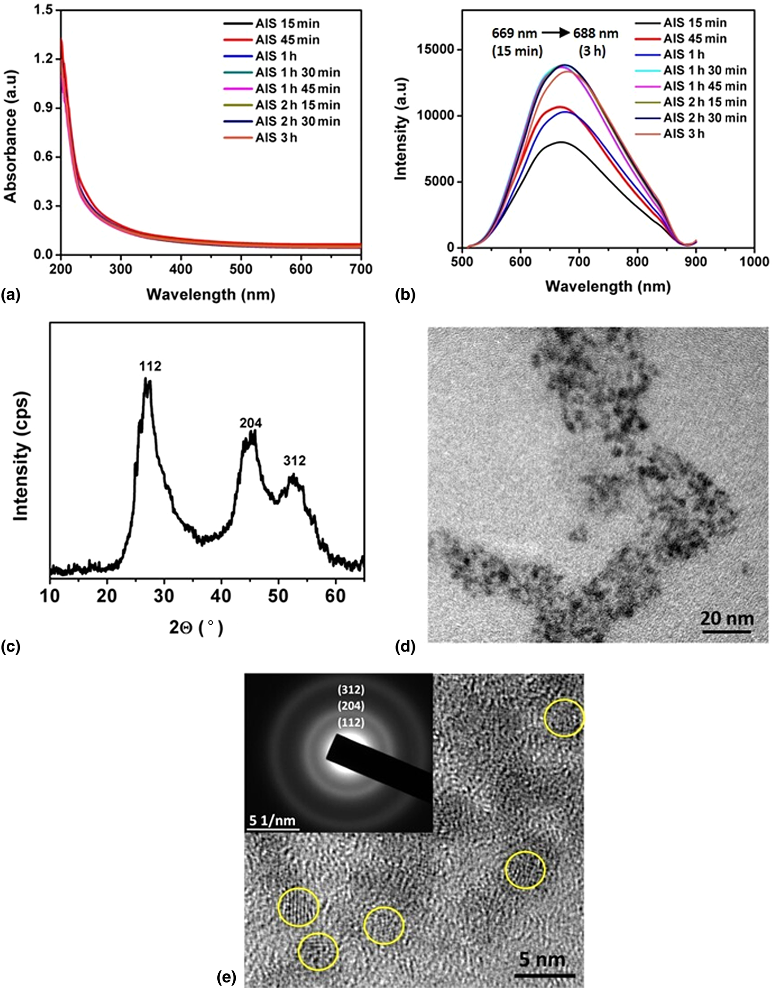
The mixture of Ag/In/stabilizers resulted in a colorless solution. The addition of Na2S revealed a sudden color change from colorless to yellow, indicating the formation of QDs. The temperature was increased to 95 °C and aliquots were taken at different reaction times to monitor the growth of the AIS QDs. Figures 1(a) and 1(b) shows absorption and PL spectra of the as-synthesized AIS QDs, respectively. There are no obvious excitonic peaks detected in the absorbance spectra as reported in previous studies[Reference Mir, Radhakrishanan, Rawat, Prasad and Bohidar15,Reference Chen, Ahmadiantehrani, Zhao, Zhu, Mamalis and Zhu16] , but a broad emission peak, a characteristic of ternary QDs, is observed within 15 min. As the reaction continues, the emission intensity increased and started decreasing at 3 h while the peak position gradually red-shifted from 669 to 688 nm indicating the growth of the QDs. The XRD patterns of the QDs consisted of three major peaks at 2θ values of 27.0°, 45.1°, and 52.6°, corresponding to the (112), (204), and (312) indices which confirmed a tetragonal crystal structure [Fig. 1(c)]. The TEM images show that the QDs are spherical in shape with an average particle diameter of ~3.1 nm [Figs. 1(d) and 1(e)]. Furthermore, the selected area electron diffraction [SAED, Fig 1(e), inset], the pattern of the QDs shows the ring pattern which can be correlated to XRD peaks.
Temporal evolution of (a) absorption spectra, (b) PL spectra, (c) XRD patterns, (d) TEM image, (e) HRTEM image of AgInS2 QDs. Inset: corresponding SAED patterns.
FA detection
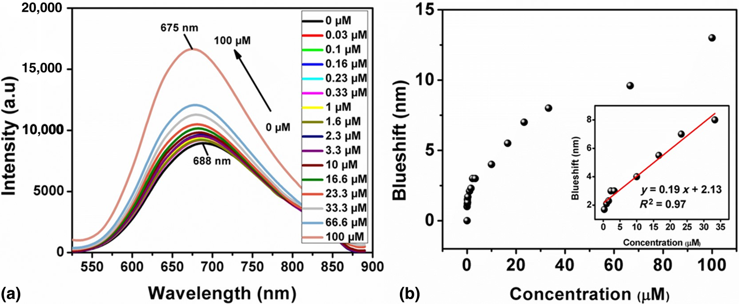
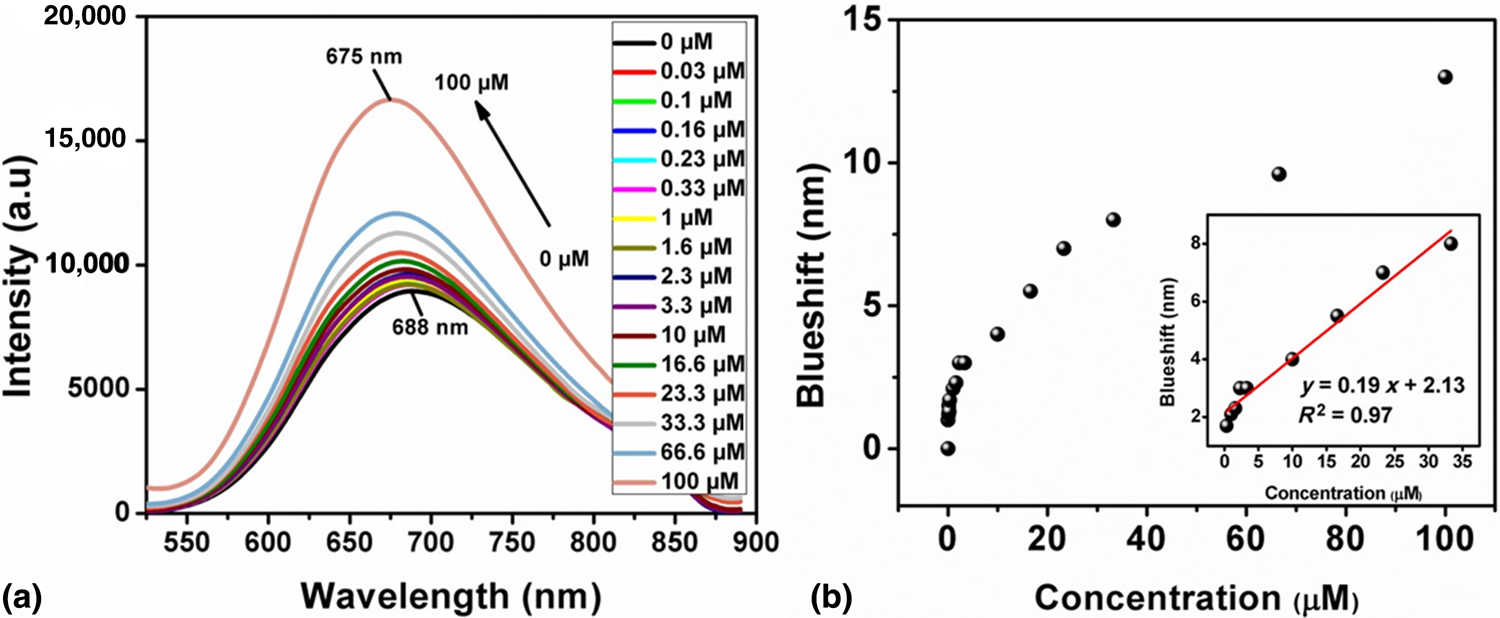
Figure 2(a) shows the PL spectra of AIS QDs after the addition of different concentrations of FA within 30 s reaction time. The results revealed continuous blueshift in the emission peak position from 688 to 675 nm with an increase in intensity as the FA concentration increased from 0 to 100 µM. A linear plot was obtained in the concentration range from 0.03 to 33 µM with the regression coefficient R 2 of 0.97. Such a long linear range of emission against FA concentration is highly advantageous for FA detection. The LOD of FA using AIS QDs as the fluorescent probe was calculated to be 52 nM. Compared to many other fluorescent probes that have been reported in the literature,[Reference Chakravarty, Dutta, Kalita and Sarma5,Reference Yu, Cui, Chen, Gao, Wei and Ding9–Reference Ensafi, Nasr-Esfahani and Rezaei12] the present AIS QDs probe composed of less toxic elements and exhibited excellent LOD towards FA.
(a) Effect of FA on the PL spectra of AgInS2 QDs, (b) plot of concentration of FA and blueshift in the PL peak position of AIS QDs. Inset: corresponding linear range.
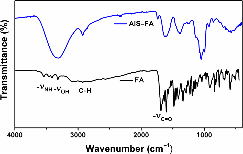
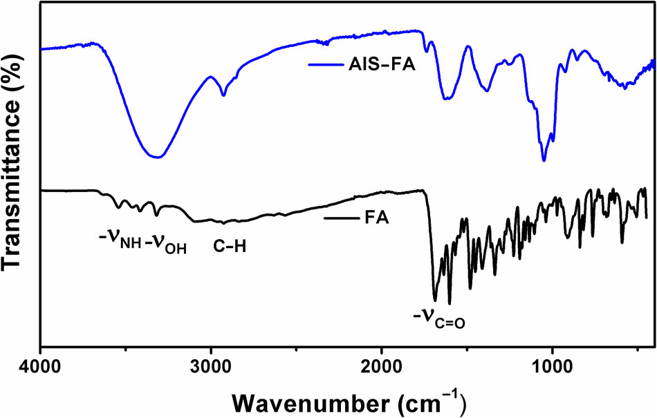
Literature studies have demonstrated that the blueshift with the increase in the emission intensity of ternary QDs is generally observed when there is a decrease in Ag:In precursors ratio.[Reference Chen, Ahmadiantehrani, Zhao, Zhu, Mamalis and Zhu16–Reference Raevskaya, Ivanchenko, Skoryk and Stroyuk18] FA is known to form an immediate complex with Ag and hence the blueshift in the emission can be attributed to the removal of surface Ag atoms by FA followed by the decrease in Ag content of the AIS QDs.[Reference Hekmatimoghaddam, Jebali and Dargahi19–Reference Su, Yang, Xia, Zhang, Chai, Wang and Qu21] In addition, excess FA eliminates the defects by satisfying the dangling bonds at the surface with its multifunctional groups thus increasing the PL intensity. Both the removal of Ag and capping the QDs by FA were simultaneous; hence, the blueshift and increase in the PL intensity were observed. Figure 3 shows the FTIR spectra of AIS QDs, FA, and AIS–FA mixture. FA has amine and carboxylic acids as the main functional groups. The characteristic –NH2 stretching of the FA was observed at 3415 and 3544 cm−1 which disappeared in the mixture of AIS–FA. This might be attributed to the formation of metal (possibly Ag)–FA complex at the surface via amine molecules of FA.
FTIR spectra of the FA and AIS QDs–FA mixture.
Selectivity study
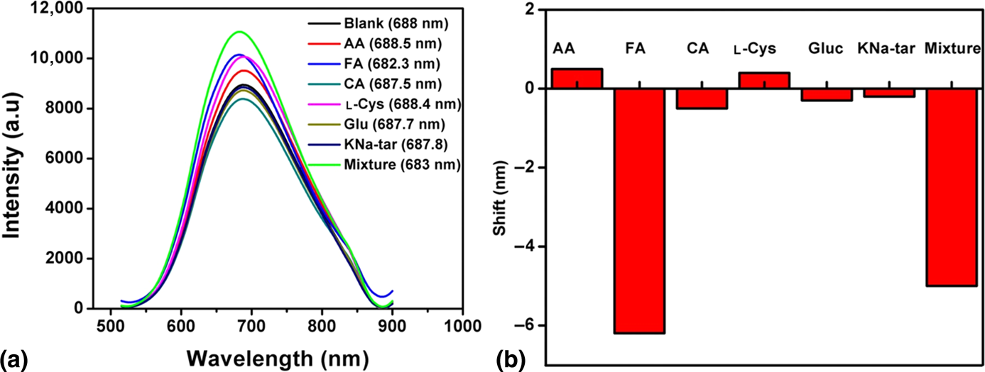
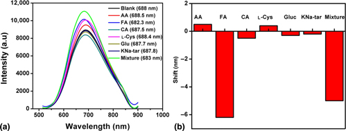
To demonstrate the selectivity of QDs towards FA, different biomolecules at the concentration of 16.6 µM were added to the as-prepared AIS QDs solution and the PL spectra were recorded after 30 s (Fig. 4). The results show that the addition of FA to AIS QDs revealed huge blue-shifted emission while a little red- or blue-shifted emission was observed for all the other biomolecules. To investigate the effect of the presence of other amino acids when detecting FA, an interference study was carried out by adding a mixture of all the amino acids to the QDs. The result shows reduced blue-shifted emission which is similar to the result obtained after the addition of only FA to the QDs. This indicates the ability of the as-synthesized QDs to detect FA in the midst of other amino acids. Hence, it can be used in the development of FA sensor.
(a) PL spectra of AIS QDs after the addition of different biomolecules. Emission maximum is given in brackets. (b) Bar graph showing the wavelength shift in the PL spectra of AIS QDs with the addition of biomolecules. AA, ascorbic acid; FA, folic acid; CA, citric acid; l-Cys, l-Cysteine; Glu, glucose; KNa-tar, potassium sodium tartrate.

Conclusions
Water-soluble, fluorescent AgInS2 QDs was synthesized via a simple method and it was used as a fluorescent probe for the detection of FA. The fluorescence sensitivity of AIS QDs towards FA resulted in blueshifting of the emission position of QDs. A linear plot of the blueshift in the PL wavelength position against FA concentration was obtained in the range of 0.03–33 µM with the detection limit of 52 nM. The AIS QDs demonstrated high selectivity towards FA while interference studies indicated that the QDs could detect FA in the midst of other biomolecules. Thus, AIS-based QDs can be used for the development of a simple and nontoxic FA sensor.
Acknowledgments
The authors would like to thank UJ-Global Excellence Stature, National Research Foundation (N.R.F.) under Scarce Skills and Innovation scholarship, Competitive Programme for Rated Researchers (CPRR), Grant No. 106060, and University of Johannesburg research committee (URC) and Faculty of Science research committee (FRC) for financial support.