Introduction
Suspected fish bones in the upper aerodigestive tract are a common otolaryngological presentation. The majority of these are managed simply, although rarely they can lead to severe complications including retropharyngeal abscess, oesophageal perforation and mediastinitis.Reference Venkatesh and Venkatanarasimha Karaddi1
Patients often present with pain and discomfort after a history of eating fish. Traditional investigation of such patients involves clinical examination, including flexible nasendoscopy, in combination with a lateral soft tissue radiograph of the neck. Management can be conservative, in the absence of an impacted fish bone, or consist of removal under local or general anaesthesia, with the addition of more detailed radiological investigations, including computed tomography (CT), in a select number of patients.
At present, there are no clear guidelines for the management of patients presenting with impacted fish bones in the upper aerodigestive tract. There are conflicting views on the usefulness of plain radiographs in the management of such cases, especially given the poor bone radio-opacity of many fish species.Reference Ell and Parker2
In order to clarify the usefulness of plain radiographs in the management of patients presenting with retained fish bones, we undertook an audit of clinical practice. A pragmatic treatment algorithm was subsequently constructed, to aid in the future management of such patients.
Materials and methods
Ethical considerations
A retrospective study was undertaken, registered and approved by the University Hospital Southampton NHS Foundation Trust audit and clinical research department.
Study design
Suitable patients were identified by searching all electronic patient records, using the terms ‘fishbone’ or ‘fish bone’, between April 2008 and February 2018. This yielded 140 patients, of which 86 were included, with the remainder excluded because of the non-availability of notes. Eligible patients presented via the emergency department or their general practitioner. Data were anonymised throughout.
Study parameters included patient age, gender, relevant investigations, foreign body location (if found) and means of retrieval. Any planned follow up, as well as emergency re-admission rates, were also analysed for these patients. Lateral neck radiographs were reviewed by the admitting doctor, as detailed in the medical notes, and checked by the study authors for accuracy (TPT and TCB).
Results
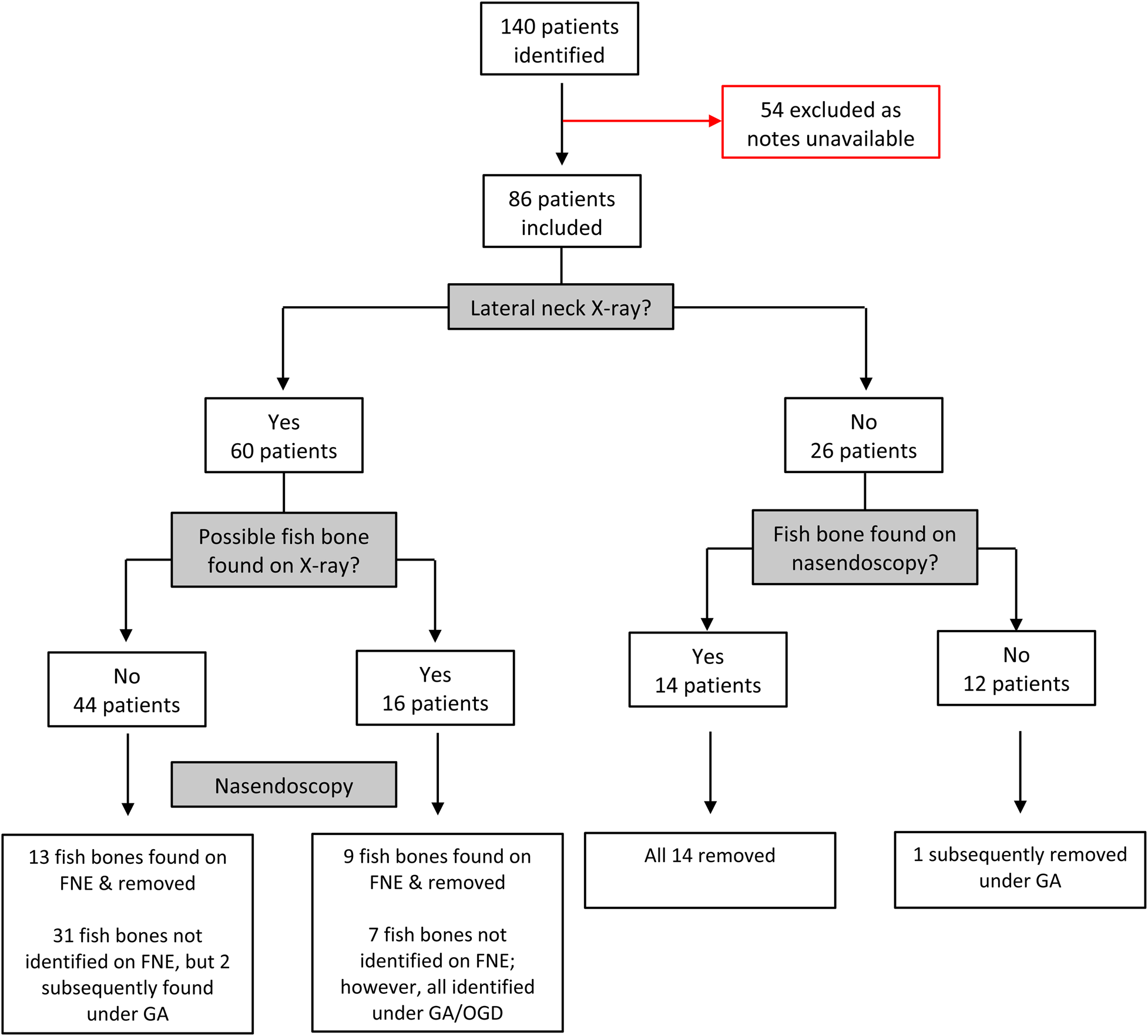
Mean patient age was 46 years, with a male to female ratio of 45:55. During admission, 60 patients (70 per cent) underwent lateral soft tissue radiographs. Of the 60 radiographs performed, 16 had features suggestive of fish bone impaction (26.7 per cent), and all these patients were subsequently found to have a fish bone present, although some required a general anaesthetic for identification and removal. Of the 44 patients with a normal radiograph, 15 subsequently had fish bones removed under local or general anaesthesia (34 per cent). These results have been summarised in Figure 1.
Flow diagram of patients presenting with suspected fish bone impaction. FNE = flexible nasendoscopy; GA = general anaesthesia; OGD = oesophagogastroduodenoscopy

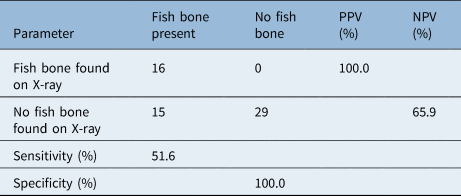
Table 1 highlights the predictive values of the radiograph results compared to the presence of fish bones found on clinical examination. The sensitivity of radiographs in the detection of fish bones was found to be 51.6 per cent. The specificity was significantly higher, at 100 per cent.
Lateral neck radiograph predictive values

Data represent numbers of cases unless indicated otherwise. PPV = positive predictive value; NPV = negative predictive value
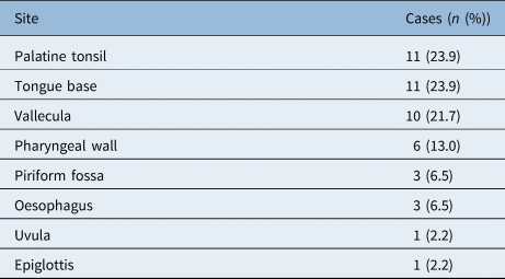
Table 2 outlines the locations of fish bones found. The majority of fish bones were found in relatively easily accessible areas, including the tonsil, tongue base, vallecula and pharyngeal wall.
Anatomical sites where fish bones were found

In total, 21 patients (24.4 per cent) had an ENT follow-up appointment scheduled, either for abnormalities picked up incidentally on flexible nasendoscopy or for persistent symptoms. No fish bones were subsequently found in returning patients. There were three unplanned re-presentations (3.5 per cent). These patients received a repeat flexible nasendoscopy as their symptoms were only mild. The three patients were once again discharged with reassurance, without a fish bone being found.
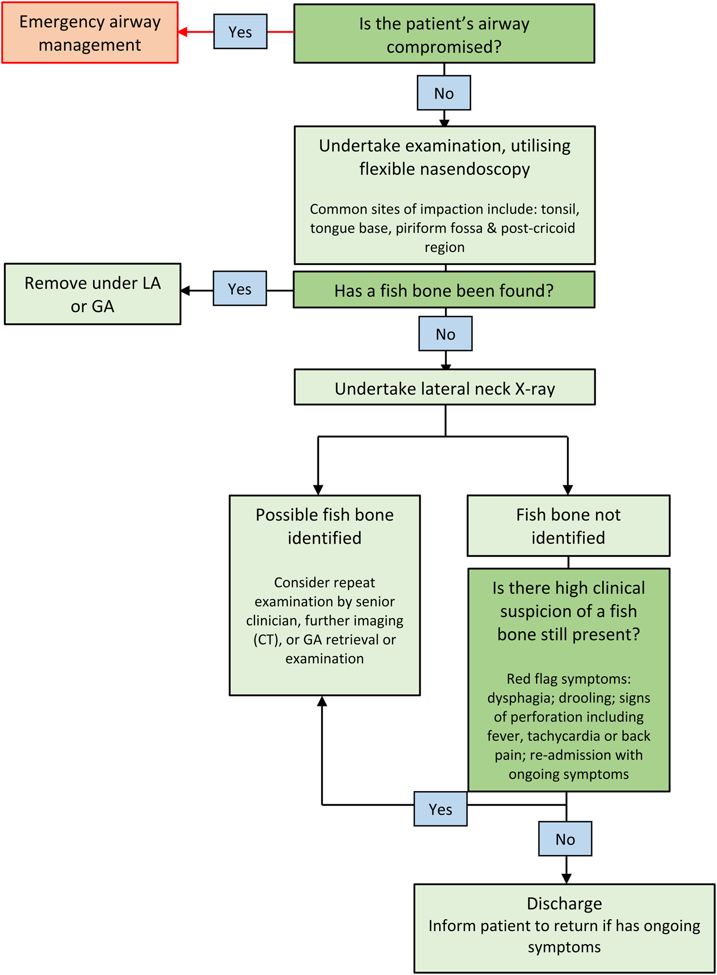
Figure 2 highlights our proposed algorithm for the management of suspected fish bone impaction in the upper aerodigestive tract.
Algorithm for the management of suspected fish bone impaction in the upper aerodigestive tract. LA = local anaesthesia; GA = general anaesthesia; CT = computed tomography

Discussion
This paper has suggested a treatment algorithm to aid in the management of fish bone impaction in the upper aerodigestive tract. The algorithm emphasises the importance of a thorough clinical examination, utilising flexible nasendoscopy, prior to radiological imaging. If patients have ongoing symptoms, or severe symptoms including dysphagia, then detailed examination under general anaesthesia is warranted, given the relative insensitivity of radiographs in delineating the presence of fish bones in the upper aerodigestive tract.
This study is the first in the literature to provide a treatment algorithm for the management of suspected fish bone impaction within the upper aerodigestive tract. Similar to previous studies,Reference Ballivet-de-Regloix, Crambert, Maurin, Bonfort, Marty and Pons3 the rate of fish bone retrieval in patients was low, at only 53 per cent. This suggests that symptoms are often due to mucosal irritation from fish bones that have already passed spontaneously. Twenty of the 46 fish bones found (43 per cent) were retrieved using foreign body forceps under local anaesthesia. The remainder required retrieval under general anaesthesia. This reflects the common sites of fish bone impaction as listed in the results.
Previous studies assessing the sensitivity and specificity of radiographs in the identification of fish bones have revealed relatively low sensitivities, of around 50 per cent, and specificities of around 70–100 per cent.Reference Sundgren, Burnett and Maly4–Reference Jahshan, Sela, Layous, Levy, Assadi and Shilo6 These results are in line with this study's findings, suggesting that lateral neck radiographs are insufficient to identify impacted fish bones. However, radiographs do provide some diagnostic value when combined with a thorough examination of the upper aerodigestive tract, preferably carried out first in order to prevent unnecessary irradiation in easily identifiable bones.
Computed tomography is a potential diagnostic tool for evaluating patients presenting with possible fish bone impaction. Studies have revealed excellent detection rates for CT in identifying impacted fish bones.Reference Jahshan, Sela, Layous, Levy, Assadi and Shilo6,Reference Akazawa, Watanabe, Nobukiyo, Iwatake, Seki and Umehara7 However, given the difficulties in access, cost and ionising radiation exposure, its use should be limited. We suggest that CT could be used in patients with a negative lateral neck radiograph and negative clinical examination findings, but with a high clinical suspicion that an impacted bone is present. General anaesthetic examination is another option favoured by many.Reference Kamath, Bhojwani, Prasannaraj and Abhijith8 Such examination has the advantage that the fish bone can be removed at the same time, but this should be weighed against the risk of perforation, on an individual case by case basis.
• Lateral neck radiographs are commonly used in the investigation and management of patients with suspected fish bone impaction
• The effectiveness of these are questioned, as many fish do not have radio-opaque bones
• This study has suggested a treatment algorithm to aid management of patients with a fish bone in upper aerodigestive tract
• The algorithm emphasises a thorough clinical examination, including flexible nasendoscopy at an early stage
• Lateral neck radiographs have limited value in management of suspected fish bone impaction, and should only be used following clinical examination of the upper aerodigestive tract
This study highlights the relative insensitivity of lateral soft tissue neck radiographs in identifying upper aerodigestive tract fish bones. When a suspected fish bone was seen on a radiograph, this was found to be a reliable indicator of an actual fish bone; however, many fish bones were found in patients with normal radiographs. Whilst a normal radiograph and normal flexible nasendoscopy findings are reassuring in patients presenting with suspected fish bone impaction, ongoing symptoms should prompt further evaluation. Two patients within our cohort (2.3 per cent) fell into this category, and proceeded to have fish bones found and retrieved under general anaesthesia. Both these patients had high levels of suspicion at the time of presentation, given the symptoms of severe pain or dysphagia. No discharged patients with mild symptoms and normal examination findings had subsequent fish bones discovered.
No patients suffered serious complications of fish bone impaction or required investigation via alternative imaging modalities. A CT scan of the neck and chest could be considered in patients presenting with ongoing symptoms, where there is a high clinical suspicion that a fish bone is present. Otherwise, examination under general anaesthesia is the procedure of choice.
In order to streamline the management of fish bone impaction, a treatment algorithm has been proposed. Using this algorithm on our patient cohort, no fish bones would have been missed and 22 patients (25.6 per cent) would have been spared unnecessary lateral neck radiographs when they subsequently had a fish bone clearly visible during oropharyngeal examination. The algorithm recognises that it is possible to have a normal lateral neck radiograph and upper airway endoscopy findings, and yet still have an occult fish bone which can be picked up through CT or general anaesthetic examination. It permits scope for clinical judgement as to how these cases should be further investigated.
Competing interests
None declared