Introduction
A link between affective disorders and dementia has been suggested (Kessing and Nilsson, Reference Kessing and Nilsson2003). In particular, much debate has centered on whether late onset depression may be a prodrome to dementia in elderly patients. However, few reports have described a manic episode as a prodrome to dementia. To the best of our knowledge, only one case of an elderly patient with dementia preceded by manic episodes has been presented in the literature (Appleby and Rosenberg, Reference Appleby and Rosenberg2007). In that article, the patient had just undergone the removal of a basal cell carcinoma, for which he received a 2–3-week course of treatment with corticosteroids. Non-psychiatric conditions, especially steroid treatment which induced manic symptoms in the above case, might be of significance. Furthermore, permanent cognitive deficits have also been associated with steroid treatment (Sirois, Reference Sirois2003). We now present a case who suffered from his first manic episode aged 63 years. Although the patient was compliant, the second manic episode happened one year later. Two computed tomography (CT) scans were performed for each episode and demonstrated a significant loss of brain parenchyma and mild deterioration of cognitive function. The patient gave written informed consent for the publication of this case report.
Case report
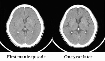
Mr. C, aged 63 years old, had retired at the age of 60. He was an excellent scientist with a specialty in mechanical engineering and was otherwise previously healthy without any psychosocial stress. There was no personal or family psychiatric history and his medical history was also unremarkable. He visited our hospital at the age of 62 due to a manic episode. Manic symptoms included expansive mood associated with decreased need for sleep, hypertalkativity, hyperactivity, hypersexuality, grandiosity and sexual indiscretion lasting about two weeks, which caused great distress to his family. After admission, he received routine physical and laboratory examinations and other non-routine examinations such as testing for syphilis and a brain CT scan (Figure 1A). All clinical data revealed no significant abnormalities. His first manic episode was thus diagnosed and he was started on valproic acid 200 mg and risperidone 2 mg per day for the treatment of manic symptoms. Three weeks later, he was discharged in a stable condition. Thereafter, he was stable and received regular follow-ups in outpatient clinics and continued taking valproic acid 200 mg per day. He felt that he was well but his cognitive function had progressively deteriorated, especially his short-term memory. A year later he suffered from manic symptoms again including reduced need for sleep, hypertalkativity, hyperactivity, mood elevation and increased sexual desire, whereupon he was readmitted to the acute psychiatric ward. Physical examination and laboratory tests showed no significant findings. However, a brain CT scan revealed significant loss of brain parenchyma (Figure 1B) compared with the first CT scan. A further brain magnetic resonance image was performed which confirmed this finding. Psychological testing revealed mild deterioration according to the Mini-mental State Examination (MMSE). The patient's total score was 29 at the first admission and was 23 at the second admission. He received valproic acid 400 mg per day for two weeks and his manic symptoms totally disappeared during this two-week period.
Computed tomography (CT) image at baseline (left) and at the one-year follow-up (right).

Discussion
It has been reported that patients with bipolar disorder could have persistent impaired cognitive function (Young et al., Reference Young, Murphy, Heo, Schulberg and Alexopoulos2006) and brain tissue loss (McIntosh et al., Reference McIntosh2007). This idea was further supported by several recent brain image studies that suggested the presence of neurofunctional abnormalities in patients with bipolar disorder and these functional abnormalities may stem from structural pathology in these or connected brain regions (Adler et al., Reference Adler, DelBello, Jarvis, Levine, Adams and Strakowski2007). Importantly, the results from these studies showed that the changes of brain parenchyma were not pervasive but region specific, such as in prefrontal or hippocampal regions (Adler et al., Reference Adler, Levine, DelBello and Strakowski2005; Devanand et al., Reference Devanand2007). In our case, we found, by visual inspection, that there was a significant loss of brain parenchyma in the frontal and temporal regions. These findings seem consistent with previous studies.
In contrast to previous studies we found that the significant loss of brain parenchyma seen within one year was quite rapid. Previous reports have shown that progressive gray matter loss may occur in bipolar patients (Moorhead et al., Reference Moorhead2007), and the speed of brain tissue loss was slow and well correlated with the deterioration of cognitive function and mood episodes. The fast decline of brain tissue in this case was consistent with a previous study which discussed the dynamics of gray matter loss in Alzheimer's disease (AD) (Thompson et al., Reference Thompson2003). The loss rate of brain tissue in AD compared with age-matched controls was found to be 5.3% ± 2.3% per year in AD vs. 0.9% ± 0.9% per year in controls. Although we did not have sophisticated imaging software to measure exact brain volume in this case, the loss of brain parenchyma by visual inspection on CT scan was at least 5%. Therefore, our case is likely to be a case of early dementia.
A recent case report suggested that valproic acid could induce reversible pseudoatrophy of the brain in a bipolar patient (Abreu et al., Reference Abreu, Issler and Lafer2009). We cannot exclude the loss of brain parenchyma as a medication effect because our patient was compliant with the use of valproic acid. A larger scale clinical study to explore the relationship between valproic acid and brain volume has demonstrated that there was no effect of valproic acid on brain volume after 12 weeks of valproic acid treatment (Chang et al., Reference Chang2009). We also followed up this case with a third CT scan which showed a similar picture to the second CT scan (image not shown). Therefore, it is unlikely that this loss of brain parenchyma was due to a medication effect.
In conclusion, we observed a case of a late onset manic episode. A good response to the use of the monotherapy – valproic acid – was observed. Early dementia is a possibility in this case based on the information from CT scans and clinical evaluation of cognitive function. This finding supports the idea that an increased risk of developing dementia may exist in patients with major affective disorders.
Conflict of interest
None
Description of authors’ roles
Dr. Hu, in his role as resident of psychiatry, treated the patient in his inpatient clinic. Dr. Chou supervised Dr. Hu and followed up this case in his outpatient clinic.