




No CrossRef data available.
Article contents
Atrioventricular septal defect associated with type A postaxial polydactyly
Published online by Cambridge University Press: 20 January 2005
Abstract
We report two more patients with the association of postaxial polydactyly and atrioventricular septal defects. Additional cardiac findings, not previously described, were tetralogy of Fallot in the first case, and double orifice right atrioventricular valve in the second child.
Keywords
Information
- Type
- Brief Report
- Information
- Copyright
- © 2004 Cambridge University Press
In 1995, we reported four children with type A postaxial polydactyly of their hands and feet, in all of whom there was an atrioventricular septal defect with common atrioventricular junction and either common or separate right and left valvar orifices.1 Since then, we have seen another two patients with this syndrome, both of whom have additional cardiac findings not previously reported. In the first patient, anatomy similar to that of tetralogy of Fallot was found together with the atrioventricular septal defect. In the second child, dual orifices were noted in the right atrioventricular valve at the time of surgical correction of the atrioventricular septal defect.
Case reports
Case 1
This black boy was initially investigated, at the age of 4 years, at another hospital. He was found to have an atrioventricular septal defect with the common atrioventricular junction guarded by a common valve in the presence of a common atrium. Obstruction was also noted across the right ventricular outflow tract, with a systolic gradient of 94 mmHg. Because of the cyanosis, he underwent a classical Blalock-Taussig anastomosis.
He was first seen at our hospital when aged 4½ years, and was followed-up thereafter for another 6 years. The Blalock shunt functioned well, with peripheral saturations of oxygen ranging between 84% and 88%. There was a history of delayed motor and speech milestones. He had extra digits on the ulnar aspects of both hands, but only 5 toes on each foot. There was an anti-mongoloid slant of the eyes, the nose was bulbous, and the “draw a man” test suggested a developmental age of 6 years when his chronological age was 10 years and 2 months. Both his height and weight remained between the 3rd and 10th centiles (Table 1).
Table 1. Summary of findings in six patients.

Case 2
This 5-year-old male was brought to our hospital for assessment of a cardiac problem. Clinical examination, together with the chest radiograph, electrocardiogram, and echocardiography revealed a common atrium with a common atrioventricular junction. There was mild incompetence of both the right and left components of the atrioventricular valves. Cardiac catheterization confirmed a common atrium, with right ventricular and pulmonary arterial pressures at the upper limit of normal. The ratio of pulmonary to systemic flow was calculated at 2.3 to 1, and the pulmonary vascular resistance was 2.1 Wood units.
Dysmorphic features included well formed extra digits on the postaxial side of the hands and feet, low set and small ears, widely spaced nipples, and bilateral Harrison’s sulcuses (Table 1). At operation to correct his cardiac lesion, there was a dilated common atrium, a common atrioventricular junction, but no potential for ventricular shunting. The surprise was finding that the right atrioventricular valve had two orifices. The superior one was the larger of the two, and showed some regurgitation (Fig. 1). A fibrous bridge separated the two openings, with four and three leaflets respectively in the superior and inferior orifices. There was a large antero-superior papillary muscle to which the edges of both adjacent leaflets were adherent. The left atrioventricular valve showed the typical trifoliate arrangement. The common atrium was septated so that the coronary sinus was incorporated on the left side. A limited annuloplasty was performed on the large superior orifice of the right atrioventricular valve. The coapting surfaces of the superior and inferior leaflets of the left atrioventricular valve were closed up to the strut cord of the inferior leaflet. Apart from a short period when he was in nodal rhythm, his postoperative recovery was uncomplicated. When we reviewed the admission echocardiogram, it was possible retrospectively to identify the dual orifices of the right atrioventricular valve in both the subcostal short axis and four chamber views (Fig. 2).

Figure 1. Artist's impression of surgical findings. 1 – septal leaflet of the larger right atrioventricular valve adherent to the ventricular septum; 2 – septal leaflet of the smaller right atrioventricular valve not adherent to the ventricular septum; 3 – fibrous bridge separating the 2 orifices of the right atrioventricular valve; 4 – large papillary muscle to which the adjacent edges of the leaflets were adherent. CS: coronary sinus; INFO: inferior orifice of right atrioventricular valve; LIL: left inferior leaflet; LSL: left superior leaflet; MV: left atrioventricular valve; SPO: superior orifice of right atrioventricular valve; TANN: right atrioventricular valve annuloplasty indicated by dotted lines; TV: right atrioventricular valve.

Figure 2. Two dimensional echocardiograms in Case 2. Top – subcostal short axis view of the atrioventricular valves; Bottom – subcostal view of right ventricular inlet and outlet. LO: large orifice of right atrioventricular valve; MV: left atrioventricular valve; RA: right atrium; RVOT: right ventricular outflow tract; SO: small orifice of right atrioventricular valve; TV: right atrioventricular valve.
Discussion
Polydactyly is defined as preaxial when there is duplication of digits on the radial side of the hand, and the tibial side of the feet. Postaxial polydactyly is present when the extra digits are on the ulnar side of the hands, and the fibular sides of the feet. If the extra digit is well developed, it is classified as type A, with type B being described when the digit is poorly differentiated.1
There are a number of syndromes where children manifest with pre- or post-axial polydactyly and congenital cardiac disease, albeit that almost all have other major abnormalities, such as Ellis van Creveld syndrome, Trisomy 13, and Lawrence-Moon-Biedl-Bardet syndrome. There are also even rarer conditions that are lethal soon after birth.1 Some of our patients showed minor dysmorphic features, while in our first patient reported here, there was a degree of neurodevelopmental delay. All of the six children that we have seen to date have had post-axial polydactyly, and an atrioventricular septal defect with common atrioventricular junction, albeit that some have had common valvar orifices, and others separate right and left valvar orifices with shunting at atrial and/or ventricular levels (Table 1). That the association between atrioventricular septal defect and polydactyly constitutes a true syndrome has received confirmation from two additional reports from Turkey2 and Italy.3 In the first, Onat2 described a patient in detail in which there was a family history of consanguinity. Mention is also made of a sporadic case seen 15 years previously. In the other communication, Digilio et al.3 observed three patients with the association of hexadactyly and an atrioventricular septal defect. This included a sporadic case, together with the familial recurrence of the complex in a father and daughter. The great-grandfathers in this case were first cousins. Thus, in some cases there might be a recessive mode of inheritance in this syndrome.
Of interest in our first patient was the finding of pulmonary stenosis in addition to the atrioventricular septal defect, which was treated by construction of a Blalock-Taussing shunt for relief of the cyanosis. This situation is very similar to a cyanosed child with Down’s syndrome who could have the identical combination of cardiac abnormalities. An even more unusual finding in our second patient was the presence of a dual orifice in the right atrioventricular valve in association with a common atrium and no potential for ventricular shunting. This abnormality, or dual orifices in a morphologically tricuspid valve, has been reported previously on only 7 separate occasions.4–10 Radermecker and co-authors8 discussed the anatomic basis for its formation in detail. The more common association of double orifice left atrioventricular valve in the setting of a common atrioventricular junction is well recognised, and occurs in up to one-tenth of cases.9 Two of the patients with atrioventricular septal defect and dual orifices in the right atrioventricular valve also had dual orifices in their left-sided valves.5, 9 The most recent documented case with dual orifices in a morphologically tricuspid valve had an associated large perimembranous ventricular septal defect.10
Our combined series of six patients has been observed over a period of 27 years. Thus, these cases are seen infrequently, albeit that they are of great diagnostic import. The exact genetic basis for the syndrome has yet to be determined.
Acknowledgements
We thank Dr S. Davis for his assistance at the operation on our second patient, and Mrs. Vivienne Hunter for her help in the preparation of this manuscript.
We are also indebted to Dr JR Harrisberg for reviewing the pre-operative echocardiogram in the second patient, and to Terry Borain, Photo and Illustration Unit, Faculty of Health Sciences for preparation of Figures 1 and 2.
References
Levin SE, Dansky R, Milner S, Benatar A, Govendrageloo K, du Plessis J. Atrioventricular septal defect and type A postaxial polydactyly without other major associated anomalies; a specific association. Pediatr Cardiol 1995; 16: 242–246.Google Scholar
Onat T. Post-axial hexodactyly and single atrium: a new syndrome? Hum Genet 1994; 94: 104–106.Google Scholar
Digilio MC, Marino B, Giannotti A. Single atrium, atrioventricular canal/postaxial hexodactyly indicating Ellis-van Creveld syndrome. Hum Genet 1995; 96: 251–253.Google Scholar
Yoo SJ, Houde C, Moes CA, Perrin DG, Freedom RM, Burrows PE. A case report of double-orifice tricuspid valve. Int J Cardiol 1993; 39: 85–87.Google Scholar
Honnekeri ST, Tendolkar AG, Lokhandwala YY. Double orifice mitral and tricuspid valves in association with Raghib complex. Ann Thorac Surg 1993; 55: 1001–1002.Google Scholar
Goldberg N, Schifter D, Aron M, Shapiro RS, Krasnow N, Stein RA. Double orifice mitral and tricuspid valves. Echocardiography 1996; 13: 85–90.Google Scholar
Prendergast B, Tometzki A, Mankad PS. Double-orifice right atrioventricular valve associated with partial atrioventricular septal defect. Ann Thorac Surg 1996; 62: 893–895.Google Scholar
Radermecker MA, Somerville J, Li W, Anderson RH, de Leval MR. Double orifice right atrioventricular valve in atrioventricular septal defect: morphology and extension of the concept of fusion of leaflets. Ann Thorac Surg 2001; 71: 358–360.Google Scholar
Nakano T, Kado H, Shiokawa Y-I, Fukae K. Surgical results of double orifice left atrioventricular valve associated with atrioventricular septal defects. Ann Thorac Surg 2002; 73: 69–75.Google Scholar
Anil SR, Rao SG, Kumar RK. Duplication of the tension apparatus of the tricuspid valve. Cardiol Young 2003; 13: 106–108.Google Scholar
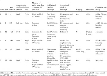
Table 1.

Artist's impression of surgical findings. 1 – septal leaflet of the larger right atrioventricular valve adherent to the ventricular septum; 2 – septal leaflet of the smaller right atrioventricular valve not adherent to the ventricular septum; 3 – fibrous bridge separating the 2 orifices of the right atrioventricular valve; 4 – large papillary muscle to which the adjacent edges of the leaflets were adherent. CS: coronary sinus; INFO: inferior orifice of right atrioventricular valve; LIL: left inferior leaflet; LSL: left superior leaflet; MV: left atrioventricular valve; SPO: superior orifice of right atrioventricular valve; TANN: right atrioventricular valve annuloplasty indicated by dotted lines; TV: right atrioventricular valve.
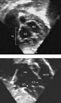
Two dimensional echocardiograms in Case 2. Top – subcostal short axis view of the atrioventricular valves; Bottom – subcostal view of right ventricular inlet and outlet. LO: large orifice of right atrioventricular valve; MV: left atrioventricular valve; RA: right atrium; RVOT: right ventricular outflow tract; SO: small orifice of right atrioventricular valve; TV: right atrioventricular valve.
 You have
Access
- 2
- Cited by