Introduction
Depression is one of the most common mental health disorders and a leading contributor to the global burden of disability (Murray et al., Reference Murray, Vos, Lozano, Naghavi, Flaxman, Michaud and Memish2012; World Health Organization, 2017). Due to several risk factors (Serafini et al., Reference Serafini, Canepa, Adavastro, Nebbia, Murri, Erbuto and Amore2017a; Thapar, Collishaw, Pine, & Thapar, Reference Thapar, Collishaw, Pine and Thapar2012), the prevalence of depression rises rapidly during adolescence and early adulthood (Gore et al., Reference Gore, Bloem, Patton, Ferguson, Joseph, Coffey and Mathers2011). This can lead to a lifetime of social, economic and physical impairment for many (Kessler, Reference Kessler2012). Cognitive models of depression suggest that a mood-congruent negative bias in processing and attending to emotional information contributes to the manifestation of its symptoms (Beck, Reference Beck2008; Bower, Reference Bower1981; Roiser, Elliott, & Sahakian, Reference Roiser, Elliott and Sahakian2012). This processing bias precedes and reinforces negative beliefs, thereby increasing an individual's likelihood of entering and perpetuating depressive mood states (Disner, Beevers, Haigh, & Beck, Reference Disner, Beevers, Haigh and Beck2011; Foland-Ross & Gotlib, Reference Foland-Ross and Gotlib2012; Leppanen, Reference Leppanen2006). Furthermore, altered sensory processing patterns of depressed individuals have been associated with a range of unfavorable outcomes (Harrison, Kats, Williams, & Aziz-Zadeh, Reference Harrison, Kats, Williams and Aziz-Zadeh2019; Serafini et al., Reference Serafini, Gonda, Canepa, Pompili, Rihmer, Amore and Engel-Yeger2017b).
The explicit processing of negatively valenced facial expressions is particularly influenced by this bias (Hankin, Gibb, Abela, & Flory, Reference Hankin, Gibb, Abela and Flory2010; Van Vleet et al., Reference Van Vleet, Stark-Inbar, Merzenich, Jordan, Wallace, Lee and Nahum2019). Those with depression commonly demonstrate abnormal activity in the amygdala, insula, anterior cingulate (ACC), dorsolateral prefrontal (dlPFC), and orbitofrontal cortices during the processing of these stimuli (Hamilton et al., Reference Hamilton, Etkin, Furman, Lemus, Johnson and Gotlib2012; Stuhrmann, Suslow, & Dannlowski, Reference Stuhrmann, Suslow and Dannlowski2011). However, rather than stemming from alterations in specific brain regions, more recent work has hypothesized that negative processing biases and their associated depressive symptoms result from widespread intra- and inter-regional (i.e. network) alterations (Li et al., Reference Li, Friston, Mody, Wang, Lu and Hu2018; Rayner, Jackson, & Wilson, Reference Rayner, Jackson and Wilson2016). Hence, changes to the interactions between these regions, rather than alterations in the regions themselves, are likely to contribute to illness progression (Disner et al., Reference Disner, Beevers, Haigh and Beck2011; MacNamara, Klumpp, Kennedy, Langenecker, & Phan, Reference MacNamara, Klumpp, Kennedy, Langenecker and Phan2017).
Functional connectivity studies have commonly investigated how depression alters connectivity between the amygdala and prefrontal regions (Helm et al., Reference Helm, Viol, Weiger, Tass, Grefkes, Del Monte and Schiepek2018). During the processing of faces, depressed patients have shown reduced connectivity between the amygdala and dlPFC (Dannlowski et al., Reference Dannlowski, Ohrmann, Konrad, Domschke, Bauer, Kugel and Suslow2009). While this has been suggested to result from amygdala hyperactivation and dlPFC hypoactivation, functional connectivity does not assess the directionality of these interactions (Fales et al., Reference Fales, Barch, Rundle, Mintun, Snyder, Cohen and Sheline2008; Siegle, Thompson, Carter, Steinhauer, & Thase, Reference Siegle, Thompson, Carter, Steinhauer and Thase2007; Zhong et al., Reference Zhong, Wang, Xiao, Yi, Zhu, Liao and Yao2011). In contrast, effective connectivity, or the directional influences between regions, has shown conflicting evidence across modalities as to the directionality of this effect (Carballedo et al., Reference Carballedo, Scheuerecker, Meisenzahl, Schoepf, Bokde, Moller and Frodl2011; Lu et al., Reference Lu, Li, Luo, Wang, Tang, Han and Yao2012). Depression has also been associated with reductions in functional connectivity between the vmPFC and the amygdala (Kong et al., Reference Kong, Chen, Tang, Wu, Driesen, Womer and Wang2013). Due to sparse anatomical connectivity between the dlPFC and amygdala, the vmPFC may play an important mediatory role in this relationship (Phillips, Drevets, Rauch, & Lane, Reference Phillips, Drevets, Rauch and Lane2003; Ray & Zald, Reference Ray and Zald2012). Given the vmPFC/rACC's role in implicit emotional regulation (Fitzgerald, Kinney, Phan, & Klumpp, Reference Fitzgerald, Kinney, Phan and Klumpp2018; Rive et al., Reference Rive, van Rooijen, Veltman, Phillips, Schene and Ruhe2013; Zotev, Phillips, Young, Drevets, & Bodurka, Reference Zotev, Phillips, Young, Drevets and Bodurka2013), reduced connectivity between these regions is hypothesized to result from greater regulation from the vmPFC in those with depression (Almeida et al., Reference Almeida, Versace, Mechelli, Hassel, Quevedo, Kupfer and Phillips2009). However, the precise manner in which the amygdala, dlPFC and vmPFC interact during the processing of these negatively valenced stimuli in those with depression remains largely unclarified due to the correlational nature of these analyses.
In addition, other research has attempted to investigate whether similar regions contribute to differences in treatment responsivity (Dichter, Gibbs, & Smoski, Reference Dichter, Gibbs and Smoski2015; Fonseka, MacQueen, & Kennedy, Reference Fonseka, MacQueen and Kennedy2018). Treatment response has been associated with increased vmPFC and dlPFC activity as well as decreased amygdala activity during the processing of emotional faces, particularly sad expressions (Costafreda, Khanna, Mourao-Miranda, & Fu, Reference Costafreda, Khanna, Mourao-Miranda and Fu2009; Fu et al., Reference Fu, Williams, Cleare, Scott, Mitterschiffthaler, Walsh and Murray2008; Ritchey, Dolcos, Eddington, Strauman, & Cabeza, Reference Ritchey, Dolcos, Eddington, Strauman and Cabeza2011; Williams et al., Reference Williams, Korgaonkar, Song, Paton, Eagles, Goldstein-Piekarski and Etkin2015). These baseline alterations have been suggested to indicate an underlying sensitivity in the neurobiological correlates of the negative processing bias which is particularly receptive to psychotherapeutic and pharmacological interventions (Harmer et al., Reference Harmer, O'Sullivan, Favaron, Massey-Chase, Ayres, Reinecke and Cowen2009; Roiser et al., Reference Roiser, Elliott and Sahakian2012; Victor, Furey, Fromm, Ohman, & Drevets, Reference Victor, Furey, Fromm, Ohman and Drevets2010). Given the high within-subject variability of activity within regions these regions, however, the utility of these regions as functional biomarkers has been questioned (McDermott et al., Reference McDermott, Kirlic, Akeman, Touthang, Cosgrove, DeVille and Aupperle2020; Nord, Gray, Charpentier, Robinson, & Roiser, Reference Nord, Gray, Charpentier, Robinson and Roiser2017). As such, focusing on connectivity parameters which have been shown to be more reliable (Nord, Gray, Robinson, & Roiser, Reference Nord, Gray, Robinson and Roiser2019), may enable more efficacious diagnostic and prognostic biomarkers (Dunlop & Mayberg, Reference Dunlop and Mayberg2014).
The current study aimed to provide a more comprehensive investigation of depression associated alterations in the face processing network and their relationship with treatment response. To assess effective connectivity, we implemented dynamic causal modelling (DCM; Friston, Reference Friston2011; Friston, Harrison, & Penny, Reference Friston, Harrison and Penny2003) and recent advances in DCM methodology, which includes a Bayesian approach to between-group analysis (Zeidman et al., Reference Zeidman, Jafarian, Seghier, Litvak, Cagnan, Price and Friston2019b), to explore effective connectivity in the Youth Depression Alleviation (YoDA-C) trial sample (Davey et al., Reference Davey, Chanen, Hetrick, Cotton, Ratheesh, Amminger and Berk2019). Adolescents and young adults were examined in this study due to this being a period of continuous development of social−emotional and underlying brain processes (Blakemore, Reference Blakemore2008) and the peak period for risk of developing MDD (Thapar et al., Reference Thapar, Collishaw, Pine and Thapar2012). We hypothesized that compared with controls, MDD patients would demonstrate (1) altered bidirectional modulation of the connectivity between the dlPFC and amygdala, consistent with the excitatory and inhibitory influences of these connections; and (2) greater inhibitory modulation from the vmPFC to amygdala. Additionally, we expected that (3) differences in the connectivity between responders and non-responders will involve alterations between these areas of the extended face processing system, and the size of this effect will be sufficiently large to predict treatment response.
Methods
Participants
One-hundred and five unmedicated, help-seeking depressed participants, aged 15–25 years, were recruited through specialist mental health clinics located in the northern and western suburbs of Melbourne, Australia. These participants were enrolled in the YoDA-C trial (Davey et al., Reference Davey, Chanen, Hetrick, Cotton, Ratheesh, Amminger and Berk2019). This was a randomized, double-blind, placebo-controlled, multicenter clinical trial comparing the efficacy of 12 weeks of cognitive behavioral therapy (CBT), plus either fluoxetine or a placebo. Participants commenced with either a daily 20-mg capsule of fluoxetine or one placebo pill. This was increased to fluoxetine 40-mg or two placebo pills, if insufficient clinical response was observed after the first four weeks. CBT was delivered by therapists in weekly 50-min sessions. Patients had been diagnosed with MDD, as assessed by the Structured Clinical Interview for DSM-IV Axis I Disorders (SCID; First, Spitzer, Gibbon, and Williams, Reference First, Spitzer, Gibbon and Williams1997). Their depressive symptoms were of at least a moderate level of severity, as indicated by Montgomery−Åsberg Depression Rating Scale (MADRS) score of ⩾ 20 (Montgomery & Asberg, Reference Montgomery and Asberg1979). Response from depression was defined as a MADRS symptom score reduction of 50% or greater following 12 weeks of treatment (Riedel et al., Reference Riedel, Moller, Obermeier, Schennach-Wolff, Bauer, Adli and Seemuller2010). Analyses were conducted on an intention-to-treat basis. For response status, we used the last post-baseline MADRS assessment carried forward. Further information concerning trial and assessments used are found in the online Supplementary Materials. Participants had no lifetime or current diagnosis of a psychotic or bipolar disorder, no current treatment with antidepressant medication, were not pregnant, and had an estimated IQ greater than 85 as determined by the Wechsler Test of Adult Reading (Wechsler, Reference Wechsler2001). Ninety-eight healthy participants were also recruited through online advertisements and were age and sex-matched with patients at a group level. They had no past mental health disorder diagnoses as assessed through the SCID criteria and did not have an IQ lower than 85.
All participants were provided with, and signed, an informed consent form to participate in the study. For those under 18 years, both participant assent and parent consent were required. This study had been approved by the Melbourne Health Human Research and Ethics Committee. Of the total number of participants who underwent scanning a total of 23 were omitted due to: incidental findings (2 controls, 1 patient), excessive within-scanner head motion (2 controls, 8 patients; see further), or poor task performance (defined as less than 80% accuracy for both of the face matching conditions; 2 controls, 8 patients). As a result, 92 health controls and 88 MDD participants were included in our analyses.
Implicit emotional face matching task
Our paradigm is detailed in full in the online Supplementary Materials and Jamieson, Davey, and Harrison (Reference Jamieson, Davey and Harrison2021). In short, the task was a blocked-designed task with three conditions: one shape matching and two implicit face processing conditions, involving either fearful or sad facial expressions. Participants were required to either match the orientation of the circular shape or the gender of the face presented in the top half of the screen to one of the two circular shapes or faces presented on the left and right in the bottom half of the screen (online Supplementary Fig. S1). Depending on the block, all faces presented would convey either a sad or fearful facial expression.
Each session involved six blocks for each of the three conditions (18 blocks total); a 10 s white fixation cross was also presented between each block, and before the first and after the final block. Each block consisted of six trials, with each trial having a duration of 3.75 s followed by a 0.25 s inter-trial interval.
General linear modelling
Image acquisition and preprocessing details can be found in the online Supplementary Materials. Each participant's preprocessed timeseries was included in a first-level general linear model analysis in SPM12. This was done by specifying the durations and onsets of each shape, sad, and fearful face matching blocks, respectively, to be convolved with a canonical hemodynamic response function. Each condition was modeled separately, with rest-fixation blocks forming the implicit baseline. A high-pass filter (1/128 s) accounted for low-frequency noise, while temporal autocorrelations were estimated using a first-order autoregressive model. Primary contrast images were estimated to examine responses to fearful (fearful faces > shapes) and sad faces (sad faces > shapes), as well as overall responses to these faces (sad and fearful faces > shapes), and were carried forward to the group-level using the summary statistics approach to random-effects analyses (online Supplementary Materials).
Dynamic causal modelling
DCM estimates the directional coupling between brain regions which are likely to underpin observed neuroimaging data (Friston et al., Reference Friston, Harrison and Penny2003). For fMRI tasks, this estimates how these relationships behave intrinsically (invariant connectivity in the absence of task modulation) and due to modulation to these connections by experimental stimuli (Friston et al., Reference Friston, Preller, Mathys, Cagnan, Heinzle, Razi and Zeidman2019; Stephan et al., Reference Stephan, Penny, Moran, den Ouden, Daunizeau and Friston2010). This coupling is measured in hertz (Hz) for between regions connectivity, with positive values indicating excitation and negative values indicating inhibition. For self-connections these parameter estimates are unitless log scaling parameters which multiply the default self-inhibition (−0.5 Hz), thus positive values indicate greater inhibition while negative values indicate reduced inhibition (Zeidman et al., Reference Zeidman, Jafarian, Corbin, Seghier, Razi, Price and Friston2019a).
Timeseries extraction
To construct a candidate model space requires the specification and extraction of chosen volumes of interest (VOIs) at an individual subject level. Our chosen VOIs were based on previous studies and included the inferior occipital gyrus (OFA), fusiform gyrus (FFA), amygdala, dlPFC and vmPFC as to model both the core and extended systems of the face processing network (Goulden et al., Reference Goulden, McKie, Thomas, Downey, Juhasz, Williams and Elliott2012; Vai et al., Reference Vai, Bulgarelli, Godlewska, Cowen, Benedetti and Harmer2016). The OFA, FFA, amygdala, and dlPFC were defined by the sad and fearful faces > shapes contrast, while the vmPFC was defined by the inverse of this contrast. Following previous studies, these regions were also constrained to the right side of the brain to allow for the exclusion of fewer participants due to inadequate activation (Rui de Moraes, Bruno Marinho, & Sérgio, Reference Rui de Moraes, Bruno Marinho and Sérgio2014; Sorger, Goebel, Schiltz, & Rossion, Reference Sorger, Goebel, Schiltz and Rossion2007).
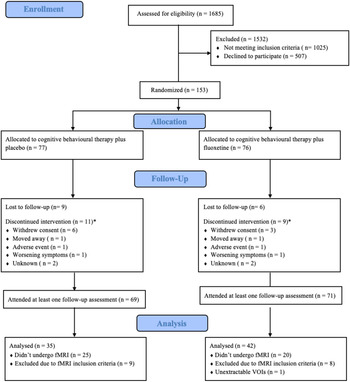
Specific coordinates for each of these regions were centered around group peaks. Center coordinates for each individual's VOIs were personalized to their local maximum, which were required to occur within 8 mm of the group level peak (see online Supplementary Table S1). The principal eigenvariate for each of these VOIs was extracted as per recently published guidelines (Zeidman et al., Reference Zeidman, Jafarian, Corbin, Seghier, Razi, Price and Friston2019a). As such, this included all voxels in a sphere with a radius of 4 mm from the center coordinate and present at a threshold of p < 0.05, uncorrected. If at this threshold a VOI for an individual still had inadequate activation of all VOIs, the threshold incrementally relaxed up to p < 0.5 (Zeidman et al., Reference Zeidman, Jafarian, Corbin, Seghier, Razi, Price and Friston2019a). After exclusion due to missing patient follow-up data (10 patients), this resulted in 89 healthy controls and 77 MDD participants having a full set of extractable VOIs and thus comprised our final sample (Fig. 1).
CONSORT flow diagram for participants included in the YoDA-C trial. *Three participants in each group continued their scheduled assessments after discontinuing the study intervention.

Model specification, estimation and parametric empirical Bayes
The candidate model was specified and estimated with DCM12.5. The intrinsic connectivity of our model was informed by previous DCM research (Dima, Stephan, Roiser, Friston, & Frangou, Reference Dima, Stephan, Roiser, Friston and Frangou2011; Goulden et al., Reference Goulden, McKie, Thomas, Downey, Juhasz, Williams and Elliott2012; Herrington, Taylor, Grupe, Curby, & Schultz, Reference Herrington, Taylor, Grupe, Curby and Schultz2011; Vai et al., Reference Vai, Bulgarelli, Godlewska, Cowen, Benedetti and Harmer2016; Willinger et al., Reference Willinger, Karipidis, Beltrani, Di Pietro, Sladky, Walitza and Brem2019). As such, it was defined with bidirectional connections from the FFA to OFA, amygdala, dlPFC and vmPFC, from the amygdala to OFA, dlPFC and vmPFC, and between dlPFC and vmPFC (for diagram see online Supplementary Fig. S2). Modulation by both fearful and sad expressions occurred for all connections. Direct external input into the network was modeled using the overall negative facial expression (fearful + sad) into both the OFA and amygdala. This full model was then estimated for each subject.
Parameteric Empirical Bayes (PEB) was used to examine between-group effects on within-subjects' parameters (Friston et al., Reference Friston, Litvak, Oswal, Razi, Stephan, van Wijk and Zeidman2016). Using classical tests to calculate these between group-differences results in the exclusion of the estimated variation of each parameter. PEB allows for the inclusion of this variance when investigating between-group effects (Friston et al., Reference Friston, Litvak, Oswal, Razi, Stephan, van Wijk and Zeidman2016). A posterior probability (PP) greater than 0.95 is typically used to determine whether a parameter demonstrates sufficient evidence to represent a non-zero effect (Zeidman et al., Reference Zeidman, Jafarian, Seghier, Litvak, Cagnan, Price and Friston2019b). When specifying our second level PEB model we investigated the effects of six regressors. These were as follows: (1) the overall mean connectivity, (2) the difference between diagnostic groups, (3) the difference at baseline between patients who responded to treatment compared with those who did not, (4) differences at baseline between treatment groups, (5) the interaction of response and treatment type, (6) the effects of age. As our regressors were mean centered, the between-subject effects (regressors 2–6) added or subtracted from the commonalities between subjects (regressor 1). Having estimated a group level PEB model including all parameters, we then searched over nested PEB models, pruning parameters that did not contribute to overall model evidence (Friston & Penny, Reference Friston and Penny2011; Rosa, Friston, & Penny, Reference Rosa, Friston and Penny2012). This was conducted separately for our A, B and C matrices, as including many parameters can result in a dilution of evidence effect (Zeidman et al., Reference Zeidman, Jafarian, Seghier, Litvak, Cagnan, Price and Friston2019b). A Bayesian model average was then performed on these models after the final iteration to determine the strength of these connections.
Leave-one-out cross-validation
The implementation of leave-one-out cross-validation in the PEB framework allows for us to determine whether the size of the effect of treatment response on these connections was sufficiently large to predict response group allocation (Zeidman et al., Reference Zeidman, Jafarian, Seghier, Litvak, Cagnan, Price and Friston2019b). This does so by estimating a PEB model without the inclusion of one subject and then using those parameters which were different between these groups to predict the allocation of the left out subject. This process is then repeated for every subject. This predicted allocation is then correlated with the observed allocation (whether these individuals responded following treatment or not) to determine the accuracy of this prediction.
Results
Clinical results
Controls and patients differed significantly on MADRS symptoms at baseline (t(124.30) = −46.48, p < 0.001; Table 1). The two treatment arms significantly differed in age (t(75) = −3.07, p = 0.015), with those in the CBT and fluoxetine group being significantly older than those treated with CBT and a placebo (an artifact of the process of randomization and optional consent to participate in MRI scanning; online Supplementary Table S2). Treatment response rates were similar for both treatment arms (CBT and placebo = 49%; CBT and fluoxetine groups = 48%, respectively). No differences were observed between treatment responders and non-responders (Table 2).
Comparison of participants' demographic and behavioral characteristics between controls and patients

Note. *Significant at p < 0.05.
Comparison of characteristics between patients that responded following treatment and non-responders

Note. *Significant at p < 0.05
Within scanner behavioral results
Neither reaction time nor accuracy differed significantly between patients and controls for either shape, sad or fearful face matching (Table 1). For differences between conditions see online Supplementary Materials. No behavioral differences were observed between the two treatment arms (online Supplementary Table S2) nor between responders and non-responders (Table 2).
Differences in effective connectivity between patients and controls
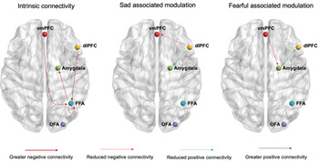
As illustrated in Fig. 2, depressed participants demonstrated strong evidence for reduced negative modulation from the dlPFC to vmPFC (expected value = 0.06 Hz, PP = 0.97) and reduced positive modulation from the dlPFC to the amygdala during the processing of sad faces (expected value = −0.08 Hz, PP = 0.99).
Group differences between controls and depressed participants in their intrinsic connectivity and the modulation by sad and fearful faces. Results are depicted with controls representing the reference group to highlight the effect of Major Depressive Disorder on these connections. Render visualized using BrainNet Viewer (Xia, Wang, & He, Reference Xia, Wang and He2013).

During fearful face processing, patients demonstrated strong evidence for reduced positive modulation from the amygdala to dlPFC (expected value = −0.09 Hz, PP = 0.99) and reduced negative modulation from the vmPFC to amygdala (expected value = 0.08 Hz, PP = 0.98; Fig. 2, online Supplementary Table S3).
Patients also showed strong evidence for greater negative intrinsic connectivity from the FFA to amygdala (expected value = −0.04 Hz, PP = 1.00), dlPFC to FFA (expected value = −0.08 Hz, PP = 1.00), and vmPFC to FFA (expected value = −0.07 Hz, PP = 1.00) and reduced inhibition from the FFA to itself (expected value = −0.07, PP = 1.00). The expected values and PP for all between group effect and connections are reported in online Supplementary Table S3.
Differences in effective connectivity between responders and non-responders
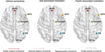
Patients who responded following treatment demonstrated meaningful differences in effective connectivity at baseline compared with non-responders. Responders demonstrated strong evidence for greater positive modulation from the amygdala to dlPFC (expected value = 0.10 Hz, PP = 0.96) during sad face processing and reduced positive modulation from the amygdala to vmPFC during fearful face processing (expected value = −0.12 Hz, PP = 0.98; Fig. 3).
Group differences at baseline between responders and non-responders in their intrinsic connectivity and the modulation by sad and fearful faces. Results are depicted with non-responders representing the reference group to highlight the effect of treatment response on these connections. Render visualized using BrainNet Viewer (Xia et al., Reference Xia, Wang and He2013).

Responders also showed strong evidence for greater positive intrinsic connectivity from the FFA to dlPFC (expected value = 0.10 Hz, PP = 1.00). The intrinsic inhibitory self-connection of the vmPFC illustrated strong evidence for being reduced in responders (expected value = −0.13, PP = 1.00), meaning that for these individuals the vmPFC exhibited reduced regulation of its own activity and thus responded more to inputs from the network (Zeidman et al., Reference Zeidman, Jafarian, Corbin, Seghier, Razi, Price and Friston2019a).
Leave-one-out cross-validation
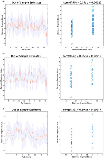
Inclusion of patients from both treatment arms resulted in an out-of-sample correlation between the predicted and observed response status of r = 0.39, p < 0.001 (Fig. 4a), corresponding to an accuracy of 60% (sensitivity = 0.81, specificity = 0.40). Subsequent analyses for these groups separately indicated a correlation of r = 0.35, p = 0.01 for the CBT and fluoxetine group (Fig. 4B) and r = 0.59, p < 0.001 for the CBT and placebo group (Fig. 4c). This indicated that the effect size of these parameters for the CBT and placebo group was large enough to predict the response of left-out subjects above chance, and had corresponding accuracy of 77% (sensitivity = 0.88, specificity = 0.66). For the CBT and fluoxetine group this corresponded to an accuracy of 60% (sensitivity = 0.84, specificity = 0.39).
Leave-one-out cross-validation predicting response status after treatment for depression. Left: The out-of-sample estimate of the treatment response status (whether after treatment individuals had a MADRS score reduction of 50% or greater) with 90% confidence interval (shaded area) for each participant. Right: The correlation between observed scores and the expected values for each individual. For (a) both treatment arms, (b) only those treated with CBT and fluoxetine, and (c) only those treated with CBT and a placebo.

Discussion
Our study showed reduced modulation of the connectivity from the dlPFC to amygdala and amygdala to dlPFC in depressed patients, supporting our first hypothesis. Notably, the altered modulation of the dlPFC to amygdala connectivity occurred during sad face processing, while the amygdala to dlPFC changes occurred during fearful face processing. Our second hypothesis was also supported, as differences in the modulation from the vmPFC to amygdala were also evident in patients, albeit limited to fearful face processing. Overall, the effect of response on the parameters of this network was sufficiently large for these to be significant predictors of treatment response, with particular utility demonstrated in those who received combined treatment with CBT and a placebo.
Differences in effective connectivity between patients and controls
In past imaging studies, emotional stimuli processing has typically been associated with increased functional connectivity between the amygdala and dlPFC (Diano et al., Reference Diano, Tamietto, Celeghin, Weiskrantz, Tatu, Bagnis and Costa2017; Gold, Morey, & McCarthy, Reference Gold, Morey and McCarthy2015). In depressed patients, however, this connectivity has been observed to be reduced, particularly in response to sad stimuli (Dannlowski et al., Reference Dannlowski, Ohrmann, Konrad, Domschke, Bauer, Kugel and Suslow2009; Lu et al., Reference Lu, Li, Luo, Wang, Tang, Han and Yao2012; Stuhrmann et al., Reference Stuhrmann, Suslow and Dannlowski2011; Tang et al., Reference Tang, Lu, Zhang, Hu, Bu, Li and Huang2018). This is consistent with our finding of reduced dlPFC to amygdala connectivity during sad face processing. The dlPFC is thought to be recruited mainly during effortful, explicit emotional regulation of the amygdala (Etkin, Buchel, & Gross, Reference Etkin, Buchel and Gross2015). As the explicit aspect of this task involved matching faces on their gender, it is expected that this interaction would typically have minimal involvement in regulating emotional states (Braunstein, Gross, & Ochsner, Reference Braunstein, Gross and Ochsner2017). In depression, however, there is evidence for the abnormal recruitment of lateral prefrontal regions during implicit regulation (Rive et al., Reference Rive, van Rooijen, Veltman, Phillips, Schene and Ruhe2013). It is therefore likely that the effect of implicitly processing sad stimuli while completing the cognitive matching task results in greater conflict between these processes for depressed patients, and requires more cognitive effort to suppress engagement with the sad stimuli to complete the cognitive aspects of the task (Dichter, Felder, & Smoski, Reference Dichter, Felder and Smoski2009). As such, this reduced positive modulation may represent a compensatory mechanism acting to reduce amygdala activity and the salience of these stimuli (Jacob, Bruck, Domin, Lotze, & Wildgruber, Reference Jacob, Bruck, Domin, Lotze and Wildgruber2014).
Unlike the altered modulation associated with sad face processing, depression was associated with changes to both vmPFC to amygdala and amygdala to dlPFC connectivity during fearful face processing. This is consistent with previous functional connectivity analyses which have illustrated reductions in the negative functional connectivity between the vmPFC and amygdala during emotional face processing (Etkin & Schatzberg, Reference Etkin and Schatzberg2011). Altered downregulation of affective subcortical circuitry has been proposed as a potential mechanism underlying depressed symptomatology (Davidson, Reference Davidson2002; Johnstone, van Reekum, Urry, Kalin, & Davidson, Reference Johnstone, van Reekum, Urry, Kalin and Davidson2007). Previous work has suggested that these changes may be in response to the depression associated amygdala hypoactivation, observed particularly in response to processing fearful expression (Beesdo et al., Reference Beesdo, Lau, Guyer, McClure-Tone, Monk, Nelson and Pine2009; Thomas et al., Reference Thomas, Drevets, Dahl, Ryan, Birmaher, Eccard and Casey2001). However, in our model, the direct input of fearful expressions into the amygdala did not demonstrate sufficient evidence to surpass our threshold. Interestingly, anxiety symptoms, which are highly comorbid in depression, have been associated with amygdala hyperactivity (Fales et al., Reference Fales, Barch, Rundle, Mintun, Snyder, Cohen and Sheline2008; van den Bulk et al., Reference van den Bulk, Meens, van Lang, de Voogd, van der Wee, Rombouts and Vermeiren2014). Due to this, the opposing effect of anxiety symptoms, may have resulted in the fearful input not meeting our threshold, despite changes to the connections surrounding it. Leave-one-out cross validation further supports the relationship between anxiety symptoms the fearful modulation from the vmPFC to amygdala, however, not from the amygdala to dlPFC. It remains unclear whether the observed altered modulation is specific to fearful faces or observable in other high arousal expressions, including those with a positive valence. To disentangle this relationship, future investigation into how this pattern is altered for other stimuli with varying levels of arousal and valence is required.
Differences in effective connectivity between responders and non-responders
While not specifically hypothesized, the relationship between response and the sad modulation from the amygdala and dlPFC is generally consistent with previous research (Dunlop & Mayberg, Reference Dunlop and Mayberg2014; Pathak, Salami, Baillet, Li, & Butson, Reference Pathak, Salami, Baillet, Li and Butson2016). Greater positive connectivity between these two areas has been associated with an increased likelihood to respond to treatment. In contrast to our findings, however, this has previously been suggested to be indicative of increased ‘top-down’ control by prefrontal regions due to these studies employing non-directional analysis techniques (DeRubeis, Siegle, & Hollon, Reference DeRubeis, Siegle and Hollon2008). Additionally, longitudinal research has suggested that reduced functional connectivity between the amygdala and dlPFC occurs following successful treatment (Arnone, Reference Arnone2019; Ruhe, Booij, Veltman, Michel, & Schene, Reference Ruhe, Booij, Veltman, Michel and Schene2012; Straub et al., Reference Straub, Metzger, Plener, Koelch, Groen and Abler2017). Thus, reductions in depressive symptomatology following treatment may be facilitated by, or even predicated on, this baseline hyperconnectivity. Previous research has suggested that the connectivity from the amygdala to dlPFC may be important in orientating conscious attention to salient stimuli (Frank & Sabatinelli, Reference Frank and Sabatinelli2012). As CBT aims to draw attention to and modify one's perception environmental stimuli (Beck, Beck, & Beck, Reference Beck, Beck and Beck2011), this may require a greater level of baseline sensitivity. If so, this interaction may define a patient subtype with an improved prognostic trajectory.
Cross-validation demonstrated that the size of the effect of the differences between responders and non-responders was large enough to significantly predict response status. The overall effect across both treatment arms is unlikely to be sufficiently large for clinical utility. Despite being associated with response generally, the individual response parameters demonstrated a greater predictive ability for the CBT and placebo arm. While this is possibly artifactual, it may reflect the effects of CBT treatment in the absence of pharmacological interventions. The antidepressant effect of serotonin selective reuptake inhibitors have been hypothesized to occur through increasing neuroplasticity (Liu, Liu, Wang, Zhang, & Li, Reference Liu, Liu, Wang, Zhang and Li2017) and normalizing the negative processing bias (Godlewska & Harmer, Reference Godlewska and Harmer2021). Thus, serotonin selective reuptake inhibitors may alter connectivity variability in such a way that the initial brain state is less informative of response than for those provided with psychotherapy alone.
Limitations
While our study has several strengths, including its sample size and method of assessing between-group differences, it is not without limitations. As the focus of this analysis was in a sample of young adults and adolescents, the generalizability of these findings to older individuals with depression remains to be examined. Further work examining these effects in older populations would aid in clarifying the specificity of this relationship. We also did not collect post-treatment scans for our participants. Such data may have allowed us to investigate whether depression associated alterations were normalized following successful treatment. While beyond the scope of this study, sex hormones are known to influence many of the brain regions explored and thus may have impacted our results. Future work examining this relationship more directly may help clarify this issue. Additionally, although our sample was relatively large, analyzing responders and non-responders in the different treatment arms resulted in much smaller subgroups. Replication of this effect in a larger sample will improve the precision, particularly for the interaction parameter estimates. In turn, this may allow for an improved disentanglement of the potential prognostic and predictive effects highlighted here.
Conclusion
The current study has demonstrated prominent depression associated alterations to the effective connectivity between the amygdala, dlPFC and vmPFC. Altered interactions from the amygdala to dlPFC were observed to be different between those who later responded following treatment and those who did not. Our results suggest that while these differences between responders and non-responder are generally prognostic, the additional effect for those treated with CBT and a placebo was sufficiently large to predict response with reasonable accuracy. This apparent specificity may be due to antidepressant pharmacotherapy altering connectivity variability, thus resulting in this baseline state being less informative of response. Further longitudinal research will necessary to confirm the generalizability of this potential biomarker in older populations as well as the specificity of this effect.
Supplementary material
The supplementary material for this article can be found at https://doi.org/10.1017/S0033291721002567
Acknowledgments
The authors thank Katerina Stephanou, Lisa Incerti and Rebecca Kerestes for contributions to data collection, as well as staff from the Sunshine Hospital Medical Imaging Department (Western Health, Melbourne).
Author contributions
AJJ: Conceptualization; Formal analysis; Methodology; Writing − original draft; Visualization. CGD: Conceptualization, Funding acquisition; Methodology; Supervision; Writing − review and editing. BJH: Conceptualization, Funding acquisition; Methodology, Supervision; Writing − review and editing.
Financial support
This work was supported by National Health and Medical Research Council of Australia (NHMRC) Project Grants (1064643) to BJH and to CGD (1024570). AJJ was supported by an Australian Government Research Training Program Scholarship. BJH was supported by a NHMRC Career Development Fellowship (1124472). CGD was supported by an NHMRC Career Development Fellowship (1061757).
Conflict of interest
The authors declare no conflicting interests.