Introduction
Classic hallucinogens like lysergic acid diethylamide (LSD) and psilocybin constitute two candidate models of psychosis that have widely been used to unravel neurobiological mechanisms of psychotic symptoms most relevant to the pathophysiology of schizophrenia (Vollenweider & Geyer, Reference Vollenweider and Geyer2001; Geyer & Vollenweider, Reference Geyer and Vollenweider2008; González-Maeso & Sealfon, Reference González-Maeso and Sealfon2009; Carhart-Harris et al. Reference Carhart-Harris2013; Halberstadt & Geyer, Reference Halberstadt and Geyer2013; De Gregorio et al. Reference De Gregorio2016). Hallucinogens such as LSD and psilocybin induce agitation, anxiety, visual hallucinations and illusion, which are reminiscent of those observed in the first episode of psychosis (Steeds et al. Reference Steeds, Carhart-Harris and Stone2015). Apart their potential to mimic some psychotic symptoms, LSD and psilocybin are particularly useful to temporally and profoundly alter an individual's visual experiences (Kometer & Vollenweider, Reference Kometer and Vollenweider2016). Activation of 5-hydroxytryptamine2A receptors (5-HT2ARs) predominantly mediates the visual hallucinations that are induced by psilocybin (Vollenweider et al. Reference Vollenweider1998; Kometer et al. Reference Kometer2013) and LSD (Preller et al. Reference Preller2017) and administration of the 5-HT2AR inverse agonist pimavanserin is effective for treatment of visual hallucinations in patients with Parkinson's disease psychosis (Meltzer et al. Reference Meltzer2010). Therefore, 5-HT2AR activation by LSD helps to study the neuropsychopharmacological mechanisms of visual hallucinations with implications for the pathophysiology of visual hallucinations in psychiatric disorders (Kometer & Vollenweider, Reference Kometer and Vollenweider2016).
Recent studies demonstrated that acute LSD administration to healthy subjects not only produces elementary and complex visual pseudo-hallucinations and perceptual illusions (Schmid et al. Reference Schmid2015; Carhart-Harris et al. Reference Carhart-Harris2016b; Preller et al. Reference Preller2017), but also impaired inhibitory processes (prepulse inhibition) (Schmid et al. Reference Schmid2015) and induced cognitive disorganization (Carhart-Harris et al. Reference Carhart-Harris2016a). These findings are in line with studies from the 1960s showing that LSD impairs inhibitory performance on the Stroop Test (Wapner & Krus, Reference Wapner and Krus1960; Krus et al. Reference Krus1963). Of note, impairments in prepulse inhibition after psilocybin administration and cognitive impairments after LSD administration were attenuated by administration of the 5-HT2AR antagonist ketanserin (Quednow et al. Reference Quednow2011; Preller et al. Reference Preller2017), indicating that 5-HT2AR activation is not only responsible for visual hallucinations but also cognitive impairments after LSD or psilocybin intake. 5-HT2ARs are indeed critically involved in different cognitive functions (Štrac et al. Reference Štrac, Pivac and Mück-Šeler2016) and abnormal 5-HT2AR activity is associated with cognitive impairments in a number of psychiatric disorders (Zhang & Stackman, Reference Zhang and Stackman2015). Furthermore, 5-HT2AR antagonists induce at least modest improvement in cognition in schizophrenia and might also help to enhance cognition in other disorders such as dementia (Roth et al. Reference Roth, Hanizavareh and Blum2004).
Cognitive impairments and visual hallucinations do often co-exist. Executive functions are involved in reality monitoring and contribute significantly to disentangling visual perception (Barnes & Boubert, Reference Barnes and Boubert2008), from determining external and internal stimuli, to the internal production of a visual image (Roth et al. Reference Roth2009). A previous study could show that Parkinson patients with visual hallucination have substantially greater impairments of inhibitory ability than those without visual hallucination (Barnes & Boubert, Reference Barnes and Boubert2008). Another study supported this result by showing that within patients with Parkinson's and Alzheimer's disease, those with visual hallucinations scored significantly lower than patients without visual hallucinations, particularly on tests evaluating prefrontal-executive functions such as inhibition (Grossi et al. Reference Grossi2011). Theoretical reflections propose that 5-HT2AR activation may cause a cognitive disability to integrate new perception that subsequently triggers the formation of aberrant feelings and visual perception (Vollenweider & Geyer, Reference Vollenweider and Geyer2001; Geyer & Vollenweider, Reference Geyer and Vollenweider2008).
In this study, we first tested the relationship between subjective LSD-induced cognitive impairments and visual imageries in healthy subjects. We then explored the acute effect of LSD on response inhibition neural networks (Go/No-Go task) and whether this effect was related to subjective feelings of impaired cognitive control and visual imageries. Based on the evidence showing a relationship between prefrontal-mediated cognitive impairments and visual hallucinations in Parkinson patients and theories proposing that 5-HT2AR stimulation by classical hallucinogens might promote visual hallucination through a cognitive failure to integrate external stimuli, we first expected a positive relationship between LSD effects on subjective feelings of cognitive control and visual imageries. Our second hypothesis was that LSD would reduce activation in prefrontal regions underlying response inhibition such as the middle, superior and inferior frontal gyrus, middle temporal gyrus, pre-supplementary motor area, anterior cingulate cortex and putamen (Simmonds et al. Reference Simmonds, Pekar and Mostofsky2008; Swick et al. Reference Swick, Ashley and Turken2011) and that this effect would be positively related to LSD-induced formation of visual hallucinations.
Methods
The study was approved by the Ethics Committee for Northwest/Central Switzerland (EKNZ) and by the Federal Office of Public Health. The study was registered at ClinicalTrials.gov prior to study start (NCT02308969).
Participants
Twenty-four subjects were recruited from the University of Basel campus by online advertisement and word of mouth. Exclusion criteria were age <25 or >65 years, physical illness (as determined by medical history and general medical examination including ECG, blood chemistry and haematology), pregnancy (as determined by urine test), nursing, use of medication that could interfere with effects of the study medication, use of illicit drugs more than 10 times a life time (except cannabis) or any time within the previous 2 months, smoking of >10 cigarettes/day, history of substance dependence, personal or first-degree relative with an axis I major psychiatric disorder (as determined by a semi-structured interview for DSM-IV). Occasional recreational use of illicit drugs (stimulants, psychedelics, opioids, etc.) in the past (<10 times) was not an exclusion criterion if no adverse reactions occurred. Subjects were asked to abstain from any illicit drug use during the study and drug screens were performed during screening and randomly before test sessions. Positive screens for stimulants, opioids or hallucinogens resulted in exclusion from the study. Moderate controlled cannabis consumption was accepted. Previous substance use is reported in the online Supplementary Table S1.
Subjects provided written informed consent and received monetary compensation for their participation. Three participants did not perform the Go/No-Go task due to too much movement in preceding functional magnetic resonance imaging (fMRI) tasks (T1 sequence, resting state fMRI, arterial spin labeling, emotional face processing; total length of MRI scanning: approximately 60 min). Three other participants had to be excluded due to too much movement during the Go/No-Go task (see fMRI data analysis for more details), resulting in a final sample of 18 subjects (nine men, nine women; mean age: 31 ± 9 years, range: 25–58).
Drug administration
Using a placebo-controlled, double-blind, cross-over design, each participant completed two study sessions, with a washout period of at least 7 days between the sessions. Placebo or 100 µg LSD were administered orally at 9:00 hours, 2.5 h before the MRI scan, taking into account the subjective and pharmacological peak effects of LSD (Schmid et al. Reference Schmid2015; Dolder et al. Reference Dolder, Liechti and Rentsch2015a, Reference Dolderb, Reference Dolder2016, Reference Dolder2017).
Subjective LSD effects on cognitive control and visual perception
We used the 5 Dimensions of Altered States of Consciousness (5D-ASC) scale (Studerus et al. Reference Studerus, Gamma and Vollenweider2010) to assess LSD effects on cognitive control (‘impaired cognition and control’) and visual perception (‘elementary/complex imagery’) 3 h after LSD intake (Liechti et al. Reference Liechti, Dolder and Schmid2016). Impaired cognition and control comprised the following subitems: ‘I felt like a marionette’, ‘I had difficulty making even the smallest decision’, ‘I had difficulty in distinguishing important from unimportant things’, ‘I felt as though I were paralysed’, ‘I felt isolated from everything and everyone’, ‘I was not able to complete a thought, my thought repeatedly became disconnected’, ‘I had the feeling that I no longer had a will of my own’. The factor elementary imagery contained items such as ‘I saw regular patterns in complete darkness or with closed eyes’, ‘I saw colours before me in total darkness or with closed eyes’, ‘I saw lights or flashes of light in total darkness or with closed eyes’, while the factor complex imagery included questions like ‘I saw scenes rolling by in total darkness or with my eyes closed’, ‘I could see pictures from my past or fantasy extremely clearly’ and ‘my imagination was extremely vivid’.
Analysis of LSD plasma concentration
The results on plasma levels have already been published (Dolder et al. Reference Dolder2017). Because we adapted these data to the subjects included in the Go/No-Go task, we report here the measuring approach as well as the result for completeness. Plasma concentrations 0, 1, 2 and 3 h after LSD administration were determined using sensitive and validated liquid-chromatography-tandem mass-spectrometry methods as reported in detail elsewhere (Dolder et al. Reference Dolder, Liechti and Rentsch2015a). The lower limit of quantification was 0.05 ng/ml (Dolder et al. Reference Dolder, Liechti and Rentsch2015a). Relationships between LSD plasma concentrations and LSD effects on ‘impaired cognition and control’ and ‘elementary/complex imagery’ 3 h after drug administration were tested with Pearson correlation analyses (Bonferroni corrected for multiple testing: p < 0.017).
The Go/No-Go task
Approximately 200 min after drug administration, all patients underwent an event-related Go/No-Go fMRI paradigm that was conducted with jittered intertrial intervals and incorporated infrequently presented oddball stimuli to optimize statistical efficiency. The task is a well-validated paradigm used in previous fMRI studies (Rubia et al. Reference Rubia2006; Borgwardt et al. Reference Borgwardt2008; Schmidt et al. Reference Schmidt2013; Daly et al. Reference Daly2014; Schmidt et al. Reference Schmidt2017) requiring either the execution or the inhibition of a motor response, depending on the visual presentation of the stimuli. The basic Go task is a choice reaction time paradigm, in which arrows point either to the left or to the right side for 500 ms, with a mean intertrial interval of 1800 ms (jitter range: 1600–2000 ms). During Go trials, subjects were instructed to press a left or right response button according to the direction of the arrow. In 12% of the trials, arrows pointing upward appeared. During these so-called ‘No-Go’ trials, participants were required to inhibit their motor response. During another 12% of the trials, arrows pointing left or right at a 22.5° angle were presented, and subjects were told to respond to these in the same way as for Go stimuli (even though they pointed obliquely). These ‘oddball’ stimuli were used to control for novelty effects associated with the low frequency and different orientation of the No-Go relative to the Go trials (stimulus-driven attention allocation). In total, there were 24 No-Go, 160 Go and 24 oddball trials, with task duration of approximately 6 min. For a graphical overview of the task, see online Supplementary Fig. S1.
Analyses of inhibitory performance and subjective drug effects
Behavioural task performance was evaluated by the probability of inhibition, correct number of Go trials and reaction time to Go trials. Treatment differences in task performance and 5D-ASC items were examined using paired t tests.
fMRI data acquisition and analysis
Scanning was performed on a 3 T scanner (Siemens Magnetom Verio; Siemens Healthcare, Erlangen, Germany), using an echo planar imaging (EPI) sequence with 2.5 s repetition time, 28 ms echo time, a matrix size of 76 × 76 and 38 slices with 0.5-mm interslice gap, providing a resolution of 3 mm × 3 mm × 3 mm and a field of view of 228 cm × 228 cm. In total, 160 volumes were acquired.
EPIs were analysed using an event-related design with SPM12 (www.fil.ion.ucl.ac.uk/spm). During preprocessing, images were realigned to the first image in the series, spatially normalized to the Montreal Neurological Institute (MNI) template and smoothed with a Gaussian kernel of 8 mm full half-width maximum. All volumes were quality checked for severe head motion and image artefacts (e.g. nose wrapping, aliasing, blurring, etc.). Furthermore, all volumes with more than 3 mm deviation from the previous volume in any dimension were excluded and replaced with the average of the neighbouring volumes. Subjects with more than 10% corrupted volumes (i.e. 3 mm deviation in any dimension) were excluded from further analysis. Following this procedure, three subjects were excluded.
Voxel-wise maximum likelihood parameter estimates were calculated during the first-level analysis using the general linear model. Our design matrix included an autoregressive AR(1) model of serial correlations and a high-pass filter with a cutoff of 128 s. Onset times for Go, No-Go and oddball trials were convolved with a canonical haemodynamic response function and motion parameters acquired during the realignment procedure were added to the individual design matrix as multiple regressors. Subject-specific condition effects during response inhibition (No-Go v. oddball trials), controlled for the attentional oddball effect due to the low-frequency occurrence of No-Go trials, were computed using t-contrasts, producing a contrast image propagated to the second-level analysis. A one-sample t test was performed to examine whole brain activation during response inhibition across all treatments (effect of task). Treatment differences between placebo and LSD were examined using a paired t test design. According to recent recommendations on cluster-extent-based thresholding in fMRI analysis (Woo et al. Reference Woo, Krishnan and Wager2014), significance was assessed at a cluster-level threshold of p < 0.05 family-wise error (FWE) corrected across the whole brain, using an uncorrected cluster-forming threshold of p < 0.001 with an extent threshold of 20 voxels.
Relationship between brain activation and task performance
To test if the relationship between brain activation during response inhibition and the degree of successful inhibition (probability of inhibition) differed between the placebo and LSD treatment, we used a SPM two-sample t test design with probability of inhibition as covariate.
Relationship between brain activation, plasma concentration, cognitive impairments and visual perception
To test if LSD-induced brain activation during response inhibition was related to subjective feelings of cognitive impairments and visual hallucinations, as well as plasma concentrations (mean of 2 and 3 h measurements), we used a SPM one-sample test designs with ‘impaired control and cognition’, ‘elementary imagery’, ‘complex imagery’ or ‘plasma concentration’ as covariates, respectively.
Results
Subjective LSD effects on cognitive control and visual perception
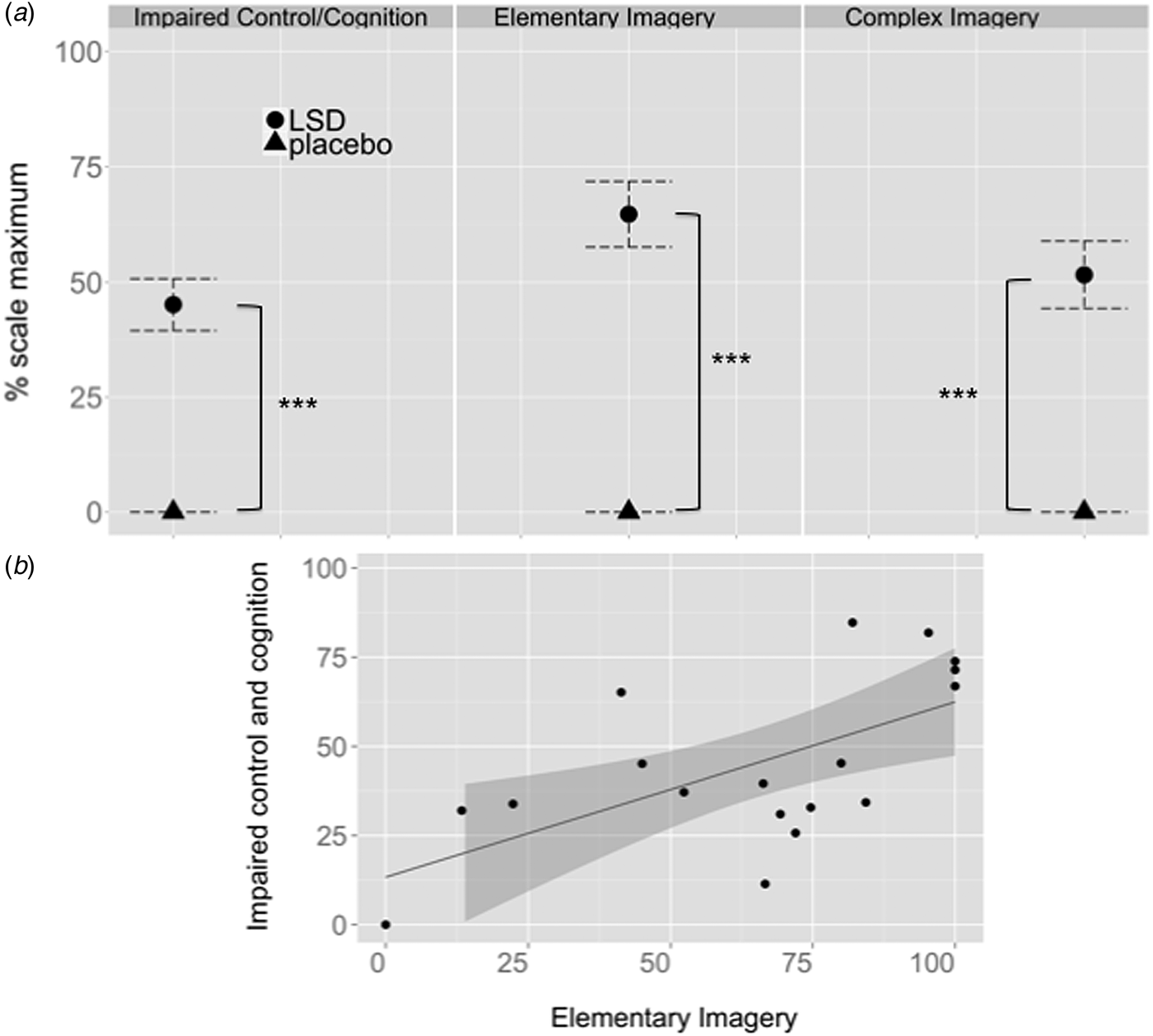
LSD produced significantly higher scores than placebo for cognitive impairments and visual hallucination as indexed by impaired control and cognition (t 17 = −8.007, p < 0.0001), elementary (t 17 = −9.099, p < 0.001) and complex imagery (t 17 = −7.052, p < 0.001), respectively (Fig. 1a). There was a significant positive correlation between impaired control/cognition and elementary (r = 0.620, p = 0.006, corrected for multiple testing) (Fig. 1b) but not complex (r = 0.235, p = 0.347) imagery after LSD intake.
(a) Subjective LSD effect on cognitive control (impaired control and cognition) and elementary and complex imagery. Error bars reflect standard errors. ***Indicates significant LSD effects at p < 0.001. (b) Significant positive correlation between impaired control and cognition and elementary imagery after LSD intake (r = 0.620, p = 0.006).

LSD plasma concentrations
LSD plasma concentrations rapidly increased to 1.29 ng/mL (s.d. 0.722) and 1.33 ng/mL (s.d. 0.59) 1 and 2 h after administration, while the concentration then decreased to 1.15 ng/mL (s.d. 0.52) 3 h after intake (online Supplementary Fig. S2). The LSD plasma concentration was positively related to subjectively experienced visual imagery 3 h after LSD administration (r = 0.56, p = 0.016, corrected for multiple testing) (online Supplementary Fig. S3), while there was also a strong trend for a positive relationship to LSD-induced cognitive impairments (r = 0.461, p = 0.054).
Behavioural task performance
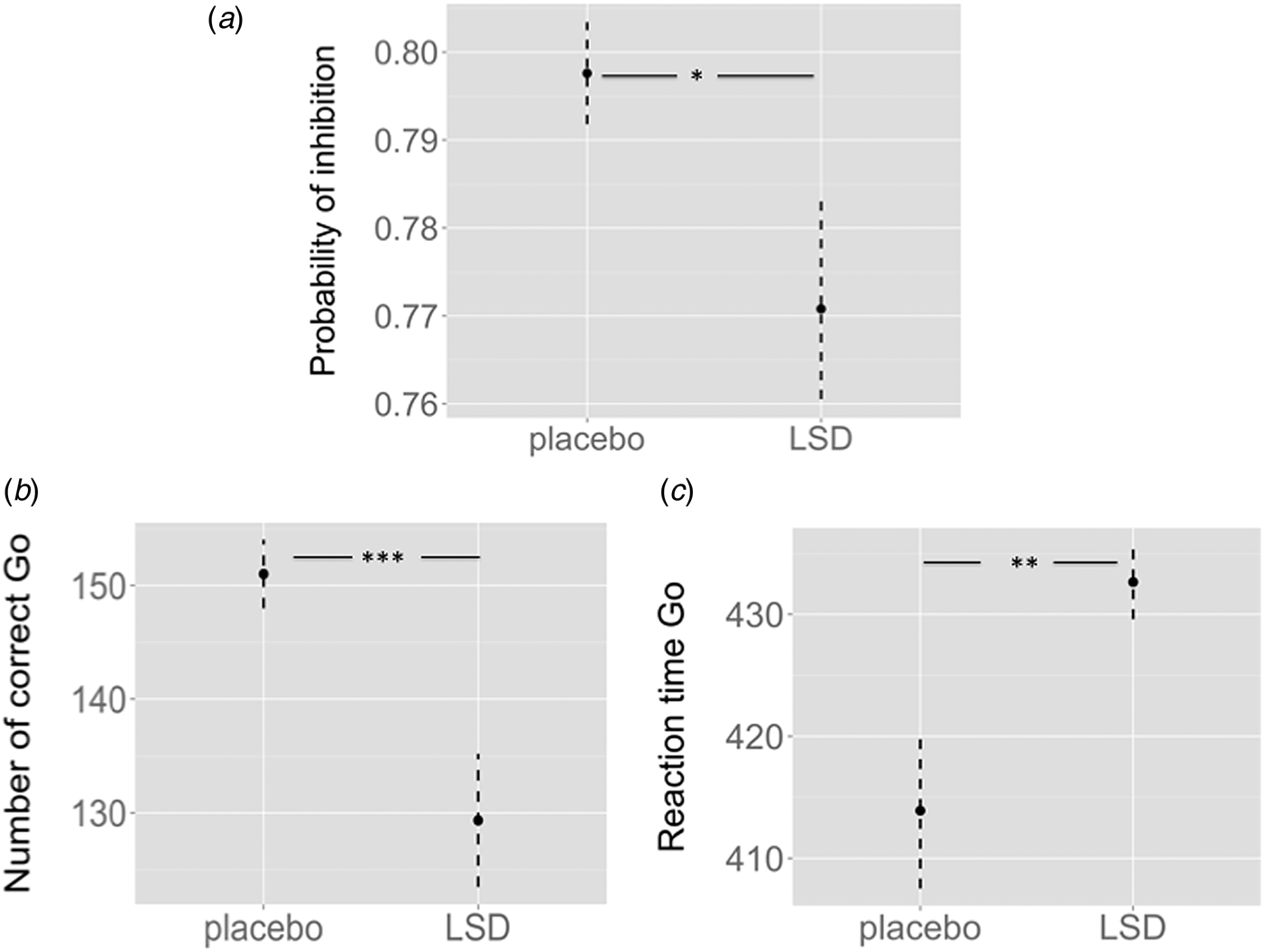
Relative to placebo (mean = 0.79, s.d. = 0.025), acute LSD administration (mean = 0.77, s.d. = 0.044) significantly reduced the probability of inhibition (t 17 = 2.19, p = 0.043) (Fig. 2a). LSD (mean = 129.33, s.d. = 24.83) reduced the number of responses to Go trials compared with placebo (mean = 151.00, s.d. = 12.84) (t 17 = 4.23, p = 0.001) (Fig. 2b). Furthermore, LSD (mean = 432.64, s.d. = 12.89) also prolonged reaction times to Go trials relative to placebo (mean = 413.90, s.d. = 27.10) (t 17 = −3.42, p = 0.003) (Fig. 2c). Notably, the probability of inhibition under LSD was negatively related to subjective LSD feelings of impaired cognition and control (r = −0.523, p = 0.026).
Task performance expressed as (a) probability of inhibition, (b) number of correct responses to Go trials and (c) reaction times to Go trials after placebo and LSD administration. Error bars reflect standard errors. *p < 0.05, **p < 0.01 and ***p < 0.001, significant differences in task performance between LSD and placebo.

Brain activation during response inhibition
Effect of task
Combined treatment maps revealed significant activation in frontal, striato-thalamic and cerebellar brain regions during response inhibition (online Supplementary Table S2).
LSD effects on brain activation during response inhibition
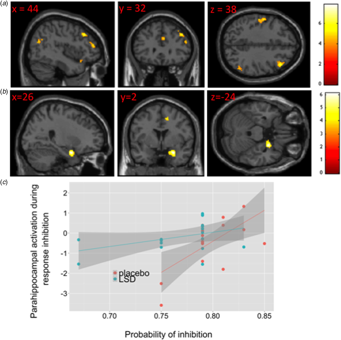
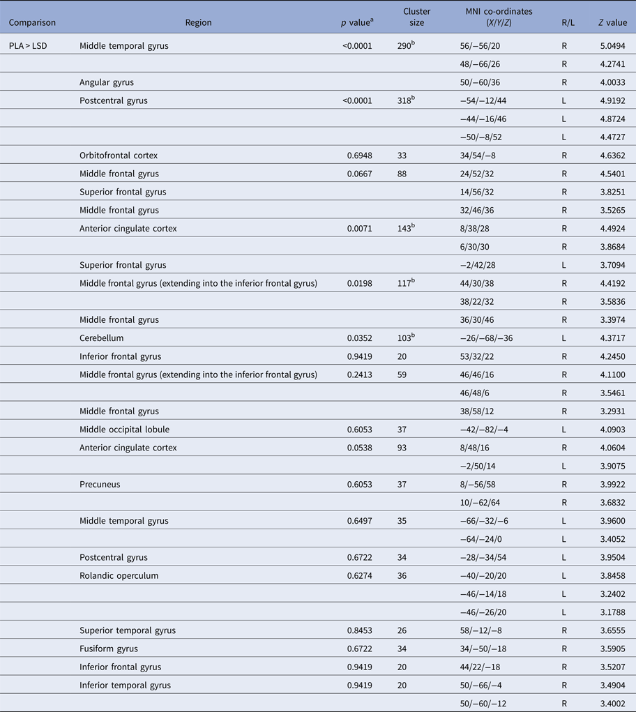
Relative to placebo, LSD significantly decreased activation in the right middle temporal, angular and inferior frontal gyrus, left postcentral gyrus, right anterior cingulate cortex and left cerebellum (Fig. 3a, Table 1).
(a) Significant difference in brain activation during response inhibition between the placebo and LSD treatment. Results are illustrated using a cluster-forming threshold p < 0.001 uncorrected, with an extent threshold of 20 voxels. (b) Significant different relationship between activation in the right parahippocampus (cluster size = 162, FWE corrected p value = 0.03, Z value = 4.95) and the probability of inhibition after placebo and LSD administration. (c) The magnitude of parahippocampal activation was positively correlated with the probability of inhibition in the placebo (r = 0.660, p = 0.003) but not LSD condition (r = 0.40, p = 0.10).

Significant differences in brain activation during response inhibition between LSD and placebo administration in 18 healthy controls

Results are reported using a cluster-forming threshold p < 0.001 uncorrected, with an extent threshold of 20 voxels.
a Cluster-level FWE-corrected.
b Survives FWE correction for multiple comparisons at the cluster level.
Relationship between brain activation and task performance
The relationship between right parahippocampal activation during response inhibition and the probability of inhibition significantly differed across the placebo and LSD treatment (Fig. 3b). While this relationship was positive under placebo exposure (r = 0.88, p < 0.001), there was no such relationship after LSD administration (r = −0.06, p = 0.8) (Fig. 3c).
Relationship between brain activation, plasma concentration, cognitive impairments and visual perception
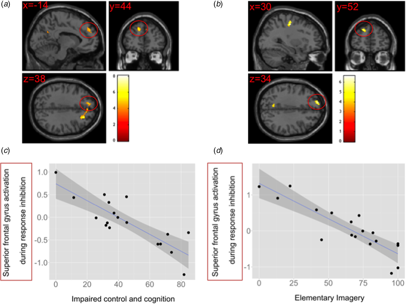
We found a significant negative relationship between the severity of subjectively experienced cognitive impairments and activation in the right middle frontal gyrus and right and left superior frontal gyrus (Figs 4a and 4c, online Supplementary Table S3). Furthermore, there was also a significant negative relationship between the severity of subjectively experienced elementary imagery and activation in the right precentral gyrus and left superior frontal gyrus (Figs 4b and 4d, online Supplementary Table S4). We found no significant relationship between LSD plasma concentration and brain activation during response inhibition.
Significant negative relationship between LSD-induced brain activation during response inhibition and (a) subjectively experienced cognitive impairments and (b) elementary imagery after LSD intake. Scatterplots showing the negative relationship between activation in the left superior frontal gyrus under LSD exposure and (c) subjectively experienced cognitive impairments (r = −0.835) and (d) elementary imagery (r = −0.875) after LSD administration.

Discussion
Our study provided the following main findings: First, there was a positive relationship between LSD effects on subjective feelings of impaired cognitive control and visual imageries. Second, acute LSD administration impaired inhibitory performance during the Go/No-Go task and reduced activation in the middle temporal gyrus, superior/middle/inferior frontal gyrus and the anterior cingulate cortex. Third, the relationship between parahippocampal activation during response inhibition and inhibitory performance was different under placebo and LSD exposure. While parahippocampal activation correlates positively with the inhibitory performance after placebo treatment, no such relationship is evident after LSD administration. Finally, activation in the left superior frontal gyrus under LSD exposure is negatively related to subjective LSD-induced cognitive impairments and visual imageries.
Acute LSD administration significantly increased subjective feelings of impaired cognitive control and visual imageries, in line with other recent LSD studies (Schmid et al. Reference Schmid2015; Liechti et al. Reference Liechti, Dolder and Schmid2016; Carhart-Harris et al. Reference Carhart-Harris2016a; Preller et al. Reference Preller2017). It has recently been shown that both of them can be blocked by the 5-HT2AR antagonist ketanserin, indicating that these subjective LSD effects are mediated via 5-HT2AR activation (Preller et al. Reference Preller2017). Consistent with our first hypothesis, these self-ratings were positively related to each other, supporting the view that 5-HT2AR activation by LSD might cause a cognitive disability to integrate new perception that triggers the formation of aberrant feelings and visual perception (Vollenweider & Geyer, Reference Vollenweider and Geyer2001; Geyer & Vollenweider, Reference Geyer and Vollenweider2008). The subjective effects of impaired cognitive control (e.g. I felt like a marionette, I felt isolated from everything and everyone, I was not able to complete a thought, my thought repeatedly became disconnected, I felt as though I were paralyzed) are phenomenologically similar to experiences of reality distortions. We have previously showed that acute LSD administration indeed increased the ‘distance to reality’ (Schmid et al. Reference Schmid2015). The here found relationship between impaired control and cognition and elementary imagery further suggest that impaired reality monitoring after LSD intake disrupts the ability to update internal representations and might thereby promote to the formation of visual imageries (e.g. I saw regular patterns in complete darkness or with closed eyes).
We also found that LSD impaired motor response inhibition during the Go/No-Go task as indexed by decreased probability of inhibition, an effect that was inversely related to the degree of subjective cognitive impairments after LSD administration. This finding might reflect an association between cognitive (inhibiting irrelevant mental processes) and behavioural (motor) inhibition (Bari & Robbins, Reference Bari and Robbins2013). In other words, LSD-induced feelings such as ‘I had difficulty in distinguishing important from unimportant things’ might have contributed to impairments in the inhibition of external stimuli (No-Go trials).
Furthermore, LSD reduced activation in key regions mediating response inhibition including the middle temporal gyrus, superior/middle/inferior frontal gyrus and the anterior cingulate cortex (Simmonds et al. Reference Simmonds, Pekar and Mostofsky2008; Swick et al. Reference Swick, Ashley and Turken2011). Reduced activation in the right inferior frontal gyrus (Ford et al. Reference Ford2004; Kaladjian et al. Reference Kaladjian2007), superior frontal gyrus (Ford et al. Reference Ford2004), middle frontal gyrus (Ford et al. Reference Ford2004; Arce et al. Reference Arce2006), anterior cingulate cortex (Rubia et al. Reference Rubia2001; Ford et al. Reference Ford2004; Arce et al. Reference Arce2006), middle temporal gyrus (Ford et al. Reference Ford2004) during response inhibition is also evident in schizophrenia patients. Furthermore, deficits in sensorimotor gating have been found to be negatively related to grey matter volumes in the right dorsolateral prefrontal cortex in schizophrenia patients (Kumari et al. Reference Kumari2008). Interestingly with the respect to the relationship between cognitive control and visual hallucinations, a longitudinal study in patients with Parkinson's disease found that the visual hallucinators had greater grey matter loss in bilateral superior and inferior frontal gyrus, anterior cingulate gyrus and limbic areas including hippocampus. Patients with Parkinson's disease without visual hallucinations at baseline did not exhibit the same pattern of atrophy at follow-up and none had developed dementia (Ibarretxe-Bilbao et al. Reference Ibarretxe-Bilbao2010).
More recently, increased connectivity has been shown between the anterior cingulate cortex and right dorsolateral prefrontal cortex during inhibition processes (Cieslik et al. Reference Cieslik2013). Previous theories of cognitive control propose that when erroneous or conflicting behaviour is detected by the anterior cingulate cortex, it signals to the lateral prefrontal cortex and other regions responsible for maintaining goal-directed behaviour that greater levels of control are necessary to successfully perform a task (Botvinick et al. Reference Botvinick2001; Brown & Braver, Reference Brown and Braver2005; Johnston et al. Reference Johnston2007). Therefore, we can speculate that the LSD-induced reduction in anterior cingulate cortex activation reflects impaired error processing what in turn led to a decreased top–down signal to the prefrontal cortex regions to adapt goal-directed behaviour (missing learning from failed inhibitions).
We also found that right parahippocampal activation correlated positively with the inhibitory performance after placebo treatment, whereas no such relationship was evident after LSD administration. Similar to the function of the anterior cingulate cortex (Rubia et al. Reference Rubia2003), activation in the parahippocampus during response has previously been reported in response to errors with the goal to engage additional top–down resources to improve behaviour (Braet et al. Reference Braet2011). Our finding might thus indicate that while people under placebo were able to learn from previous errors during the task and improve response inhibition via parahippocampal activation, acute LSD administration seemed to prevent a continuous improvement of task performance due to an impaired learning signal from the parahippocampus. The hippocampus and the anterior cingulate cortex work in concert during error processing; activation in the hippocampus correlated with error-feedback-related activation in the anterior cingulate cortex (Hester et al. Reference Hester2008). Like the anterior cingulate cortex, the hippocampus is also functionally related to the lateral prefrontal cortex during inhibitory processing (Chudasama et al. Reference Chudasama, Doobay and Liu2012), and disruption of hippocampal–prefrontal interactions have been observed in psychiatric diseases, most notably in schizophrenia (Godsil et al. Reference Godsil2013; Sigurdsson & Duvarci, Reference Sigurdsson and Duvarci2015). These findings together suggest that LSD impaired the error-related activation in the anterior cingulate cortex and parahippocampus and thereby impeded a subsequent recruitment of dorsolateral prefrontal regions (superior/middle/inferior frontal gyrus) to adjust further task performance.
Finally, there was a negative relationship between left superior frontal gyrus activation during response inhibition and subjective feelings of impaired cognitive control and visual hallucinations after LSD administration. Previous meta-analytical evidence revealed the involvement of the left superior frontal gyrus during response inhibition (Swick et al. Reference Swick, Ashley and Turken2011) and it has been shown that patients with left superior frontal gyrus lesions were globally impaired in cognitive control processes (du Boisgueheneuc et al. Reference du Boisgueheneuc2006). Decreased cortical thickness in the left superior frontal gyrus was related to decreased cognitive control in patients with schizophrenia (Tully et al. Reference Tully2014). Moreover, the superior frontal gyrus was shown to be involved in the inhibition of internally represented information (Corbetta et al. Reference Corbetta, Kincade and Shulman2002). Our results suggest that the subjective LSD-induced impairments in cognitive control (e.g. ‘I had difficulty in distinguishing important from unimportant things’, ‘I was not able to complete a thought, my thought repeatedly became disconnected’) might have contributed to an enhancement of internally generated representation and thereby impaired response inhibition via reduced activation in the superior frontal gyrus. This rekindling of internal representations (together with a neglect of external stimuli) possibly led to LSD-induced visual imageries. Supportive for this interpretation, previous studies showed that Parkinson patients with visual hallucinations had reduced grey matter volume in the left superior frontal gyrus compared to healthy controls (Gama et al. Reference Gama2014) and less activation in the left superior frontal gyrus than non-hallucinating patients during the processing of complex visual-perceptual stimuli (Ramírez-Ruiz et al. Reference Ramírez-Ruiz2008).
Taken together, our results show that LSD reduced activation in regions responsible for error detection such as the anterior cingulate cortex and parahippocampus during response inhibition, which might have led to an insufficient recruitment of dorsolateral prefrontal regions (i.e. superior/middle/inferior frontal gyrus) and in turn to an impaired learning from these errors to update goal-directed behaviour. This cascade and in particular reduced activation in the superior frontal gyrus after LSD administration finally might have shifted the focus away from external stimuli towards internally generated representation and possibly the formation of visual hallucinations. Of course, other brain areas are certainly also involved in the hallucinatory effects of LSD, in particular the visual cortex. It has been shown that LSD increased cerebral blood flow in the visual cortex and the functional connectivity to other brain regions, and both effects were correlated with complex imagery after LSD intake (Carhart-Harris et al. Reference Carhart-Harris2016b).
There are some limitations to be considered in the present study. Although we used a well-established paradigm from previous fMRI studies (Rubia et al. Reference Rubia2006; Schmitz et al. Reference Schmitz2006; Borgwardt et al. Reference Borgwardt2008; Lawrence et al. Reference Lawrence2009; Schmidt et al. Reference Schmidt2013; Bhattacharyya et al. Reference Bhattacharyya2014; Daly et al. Reference Daly2014; Bhattacharyya et al. Reference Bhattacharyya2015; Schmidt et al. Reference Schmidt2017), we were not able to disentangle neural activation in response to successful v. failed inhibitions in the present study due to the modest number of No-Go trials. This also relativizes our interpretations that LSD effects on the anterior cingulate cortex and parahippocampus are probably related to impaired error processing. Due to the fast event-related design, the short event durations and the modest number of No-Go trials, the paradigm is further not well suited to address inhibition-induced connectivity within the neural response network. The here found relationship between cognitive impairments and visual imageries after LSD intake are correlative and should thus be considered with caution. Further studies are warranted to understand the causality of this relationship. To further disentangle the relationship between cognitive impairments and visual hallucinations after 5-HT2AR stimulation, future studies might also want to conduct multiple tests to assess prefrontal-medicated cognitive control and reality monitoring processes. Having in mind that LSD did not only reduce the probability of inhibition during the task, but also decreased responses to Go trials and prolonged reaction times, a broad cognitive test battery should also help to explore if LSD specifically impairs response inhibition or rather cognitive processes in general. A further limitation of all studies using LSD is that blinding is difficult to maintain due to the subjective drug effects of the substance. We can therefore not exclude that expectations influenced the subjective drug effects of LSD, while this seems less likely for the neuronal activity patterns.
In summary, the present study showed that 5-HT2AR activation by LSD led to deficits in inhibitory processing mediated via reduced parahippocampal–prefrontal activation in healthy volunteers. Our findings further provide a neuropsychopharmacological mechanism how impaired inhibitory processing might contribute to the formation of LSD-induced visual hallucinations. This study helps to better understand the neuropsychopharmacological mechanisms of visual hallucinations in LSD-induced states and neuropsychiatric disorders.
Supplementary Material
The supplementary material for this article can be found at https://doi.org/10.1017/S0033291717002914.
Acknowledgements
This work was supported by the Swiss National Science Foundation (grant no. 320030_170249 to MEL and SB).
Declaration of Interest
None.