Introduction
Salinity stress can cause lichen dehydration, ionic imbalances and the inhibition of photosynthesis. A few works have addressed the impact of salinity stress on lichens, mostly focusing on its effects on net photosynthesis (Matthes-Sears et al. Reference Matthes-Sears, Nash and Larson1987; Nash III & Lange Reference Bates, Wibbelmann and Proctor1988; Smith & Gremmen Reference Smith and Gremmen2001). Investigations on Ramalina menziesii Tayl., found high variability in water content, CO2 exchange (Matthes-Sears et al. Reference Matthes-Sears, Nash and Larson1986) and morphology (Larson et al. Reference Bates, Wibbelmann and Proctor1985) between samples collected along a coastal to inland gradient. While the morphological variation was correlated with the amount of sea salt that could be eluted from the thallus (Larson et al. Reference Bates, Wibbelmann and Proctor1985), manipulated intrathallus salt concentrations had no effect on net photosynthesis (Matthes-Sears et al. Reference Matthes-Sears, Nash and Larson1987). On the other hand, twelve species from a coastal to inland gradient demonstrated considerable variation in salt sensitivity, proportional to the distance from the coast (Nash III & Lange Reference Bates, Wibbelmann and Proctor1988). More recently, a study with the lichen Turgidiusculum complicatulum (Nyl.) Kohlm found that salinity up to the level of undiluted sea water had no effect on chlorophyll fluorescence and gas exchange rates, thus allowing the authors to suggest that this is a non-obligate halophyte species (Smith & Gremmen Reference Smith and Gremmen2001). Despite the use of different methodologies in these works, it is clear that the response to salinity stress is highly variable between species, thus determining their distribution patterns along the coastal-inland gradient. In the Mediterranean area, work on salinity has focused mostly on Ramalina canariensis Steiner and R. calicaris (L.) Fr. with the aim of using them as biomonitors of salt deposition. However, the physiological impact of salinity stress has been neglected (Figueira et al. Reference Figueira, Branquinho, Pacheco, Sousa and Catarino1994, Reference Bates, Wibbelmann and Proctor1998, Reference Bates, Wibbelmann and Proctor1999a, b, Reference Bates, Wibbelmann and Proctor2002).
To investigate the impact of saline stress in lichens it is essential to determine the cellular location of Na+, K+, Ca2+ and Mg2+. Elements may be found in three major cellular locations: 1) deposited at the lichen surface or trapped in the intercellular spaces (surface fraction); 2) bound to exchange sites on the cell wall (cell wall fraction); and 3) accumulated in the cytoplasm (intracellular fraction) (Brown & Brown Reference Brown and Brown1991). The intracellular fraction is particularly important when studying the impacts of element accumulation on physiological parameters (Buck & Brown Reference Buck and Brown1979; Brown & Brown Reference Brown and Brown1991). The quantities of extracellular elements (surface and cell wall fraction) are dependent on physicochemical processes, such as deposition on the thallus surface or ionic binding to cell wall exchange sites, rather than to physiologically controlled phenomena (Brown & Brown Reference Brown and Brown1991; Figueira et al. Reference Figueira, Sousa, Brown, Catarino and Pacheco1999a).
In the work reported here we investigated the effect of different sea water concentrations, and the time of exposure, on the lichen Ramalina canariensis. This is a widely distributed epiphytic species, common in south-western coastal areas of Portugal (Matos Reference Matos2007). The aim of the work presented here was to study the effects of time of incubation with sea water on ion location and chlorophyll fluorescence parameters in this species.
Material and Methods
Saline contamination
Samples of R. canariensis were collected in Comporta, a coastal site located in Baixo Alentejo, Portugal (517366, 4248326 WGS 1984 UTM Zone 29), from trunks of pine trees (Pinus pinaster L.) at an average height of 1·5 m. Samples were air dried, cleaned and stored in plastic bags at 4°C for further analysis. Before each experiment, thalli of approximately 50 mg were kept for 24 h in a chamber with high relative humidity (over water in a closed box). This step is essential to reactivate physiological activity and to re-establish the integrity of cellular membranes (Buck & Brown Reference Buck and Brown1979).
Three different experiments were performed, all of them under normal laboratory light which varied from 55 to a maximum of 100 µmol m−2 s−1 at room temperature of about 20°C. In the first experiment, samples were immersed in 2 l of artificial sea water (Sigma, S-0131; 7·9 mM Ca2+, 8·3 mM K+, 36·4 mM Mg2+ and 397·4 mM Na+ in the 100% sea water solution) of different concentrations (0% – control, 1%, 10%, 20%, 50% and 100%) for 2 h. Solutions were stirred with an air pump. Samples were removed from the solutions and chlorophyll fluorescence measurements were performed and the cellular location of ions was determined. In the second experiment, samples were immersed in 2 l of artificial sea water solution of 100% (0% – control) for 30 s, 1, 5, 15, 30, 60, 90 and 120 min, and then chlorophyll fluorescence and the cellular fractions of saline elements were measured. In the third experiment, samples were pre-incubated for 120 min in 2 l of an artificial sea water solution of 100 % (0% – control) and were then transferred to deionized water to investigate whether the accumulation of saline elements and its effects on the photosynthetic apparatus were reversible. Following different incubation times in 2 l of deionized water (30 s, 1, 5, 15, 30, 60, 90 and 120 min) chlorophyll fluorescence and the cellular fraction of saline elements were determined. In all experiments, five replicates were used.
Lichen element content determination
The cellular fractions of cations (Na+, K+, Mg2+ and Ca2+) were determined following a modified version of the sequential elution technique developed by Brown & Wells (Reference Bates, Wibbelmann and Proctor1988). To extract the surface fraction, lichen samples were sequentially stirred in two plastic containers with 10 and 5 ml of deionized water for 40 and 30 min respectively, and then for 40 and 30 min with 10 and then 5 ml of a NiCl2 solution (20 mM) to obtain the cell wall fraction. The aliquots were combined before analysis. Afterwards, the dry mass of the samples was determined after drying to constant mass (c. 16 h) in an oven at 80 ºC, and then stirred for 90 min in 10 ml of a NiCl2 solution (20 mM) to extract the intracellular soluble fraction. Element concentrations were measured using a flame atomic absorption spectrophotometer (air / acetylene flame) (Varian Techtron AA59, Australia). When needed, CsCl and LaCl3 (1 g l−1) were added to samples and standards as suppressants of ionization and refractory compound formation, respectively.
Chlorophyll fluorescence measurements
Measurements were made using a Mini-Pam fluorometer (Walz, Effeltrich, Germany). In the first experiment, measurements were performed 0, 14, 24, 32 and 48 h after samples had been incubated in artificial sea water solutions of different concentrations (see above). In the second and third experiments, measurements were taken 0 and 24 h after samples had been incubated for different time-intervals (see above). Between measurements, samples were kept fully hydrated at room temperature and light (which varied from 55 to a maximum of 100 µmol m−2 s−1) over water in a closed box. The minimum level of fluorescence (F 0) was obtained under modulated red light and maximal fluorescence yields (F m) recorded following exposure to a saturating light pulse. Variable fluorescence (F v) is the difference between these two parameters. The ratio of variable to maximal fluorescence (F v/F m) was measured after 5 min dark adaptation (Branquinho et al. Reference Branquinho, Catarino, Brown, Pereira and Soares1999).
Statistical analyses
Statistical analyses were performed using SPSS (v. 14.0, SPSS Inc., Chicago, Illinois, USA). Data were first checked for normal distribution with the Kolmogorov-Smirnov test and for homogeneity of variances with Levene's test. The main effects of artificial sea water concentration and incubation time on element concentrations and F v/F m were investigated using one-way analysis of variance (ANOVA). Treatment means were then compared using Tukey's test, whenever significant differences were found. Simple linear correlation coefficients (Pearson's r) were calculated between all elements (Na+, K+, Mg2+ and Ca2+) determined in both the cell wall and intracellular fractions analyzed in samples treated with different artificial sea water concentrations and between these concentrations and the F v/F m. A t-test for independent samples was performed to detect differences between element concentrations and F v/F m values in treatment and control samples at each incubation time.
Results
Impacts of different saline concentrations
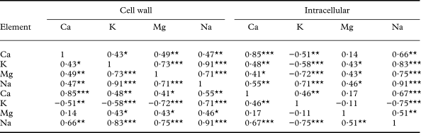
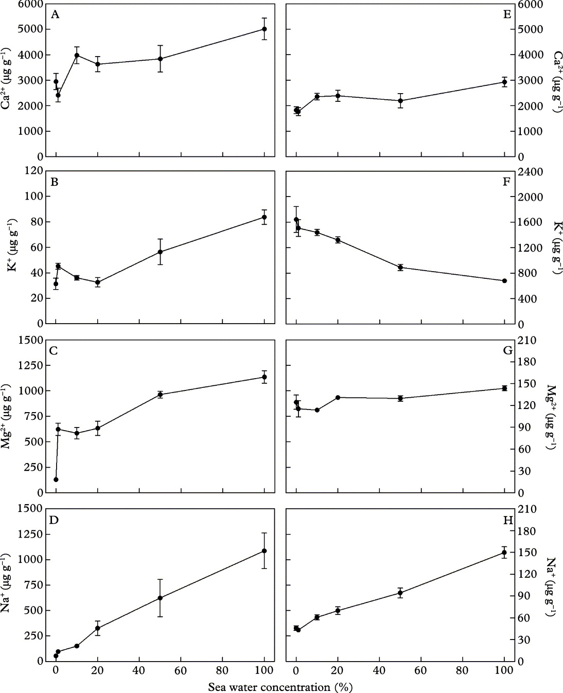
An increase in the concentration of the sea water solution resulted in higher accumulation of elements in the cell wall fraction. Samples incubated in a 100% artificial sea water solution had significantly (P <0·001) higher concentrations of all four elements in comparison to control samples, that is, those incubated in deionized water (Fig. 1A–D). This pattern was also observed for intracellular Na+ and Ca2+ concentrations (P <0·01 and P <0·001, respectively) (Fig. 1E & H). The K+ intracellular concentration significantly declined in those samples incubated in artificial sea water solution with concentrations equal or higher than 50% (Fig. 1F), while the intracellular levels of Mg2+ were not significantly affected by incubation in artificial sea water solutions (Fig. 1G). Intracellular Na+ concentration and the cell wall K+ concentration (Table 1) were positively correlated (0·83). In contrast, there was a negative correlation (−0·75) between their intracellular concentrations.
Correlation coefficients matrix of elements determined in the cell wall and intracellular fractions of Ramalina canariensis after a 2 h incubation period in artificial sea water solutions of different concentrations (0% – control, 1%, 10%, 20%, 50% and 100%)

Asterisks indicate significant correlations:
* P <0·05
** P <0·01
*** P <0·001
Concentration of Ca2+, K+, Mg2+ and Na+ (µg g−1) in cell wall (A, B, C, D) and intracellular fractions (E, F, G, H) in samples of Ramalina canariensis following a 2 h incubation period in artificial sea water solutions of different concentrations. Values represent mean ± 1 SEM (n = 5).

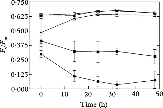
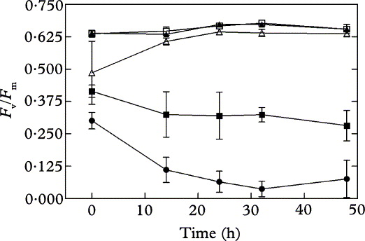
In samples incubated for 2 h in artificial sea water solutions of 20%, 50%, and 100%, F v/F m significantly declined by 24%, 35%, and 53%, respectively, in comparison to control samples (Fig. 2). Measurements taken 48 h after the end of the incubation showed no recovery in F v/F m for samples incubated in 50% and 100% artificial sea water solutions. However, the F v/F m of samples which had been incubated in a 20% artificial sea water solution recovered to control levels 48 h after incubation.
Effect of 2 h incubation in artificial sea water solutions of different concentrations (0% – ○; 1% – □; 10% –▲; 20% – △; 50% – ■; 100% – ●), on maximum photochemical efficiency of Photosystem II (F v/F m) in samples of Ramalina canariensis. Measurements were taken at 0, 14, 24, 32, and 48 h after incubation. Values represent mean ± 1 SEM (n = 5).
Impact of incubation times
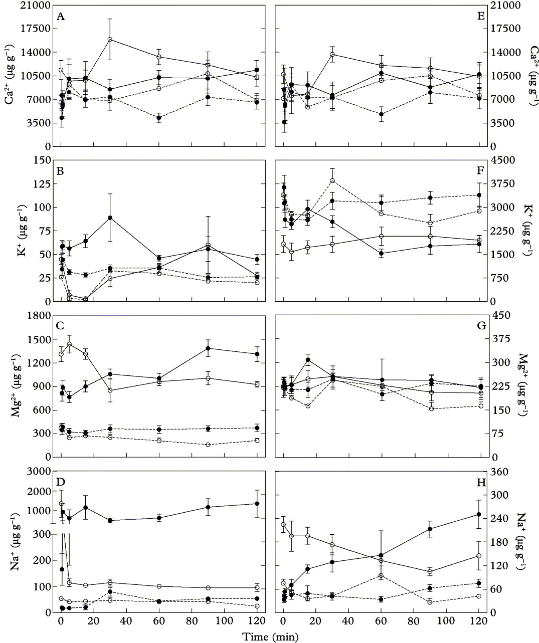
Significant shifts in the levels of K+, Na+ and Mg2+ (P <0·05, P <0·01 and P <0·05, respectively) in the cell-wall fraction (Fig. 3B–D), in comparison to controls, were detected after 30 s. The levels of Na+ and Mg2+ stabilized after 1 min, while K+ concentrations stabilized after 60 min. In addition, after 60 min, the cell wall Ca2+ concentration significantly increased (Fig. 3A).
Concentration of Ca2+, K+, Mg2+ and Na+ (µg g−1) in the cell-wall (A,B,C,D) and intracellular (E,F,G,H) fractions in samples of Ramalina canariensis. Solid symbols (●) represent samples incubated in a 100% artificial sea water solution during different incubation times (30 s, 1, 5, 15, 30, 60, 90 and 120 min). Open symbols (○) represent samples incubated in deionized water for different incubation times (0, 1, 5, 15, 30, 60, 90 and 120 min) following a 2 h pre-incubation in an artificial 100% sea water solution. Control samples were incubated in deionized water for the same periods of time as treatments and are represented by a dashed line. Values represent mean ±1 SEM (n = 5).
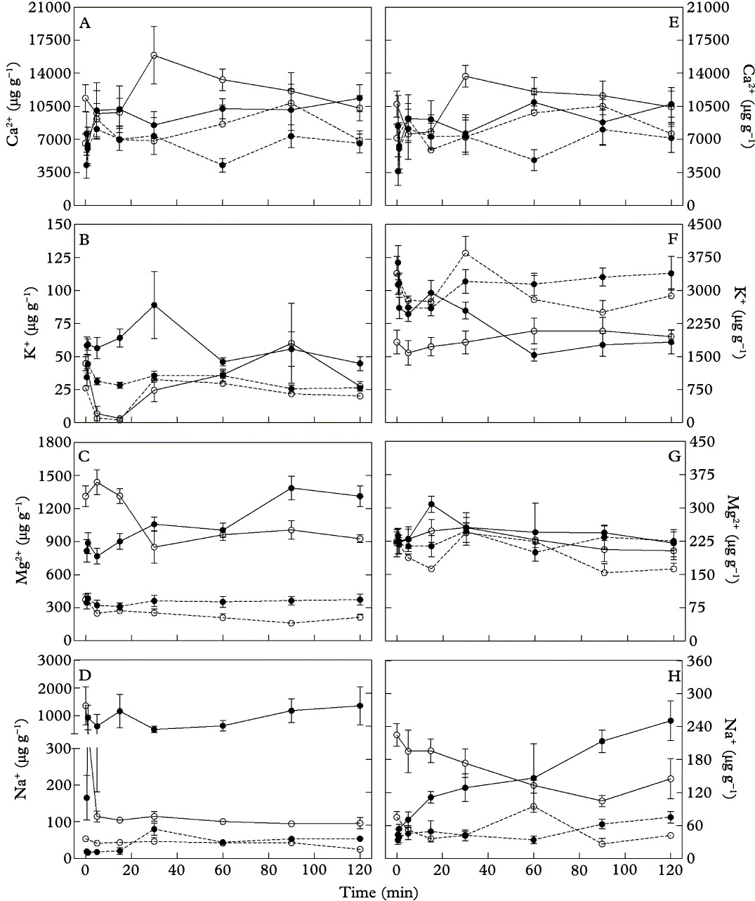
In the intracellular fraction, the Na+ concentration increased until the incubation time had reached 60 min (Fig. 3H), whereas K+ levels decreased (Fig. 3F). After 60 min, both the Na+ and K+ intracellular concentrations stabilized. The intracellular concentration of Ca2+ increased significantly after 60 min (P <0·01), but Ca2+ levels did not stabilize thereafter (Fig. 3E). No significant changes in intracellular Mg2+ levels were observed (Fig. 3G), consistent with results from the first experiment (see above).
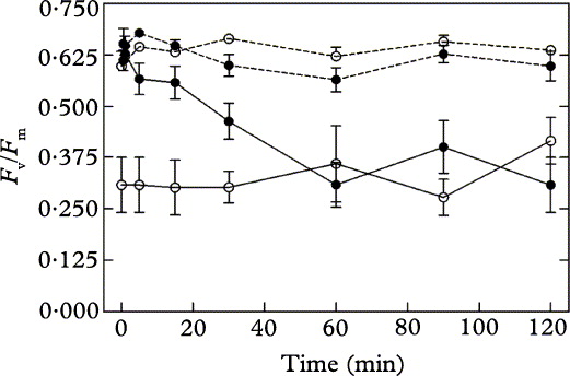
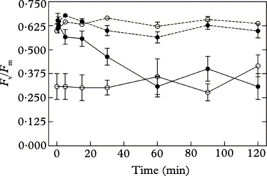
Incubating lichens in an artificial 100 % sea water solution significantly (P <0·05) decreased F v/F m after 5 min (Fig. 4). Inhibition increased with time (P <0·001), and stabilized after 60 min of incubation. Measurements taken 24 h after the end of incubation revealed that F v/F m values of lichen samples which had been incubated for 30 min or longer did not recover to control values (data not shown).
Impact of increasing incubation times on maximum photochemical efficiency of Photosystem II (F v/F m) in samples of Ramalina canariensis. Solid symbols (●) represent samples incubated in a 100% artificial sea water solution during different incubation times (30 s, 1, 5, 15, 30, 60, 90 and 120 min). Open symbols (○) represent samples incubated in deionized water for different incubation times (0, 1, 5, 15, 30, 60, 90 and 120 min) following a 2 h pre-incubation in an artificial 100% sea water solution. Control samples were incubated in deionized water for the same periods of time as treatments and are represented by a dashed line. Values represent mean ± 1 SEM (n = 5).
Reversibility of changes in saline element concentrations and F v/F m
Immersing lichens for 5 min in deionized water reduced the cell-wall Na+ concentration in samples which had been pre-incubated in a 100% artificial sea water solution (Fig. 3D). However, further immersion in deionized water resulted in no additional changes in the cell wall levels of Na+ which were still higher than those of control samples, that is samples which had been pre-incubated in deionized water. Immersion in deionized water also decreased intracellular Na+ concentration, but values remained higher than those of control samples (Fig. 3H). In the cell wall and intracellular fractions Na+ levels stabilized after 5 min and 60 min respectively. The concentration of K+ in the cell wall fraction of samples pre-incubated in a 100% artificial sea water solution returned to control values following immersion in deionized water (Fig. 3B). Although the decrease in intracellular K+ observed in pre-incubated samples was reversed following a 2 h immersion with deionized water, the K+ intracellular concentrations did not recover to values observed in control samples (Fig. 3F). The concentration of Mg2+ in the cell-wall fraction declined after a 30 min immersion in deionized water, and values remained higher than those of control samples (Fig. 3C). Similarly, the levels of both intracellular and cell-wall Ca2+ (Fig. 3A & E) did not return to values of control samples.
Immersion in deionized water did not result in an observable recovery of the F v/F m of samples which had been pre-incubated in an artificial 100% sea water solution for 2 h (Fig. 4). Furthermore, 24 h after the end of immersion in deionized water, F v/F m values were still significantly (P<0·05) lower than the F v/F m of control samples (data not shown).
Discussion
Incubating R. canariensis in sea water had a significant effect on the cellular location of Na+, K+, Mg2+ and Ca2+. This effect was time dependent both in the extra- and intracellular fractions and was accompanied by a decrease in lichen vitality, expressed as K-loss from the cell interior and a decline in F v/F m. These effects were irreversible. The determination of the intracellular concentrations of ions allowed us to explain the physiological impacts of sea water, and to detect relationships between element concentrations and effects on lichen vitality which depended predominantly on the intracellular concentrations of K+ and Na+, and not the total concentrations.
As with previous studies (Branquinho et al. Reference Branquinho, Brown, Máguas and Catarino1997a, b; Figueira et al. Reference Figueira, Sousa, Brown, Catarino and Pacheco1999a), results presented here reveal the importance of determining the cellular location of ions. During incubation in sea water, intracellular Na+ concentrations increased more slowly than those in the cell-wall, and there was no change in the Mg2+ intracellular concentration. Intracellular K+ concentrations decreased more than those in the cell-wall. A study with Ramalina calicaris reported similar findings for the effects of salinity stress on the accumulation of Na+ and Mg2+ in the cell-wall and intracellular fraction (Figueira et al. Reference Figueira, Branquinho, Pacheco, Sousa and Catarino1994), and contamination assays with Pb and Cu observed similar shifts in the intracellular concentrations of K+ and Mg2+ in Ramalina farinacea (L.) Ach. and Ramalina fastigiata (Pers.) Ach., respectively (Branquinho et al. Reference Branquinho, Brown, Máguas and Catarino1997a, b). Results therefore suggest that saline stress compromised cell membrane permeability but it did not result in generalized membrane damage. A major effect of pollution in lichens is a change in membrane permeability to ions, accompanied by electrolyte loss, namely K+, and Mg2+ (Nash Reference Bates, Wibbelmann and Proctor1996). Several investigations with lichens (Branquinho et al. Reference Branquinho, Brown, Máguas and Catarino1997a, b, Reference Bates, Wibbelmann and Proctor1999; Garty et al. Reference Garty, Kloog and Cohen1998, Reference Bates, Wibbelmann and Proctor2001; Godinho et al. Reference Godinho, Freitas and Wolterbeek2004; Hauck et al. Reference Hauck, Paul and Spribille2006) have used loss of intracellular K+ as an indicator of cell membrane integrity. However, in the event of generalized membrane damage, as in the case of desiccated lichens, loss of intracellular Mg2+ has been observed (Buck & Brown Reference Buck and Brown1979; Branquinho et al. Reference Branquinho, Brown, Máguas and Catarino1997a).
The present study is the first on the effects of sea water on F v/F m in R. canariensis, a Mediterranean species. Our results revealed that the incubation of lichens in artificial sea water solutions with a concentration equal or higher than 50% significantly reduced F v/F m. In contrast to our results, a study with Turgidiusculum complicatulum, a rocky shore lichen exposed to sea spray and even periodic sea water inundation, reported that salinity up to the level of undiluted sea water had no effect on the chlorophyll fluorescence and gas exchange rates (Smith & Gremmen Reference Smith and Gremmen2001). Smith & Gremmen (Reference Bates, Wibbelmann and Proctor2001) suggested that T. complicatulum is a salt-tolerant, but non-obligate halophyte species. In our study, the steep decline observed in the F v/F m and its irreversibility in immersions for more than 15 min suggest that R. canarinesis has some degree of tolerance to sea salt but cannot be considered a halophytic species. Periodic submersions in sea water, such as those experienced by T. complicatulum or some Verrucaria halophytic species from coastal maritime rocks, may induce critical damage to R. canarinesis, threatening its survival, but more work is needed to confirm this.
In the present study, the decline observed in F v/F m was probably not due to photo-injury. Bates et al. (Reference Bates, Wibbelmann and Proctor2009) showed that the osmotic stress posed by sea water could increase susceptibility to photo-injury in non-halophytic bryophytes, and showed that this was also detectable in halophytic species. However, under the low light conditions used in this study, photo-injury is unlikely to occur. Therefore, F v/F m can be used as a direct indicator of saline stress, as shown in previous studies (Branquinho et al. Reference Branquinho, Brown, Máguas and Catarino1997a, b; Bates et al. Reference Bates, Wibbelmann and Proctor2009).
An exposure of 30 s in 100% sea water solution did not reduce F v/F m. Although quite different in methodology, a study with the same time of exposure, but repeated every second day until a maximum of 27 days, also found no effect of salt exposure (max. 300 mol m−3 NaCl) on net photosynthesis in R. menziesii (Matthes-Sears et al. Reference Matthes-Sears, Nash and Larson1987). More recent studies (Jensen et al. Reference Jensen, Chakir and Feige1999; Hájek et al. Reference Hájek, Barták and Dubová2006) found that high concentrations of NaCl and sucrose inhibited PSII activity in several foliose lichens. In our work, the decline in the F v/F m was dependent on the time of incubation: after 5 min it was already detectable, and no further decline occurred after 60 min, the same time when no further changes in intracellular concentration of Na+ and K+ occurred. The recovery of PSII activity was dependent on the length of time the lichens were exposed to the saline stress. In the present study, for incubations up to 15 min, the damage to PSII was reversible after 24 h (data not shown). However, for samples incubated for 2 h, the damage to PSII was irreversible, as F v/F m values remained consistently lower than controls even 48 h after 2 h incubation with deionized water. Nash & Lange (Reference Bates, Wibbelmann and Proctor1988) treated three very saline-sensitive species to different solutions with osmotic values similar to a 100% sea-salt solution. They found that the reduction in net photosynthesis was a response to both salt ion and osmotic effects, but varied depending on the species.
The production of reactive oxygen species (ROS) such as superoxide (O2.−), hydroxyl radical (.OH), singlet oxygen (1O2) and hydrogen peroxide (H2O2) is enhanced by stress (Kranner et al. Reference Kranner, Beckett, Hochman and Nash2008). In the lichen Ramalina lacera (With.) J. R. Laundon desiccation induced the production of ROS (Weissman et al. Reference Weissman, Garty and Hochman2005) but no work has been done regarding saline stress. A study with moss species suggests that the generation of reactive oxygen species under saline stress may also contribute to irreversible declines in F v/F m, particularly under high light conditions (Bates et al. Reference Bates, Wibbelmann and Proctor2009). ROS generation might have been responsible for the irreversible reduction in the F v/F m observed in our work. Future work on the physiological causes of saline stress should include measurements of additional chlorophyll fluorescence parameters, such as non-photochemical quenching (NPQ) and the measurement of ROS generation to establish the precise cause of the damage.
In the present study the F v/F m measurements indicate that saline stress, that is incubation in an artificial sea water solution of 50% and above, may irreversibly impair photosynthesis, thus compromising lichen vitality. An irreversible reduction in F v/F m was detected for intracellular Na+ levels above 70 µg g−1 and for intracellular K+ concentrations below 1320 µg g−1, following a 2 h incubation period. These results give us an indication of the resistance threshold of R. canariensis in an acute exposure. At coastal sites, due to high relative humidity and frequent fog episodes, organisms can be exposed to high levels of saline stress, for salt crystals solubilize when relative humidity rises above 70%. The lichen studied, R. canariensis, is a frequent species in sites located near the sea (Matos Reference Matos2007). The distribution of the lichen in the study area (P. Matos, unpublished) indicates that the abundance of R. canariensis increases close to the coast, unlike other Ramalina species in the region, so its coastal populations might have adaptative mechanisms involving either exclusion and/or increased levels of tolerance to saline stress. Further work is needed to establish whether R. canariensis can be regarded as partially halophytic.
The authors thank Lurdes Aires for technical assistance. The work was funded by FCT, contract POCTI/MGS/41521/2001.