Introduction
Axymyiidae is a small family of semi-aquatic lower (nematocerous) Diptera. The family contains three described extant genera: Axymyia McAtee, Mesaxymyia Mamaev, and Protaxymyia Mamaev and Krivosheina (Krivosheina Reference Krivosheina2000). There are currently seven described species, of which all except Axymyia furcata McAtee are Palearctic. Axymyia furcata is widespread in eastern North America, ranging from the upper midwestern United States of America (Minnesota and Iowa) through the northeastern United States and southeastern Canada, and throughout Appalachia to South Carolina and Georgia (Wihlm and Courtney Reference Wihlm and Courtney2011). An undescribed eighth species has been recorded in Oregon (Wood Reference Wood1981; Dudley and Anderson Reference Dudley and Anderson1982; G.W. Courtney, unpublished records). In addition, three extinct genera, Sinaxymyia Zhang, Juraxymyia Zhang, and Psocites Hong, are known from Kazakhstan and China (Zhang Reference Zhang2010).
Adult and larval A. furcata were first described, respectively, by McAtee (Reference McAtee1921) and Krogstad (Reference Krogstad1959; see also Peterson Reference Peterson1960, fig. D9). Wihlm (Reference Wihlm2009) and Wihlm and Courtney (Reference Wihlm and Courtney2011) have shown that immature axymyiids, once considered rare, can be abundant in suitable habitats. These flies are dependent on woody debris in small streams and seeps, where the larvae and pupae reside in galleries formed in wet, decomposing wood. Adults emerge in early to mid spring, are short lived, and appear to remain near their natal habitat. With oviposition also occurring on prospective larval habitat, these flies are clearly dependent on a xylophilic lifestyle.
Although distinct in all life stages, the phylogenetic position of Axymyiidae has remained among the most problematic questions in lower Diptera. The family has in various classifications been associated with Bibionidae, Anisopodidae, or Pachyneuridae (see reviews by Mamaev and Krivosheina Reference Mamaev and Krivosheina1966; Wood and Borkent Reference Wood and Borkent1989). Wood and Borkent (Reference Wood and Borkent1989) identified no synapomorphies to link Axymyiidae conclusively to any other major lineages, and therefore gave the family an infraordinal rank. Subsequently, Oosterbroek and Courtney (Reference Oosterbroek and Courtney1995) suggested a sister-group relationship between Axymyiidae and Bibionomorpha sensu Wood and Borkent (Reference Wood and Borkent1989). However, the character basis for this hypothesis remained weak. Although a recent molecular phylogenetic analysis using four nuclear genes (Bertone et al. Reference Bertone, Courtney and Wiegmann2008) failed to resolve the matter, a combined analysis using multiple nuclear genes, complete mitochondrial genomes, and 371 morphological characters (Wiegmann et al. Reference Wiegmann, Trautwein, Winkler, Barr, Kim and Lambkin2011) placed Axymyiidae within a newly defined Bibionomorpha (Bibionomorpha sensu Wood and Borkent (Reference Wood and Borkent1989) plus Anisopodidae, Scatopsidae, and Canthyloscelidae). Despite these results, the phylogenetic position of Axymyiidae will likely remain a matter of further interest and debate.
This paper on axymyiid morphology is dedicated to the 30-year anniversary of the publication of Volume 1 of the Manual of Nearctic Diptera (McAlpine et al. Reference McAlpine, Peterson, Shewell, Teskey, Vockeroth and Wood1981). Published while the junior author (GWC) was an undergraduate assistant in an aquatic entomology laboratory, this volume has been not only an invaluable resource on Diptera morphology and identification, but has provided information on many fascinating and obscure groups of flies. For many young and aspiring students of Diptera, this publication was their first exposure to some families (e.g., Axymyiidae). The same can be said three decades since its publication. Volume 2 (McAlpine et al. Reference McAlpine, Peterson, Shewell, Teskey, Vockeroth and Wood1987) had a comparable influence on students of Brachycera, and the final volume (McAlpine and Wood Reference McAlpine and Wood1989) established the framework and incentive for many future investigations of Diptera phylogeny. Likewise, the series provided a template for many other regional Diptera “Manuals” (e.g., Papp and Darvas Reference Papp and Darvas1997, Reference Papp and Darvas1998, Reference Papp and Darvas2000a, Reference Papp and Darvas2000b; Brown et al. Reference Brown, Borkent, Cumming, Wood, Woodley and Zumbado2009, Reference Brown, Borkent, Cumming, Wood, Woodley and Zumbado2010) and the masterful illustrations by Ralph Idema have populated not only books on Diptera, but those on immature (e.g., Stehr Reference Stehr1991) and aquatic insects (e.g., Merritt et al. Reference Merritt, Cummins and Berg2008). In sum, the Manual of Nearctic Diptera has been and will continue to be a major influence on students of Diptera. At the core of the series is a solid understanding of Diptera morphology and homology across taxa. Following this standard, our intent in this study is to present details on the structure of all life stages of A. furcata as an exemplar of Axymyiidae, emphasizing use of scanning electron microscopy (SEM) to illustrate morphological characteristics.
Materials and methods
Eggs oviposited on wood by reared but apparently unmated females of A. furcata were collected and preserved in 70% EtOH. Except for first-instar larvae, which were obtained from the Canadian National Collection of Insects, Arachnids, and Nematodes, Ottawa, Ontario (CNC), most larvae and pupae used in this study were collected during 2005–2008. Field-collected specimens were removed from their galleries and immediately preserved in 70% or 95% ethanol. Most adult specimens were reared from larvae and pupae, following protocols described by Wihlm and Courtney (Reference Wihlm and Courtney2011). Additional fresh specimens were collected with Malaise traps. Most reared and trapped adults were preserved in 70% EtOH. Numerous dried adults were borrowed from the following research collections: CNC; Cornell University Insect Collection, Ithaca, New York (CUIC); Museum of Comparative Zoology, Cambridge, Massachusetts (MCZ); United States National Museum of Natural History, Washington, District of Columbia (USNM); University of Guelph Insect Collection, Guelph, Ontario (UGIC); University of Minnesota Insect Collection, St. Paul, Minnesota (UMIC); and University of Wisconsin Insect Research Collection, Madison, Wisconsin (UWIC). Most of our specimens were deposited in the Iowa State Insect Collection at Iowa State University, Ames, Iowa; additional material was deposited in the CNC and USNM.
Whole specimen observations of all life stages were made using Olympus SZX9, SZX12, and SZH dissecting microscopes. Light micrographs were obtained using a SPOT RT® digital camera; high-resolution images were created by combining a series of images using Helicon Focus 3.10® software.
The structure of the terminalia and reproductive tract was studied by removing the distal half of the abdomen and clearing it in dilute (∼10%) potassium hydroxide (KOH). The pharyngeal filter was prepared by clearing the larval head capsule in dilute KOH, followed by transfer to glycerin and temporary slide mounts. Observations were made using a Nikon E800 compound microscope. Light micrographs of slide mounted material were captured using a Digital Sight DS-2Mv camera mounted on a Nikon Eclipse 50i compound microscope and NIS Elements 2.3 software. Drawings were made using Adobe® Illustrator® CS3.
Material for SEM was sonicated in 70% EtOH for 10–15 s (repeated as necessary) to remove debris. Specimens were subsequently critical point dried and then sputter-coated with palladium-gold alloy 60/40. Observations and digital images were made using a JEOL JSM-5800LV SEM. SEM was performed at the Bessey Microscopy and Nanoimaging Facility (Iowa State University).
Terminology for larval and adult morphology is based primarily on Courtney et al. (Reference Courtney, Sinclair and Meier2000) and Cumming and Wood (Reference Cumming and Wood2009), respectively.
Morphology of A. furcata
Egg
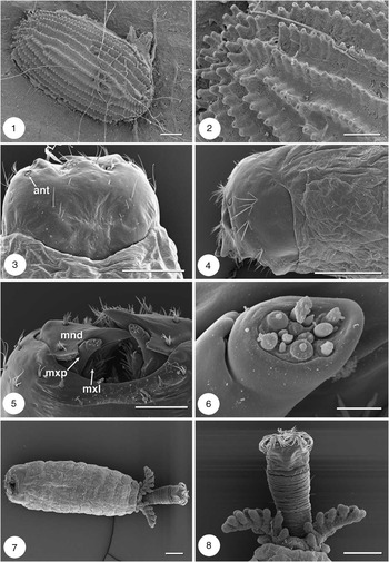
Approximately 1 mm in length, 0.5 mm in width, oval in shape, burr-like in appearance (Fig. 1). Ventral surface, which adheres to wood, somewhat smooth in texture. All other surfaces covered in rows of raised tubercles, each bearing an apical pore (Fig. 2). Surface microsculpture appearing to enhance retention of thin water film on surface.
Scanning electron micrographs of egg and first instar of Axymyia furcata. 1, Egg, dorsal view; 2, egg microsculpture; 3, first-instar head capsule, dorsal view; 4, first-instar head capsule, lateral view; 5, first-instar mouthparts, ventral view; 6, apex of first-instar right maxillary palpus; 7, first-instar habitus, ventral view; 8, first-instar respiratory siphon and anal papillae, ventral view. Abbreviations: ant = antenna, mnd = mandible, mxl = maxillary lacinia, mxp = maxillary palpus. Scale bars = 100 μm (Figs. 1, 3, 4, 7, 8), 50 μm (Figs. 2, 5), 5 μm (Fig. 6).

Larva
General (Fig. 51)
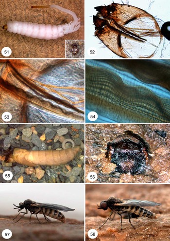
Eucephalic, head capsule partly invaginated into prothorax, sclerotization of cranium decreasing gradually toward posterior margin, mouthparts prognathous. Body cylindrical, white, divided into three thoracic and eight abdominal divisions. Eighth division (= anal division) with elongated respiratory siphon, its length in mature larvae greater than combined length of head and body (not including the anal papillae). Pair of large anal papillae at base of siphon, each elongate, branched. General cuticle translucent, fat-body easily visible. Respiratory system amphineustic, with anterior pair of spiracles located laterally on prothorax and posterior pair at end of respiratory siphon.
Cranium
First instar: Posterior three fourths of frontal plate membranous. Egg burster greatly reduced/absent. Dorsal sensilla forming two rows of three setae, progressively larger posteriorly to anterolaterally, plus two short chaetiform setae near base of clypeus; all sensilla simple (Fig. 3). Lateral surface of genae smooth, with five tufted sensilla, dorsal three with three branches, ventral, anteroventral sensilla each with four to five branches (Fig. 4). Sclerotized bar between clypeus and frontal plate less pronounced than in later instars. Clypeus short, wide with four prominent setiform sensilla (Fig. 3).
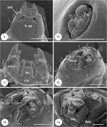
Fourth instar: Frontoclypeal apotome ovoid, elongate, narrowing posteriorly, not reaching posterior cranial margin. Anterior margin with pair of short, branched sensilla (one on each side) on heavily sclerotized ridge separating frontal plate from clypeus and spanning lateral margins at mandibular articulation. Genae meet at narrowed end of frontoclypeal apotome, forming posterior margin of cranium. Dorsal surface of gena smooth, with row of three sensilla adjacent to ecdysial suture; sensilla progressively larger posteriorly to anterolaterally, posterior sensillum setiform, others two-branched (Fig. 9). Posterolateral portion of gena less sclerotized where overlapping portion of prothoracic cuticle attached. Lateral surface of gena smooth, with five tufted sensilla, arranged as in first instar but all with four to five branches (Figs. 9, 12). Clypeus short, wide, with two pairs of tufted sensilla (Figs. 9, 11).
Ventral cranium
Genae fused ventromedially, anterior margin simple, not serrated. Postgenal bridge with scattered papillae; single large tubercle on either side of midline, three pairs of small tufted sensilla laterally (Fig. 12). Posterior margin of ventral cranium heavily sclerotized, forming postoccipital carina (Fig. 52).
Antenna
Antenna reduced, on anterior margin of gena just dorsolateral of mandible, comprising flat pad of sensilla within raised cuticular ring (Figs. 3, 9, 16). With eight blunt, peg-like sensilla, three large cushion-like sensilla, two of latter slightly smaller and bearing small bifurcated apical sensillum (Fig. 10).
Scanning electron micrographs of fourth instar of Axymyia furcata. 9, Head capsule, dorsal view; 10, left antenna; 11, fourth-instar labrum, clypeus, and mandibles, dorsal view; 12, head capsule, oblique-frontal view; 13, mouthparts, oblique-frontal view; 14, labrum and right mandible and maxilla, frontal view. Abbreviations: ant = antenna, cly = clypeus, fr ap = frontal apotome, lbm = labium, lbr = labrum, mnd = mandible, mx = maxilla. Scale bars = 500 μm (Fig. 9), 200 μm (Figs. 11, 13), 100 μm (Fig. 14), 50 μm (Fig. 14), 20 μm (Fig. 10).

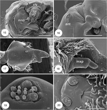
Scanning electron micrographs of larval mouthparts of Axymyia furcata. 15, Labrum and right mandible, frontal view; 16, left antenna and mandibular articulation, dorsal view; 17, right mandible, lateral view; 18, right maxilla, ventral view; 19, apex of maxillary palpus; 20, base of labium, showing labial palpi. Abbreviations: ant = antenna, cly = clypeus, mnd = mandible, mxl = maxillary lacinia, mxp = maxillary palpus. Scale bars = 100 μm (Figs. 15, 16), 20 μm (Fig. 20), 10 μm (Figs. 17, 18), 1 μm (Fig. 19).

Mouthparts
Labrum–epipharynx: Labrum somewhat conical, projecting anteriorly from clypeus. Dorsal surface mostly flat, with pair of tufted sensilla (Fig. 11). Anteroventral surface (epipharynx) densely covered with macrotrichia (Figs. 13, 14). Macrotrichia mostly simple, setiform but those in posterior region of epipharynx somewhat serrated.
Mandible: Large, heavily sclerotized, with one large, sharp, apical tooth, two smaller, lobed teeth on medial lateral surface (Fig. 11). First instar with subapical teeth reduced but with small, saw-like ventral lobe abutting maxillary palpus when mandible adducted (Fig. 5). Apical teeth overlap entirely when mandibles adducted (Figs. 5, 12, 13). Posterodorsal surface with large epicondyle functioning as fulcrum, articulating on prominent genal lobe, with deep groove between genal margin and posterior margin of clypeus (Figs. 9, 16). Anterolateral surface with three tufted sensilla (Figs. 5, 11, 13, 15, 17). Adoral surface with prominent prostheca (Fig. 17).
Maxilla: Reduced, comprising fleshy lacinia, elongate palpus (Figs. 5, 13, 14). Microtrichia on medial surface of lacinia curved, serrate, those near apex straight, smooth. Ventral surface of lacinia with prominent spiniform sensillum (Fig. 18). Length of lacinia and palpus subequal in later instars (Fig. 18); palpus much longer than lacinia in first instar (Fig. 5). Palpus biarticled, with basal article (palpiger) greater in length and circumference than distal article. Distal end of proximal article with single setiform sensillum arising near base of distal article (Fig. 18). Apex of palpus concave, bearing two large, 10 smaller sensilla; arrangement of latter varying slightly between first- and fourth instars (Figs. 6, 19).
Labium: Small, mostly hidden by maxillae, forming conical setose lobe, distal portion covered in curved serrate macrotrichia, similar in appearance to apex of lacinia (Fig. 14). Base of labium with small palpus, just ventral to opening of salivary duct (Fig. 20). Palpus short, button-like, with four apical sensilla.
Pharyngeal filter: With two, narrow, sclerotized plates extending from near anterior margin of esophagus to nearly length of frontoclypeal apotome (Fig. 52). Plates run subparallel, converging posteriorly. Each medial surface of filter irregular, bearing approximately 10 parallel longitudinal ribs. Each rib running entire length of plate, bearing continuous row of microtrichia (Figs. 53, 54).
Body
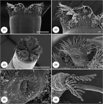
Diameter of first-instar head, thorax and abdomen subequal; thorax and abdomen of mature larva nearly twice diameter of head (Fig. 51). Prothoracic spiracles prominent, black, situated just above lateral midline of invaginated head. Seven abdominal segments present anterior to anal division (segment VIII). Dorsal surface of segments I–V each with transverse bands of spines, located centrally on each tergite; bands on segments I and V with two rows, with anterior row projecting anteriorly, posterior row projecting posteriorly; bands on segments II–IV have three rows, with anterior two rows projecting anteriorly, posterior row projecting posteriorly. Ventral surface of segments I–VI each with transverse bands of spines anteriorly; band on segment I comprising two rows, both projecting posteriorly; bands on segments II–IV each comprising four rows, anterior two rows project anteriorly, posterior two rows project posteriorly; bands on segments V and VI each comprising two rows, with anterior row projecting anteriorly, posterior row projecting posteriorly (Fig. 7). Anal papillae large, paired, arising from abdominal segment VII near base of respiratory siphon (Figs. 8, 26). Each papilla a central stem with three rows of approximately six finger-like projections along length; projections simple in first instars, moniliform in later instars. Abdominal segment VIII comprised mostly of elongate, extensile, respiratory siphon. Siphon circumference nearly constant throughout its length, with only gradual decrease from base to apex. Cuticle of anterior portion of siphon similar to that of abdomen, cuticle of terminal third more heavily sclerotized, darkly pigmented (Fig. 51). Distal end of siphon separated from remaining siphon by ring-like indentation (Fig. 23), beyond which siphon lacks compact rings of support tissue present in remaining siphon. Apex of siphon in form of spiracular disc with five prominent lobes, a variety of setae, hooks of varying sizes (Figs. 21, 22). Posterior spiracles elevated to level of lateral tubercles (Figs. 24, 25).
Scanning electron micrographs of larval abdominal features of Axymyia furcata. 21, Distal end of respiratory siphon, lateral view; 22, spines at distal end of respiratory siphon, lateral view; 23, apex of respiratory siphon, showing spiracular disc; 24, spiracular lobe; 25, spiracle; 26, anal papillae and base of respiratory siphon. Scale bars = 200 μm (Fig. 23), 100 μm (Figs. 21, 26), 10 μm (Figs. 22, 24, 25).

Pupa
General (Fig. 55)
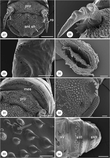
Cylindrical, blunt anteriorly, tapering posteriorly. Dorsal surface of head, prothorax, anterior mesothorax heavily sclerotized, forming circular disc covered with ornate ridges, numerous spines (Figs. 27, 31, 56). Prothoracic respiratory organs prominent, paired (Figs. 27, 28, 56).
Head
Dorsal surface of head heavily sclerotized, covered with spines, tubercles (Figs. 27, 31–33). Lateral, ventral surface of head much less sclerotized, similar in sclerotization to metathorax. Dorsolateral margin of head with large, semicircular, blade-like tubercles (Fig. 32). Antennal sheath free, heavily sclerotized, extending along anterodorsal margin of eye. Cuticle medial to antennal base with single large sensillum. Posterolateral margin of eye with prominent tubercle bearing two hair-like setae. Maxillary palpus sexually dimorphic; female palpus long, robust, male palpus short, delicate.
Scanning electron micrographs of pupae of Axymyia furcata. 27, Head and anterior part of thorax, dorsal view; 28, right thoracic respiratory organ, dorsal view; 29, microsculpture on thoracic respiratory organ, dorsal view; 30, cross-section of thoracic respiratory organ showing plastron; 31, microsculpture on dorsum of thorax, anterodorsal view; 32, microsculpture on left side of head, anterodorsal view; 33, microsculpture on left side of head; 34, abdominal segments V–VII, ventral view. Abbreviations: ant sh = antennal sheath, mes = mesonotum, pro = pronotum, ro = respiratory organ, sV–VII = sternites V–VII. Scale bars = 0.5 mm (Fig. 27), 200 μm (Figs. 31, 34), 100 μm (Figs. 28, 32), 50 μm (Figs. 29, 30, 33).

Thorax
Dorsal portion of prothorax, anterodorsal region of mesothorax heavily sclerotized, dark brown to black in color, covered with irregular, ornate microsculpture (Figs. 31, 56). Cuticle on metathorax, posterior portion of mesothorax, wing- and leg sheaths lightly sclerotized, light brown to cream colored. Abdominal cuticle mostly membranous, translucent, pale brown to deep gold in color.
Anterior respiratory organ large, produced anteriorly from dorsolateral margin of prothorax, laterally compressed, curved anteriorly over lateral margin of head (Figs. 27, 56). Dorsal surface with serpentine ridge (Fig. 28) lined with series of small, dome-like tubercles and small pores (Fig. 29). Cross-section of respiratory organ reveals plastron comprising cuticular pillars around inner margin (Fig. 30).
Ecdysial line extending along thoracic midline, bisecting heavily sclerotized ridge on anterior mesothorax (Fig. 31). Single, prominent setiform sensillum posterior to ridge, on either side of ecdysial line.
Wing- and leg sheaths tightly appressed to body. Base of each wing sheath with single setiform sensillum.
Abdomen
Cuticle translucent, membranous. Abdominal segments II–VII each with pair of dark brown to black spiracles near anterolateral margin. Small group of spines and single setiform sensillum posteroventral to each spiracle, along lateral midline. Tergites III, V, VI each with single posterior row of delicate, posteriorly curved spines, tergite IV with thick band of coarse, anteriorly curved spines. Sternites III and IV each with single row of delicate spines, sternites V–VII each with row of delicate spines and band of coarse spines (Fig. 34). All ventral spines curving posteriorly. Anal division with prominent, fleshy, dorsal lobe, presumably a remnant of larval anal siphon. Apex of abdomen sexually dimorphic; male with distinct sheaths of hypandrial lobes, female with distinct sheaths of large pad-like cerci.
Adult
General (Figs. 57, 58)
Mid-sized, heavy bodied, similar in appearance to Bibionidae, with prominent eyes and bulging thorax. Color ranging from light mahogany brown to black. Cuticle covered in dense minute microtrichia. Sexual dimorphism apparent in structure of eyes and terminalia.
Head
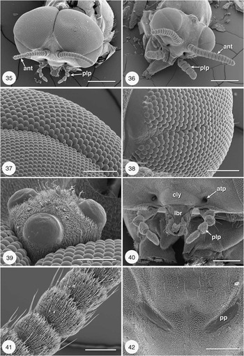
Cuticle: Large smooth tubercle at very posteroventral margin of eye. Posterodorsal margin lined with long hair-like setae. Similar setae between antennal bases. Anteriorly with two large deep tentorial pits, each between antenna and maxillary palpus (Figs. 35, 36).
Scanning electron micrographs of adults of Axymyia furcata. 35, Male head, oblique-frontal view; 36, female head, oblique-frontal view; 37, male eye at junction of upper and lower divisions; 38, female eye at junction of upper and lower divisions; 39, male ocelli, oblique-frontal view; 40, male mouthparts and tentorial pits, frontal view; 41, male antenna; 42, prescutal pits (female), anterior = top. Abbreviations: ant = antenna, atp = anterior tentorial pit, cly = clypeus, lbr = labrum, plp = palpus, pp = prescutal pit. Scale bars = 0.5 mm (Fig. 35, 36), 200 μm (Figs. 40, 42), 100 μm (Figs. 37–39), 50 μm (Fig. 41).

Eyes: Large, bulging, without ommotrichia. Male eyes holoptic, contiguous dorsally (Fig. 35). Female eyes smaller, dichoptic (Fig. 36). In both sexes divided into upper and lower hemispheres by shallow groove and scattered row of small, delicate setae (Fig. 37). In male, upper hemisphere much larger than lower hemisphere; arrangement opposite in females. In male, ommatidial facets of upper hemisphere larger, lighter in color than facets of lower hemisphere (Fig. 37). In females, facets of upper and lower hemispheres uniform in both size and color (Fig. 38).
Ocelli: Large, located on prominent, dome-like tubercle on posterodorsal surface of head (Figs. 35, 36). Tubercle with narrower base, more prominently raised in male than female, extending above dorsal margin of eyes (Fig. 39).
Antenna: Moniliform, comprising pedicel, scape, and 14 flagellomeres. Pedicel and scape similar in structure to flagellomeres, paler in color. Female antenna (Fig. 36) longer than, nearly twice as robust as that of male (Fig. 35). All flagellomeres setaceous, each with additional crown of long sensilla (Fig. 41).
Mouthparts: Clypeus prominent, sclerotized, triangular in shape. Labrum and labella greatly reduced, primarily membranous (Fig. 40). Apex of labellum with small, sclerotized, circular process covered in dark setae. Maxillary palpus short, delicate in males (Figs. 35, 40), longer, robust in females (Fig. 36). Most palpal segments with several prominent setiform sensilla. Maxillary palpus with five segments in most individuals, but with four to six segments in some individuals.
Thorax
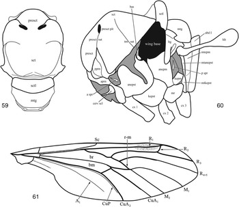
Cervical sclerite prominent but partially hidden by head; triangular in shape, with sharply acute angle pointed inward toward posteromedial margin of head. Antepronotum reduced medially but increasing in width anteroventral of forecoxa. Postpronotal lobe separated from posterior margin of prescutum and anterior margin of scutum and delineated by inverted Y-shaped suture; stem of suture (= prescutal suture) terminates at large dark spot or scar (prescutal pit) on anterior scutum (Fig. 42). Prescutum and scutum swollen, giving hunchback appearance. Transverse suture faint, slightly invaginated, arising laterally from wing base, extending toward but terminating before midline of scutum (Fig. 59). Scutellum narrowed transversely, strongly domed. Scutum and scutellum covered with long, fine, widely spaced setulae. Mediotergite nearly twice the length of scutellum, lacking setulae.
Episternum large, with distinct division between anepisternum and katepisternum. Anepisternal cleft dividing anepisternum into large anterior portion, much narrower posterior portion. Basalare prominent, dorsal to narrow portion of anepisternum. Anepimeron broad, narrowing ventrally; katepimeron subquadrate, clearly separated from meron. Meron large, subcircular, about half the size of katepisternum. Metepisternum and metepimeron reduced, represented by elongate bands below haltere (Fig. 60). Prescutum and lateral margin of scutum (anterior to transverse suture) set with numerous elongate, thin hairs; scutum dorsally with sparse short hairs. Scutellum laterally with patches of short hairs, all oriented medially. Katepisternum and anterior part of anepisternum with few short hairs. Nearly all thoracic sclerites set with dense microtrichia.
Legs
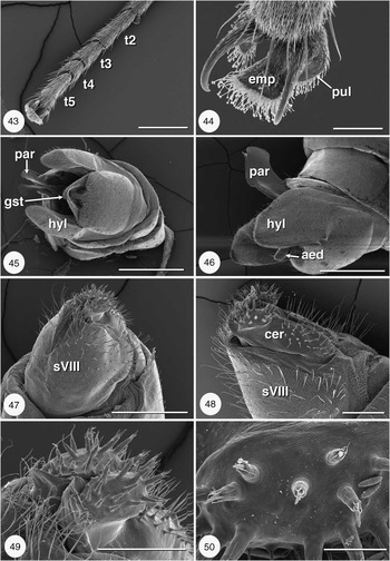
Legs somewhat short, subequal in overall length, without spurs, with microtrichia and scattered setiform sensilla (Fig. 43). Length of mid- and hind coxae approximately half-length of forecoxa. Pretarsus with prominent claws, empodium and pulvillus. Empodium spatulate, about twice the size of pulvillus; ventral surface of empodium and pulvillus densely set with long, spatulate macrotrichia (Fig. 44).
Scanning electron micrographs of adults of Axymyia furcata. 43, Female tarsus, dorsal view; 44, female pretarsus, showing claws, empodium and pulvilli; 45, male terminalia, posterior view; 46, male terminalia, lateral view; 47, female terminalia, dorsolateral view; 48, female terminalia, lateral view; 49, female cerci, oblique-posterior view; 50, sensilla on female cercus. Abbreviations: aed = aedeagus, cer = cercus, emp = empodium, gst = gonostylus, hyl = hypandrial lobe, par = paramere, pul = pulvillus, sVIII = sternite VIII, t2–t5 = tarsomeres 2–5. Scale bars = 500 μm (Figs. 45–47), 200 μm (Figs. 43, 48, 49), 50 μm (Figs. 44, 50).

Wings and halteres
Wings large, broad, about equal in length to the entire body; pale brown to slate gray, with distinct, but light, pterostigma. Membrane covered in fine microtrichia. Radial vein (R) with four branches; R2 short, nearly perpendicular to R3; R3, R4 + 5 long and subparallel. Medial vein (M) with two branches, stem quite faint, branches more prominent. Radial-medial cross vein (r-m) oblique. Posterior cubital branch (CuP) present, but faint, not reaching wing margin. Posterior branch of anal vein (A1) weakly present, not reaching wing margin (Fig. 61). Alula absent. Halter prominent, with long stem; covered with fine microtrichia; middle section of stem with cluster of long, stout setae.
Abdomen
Abdomen large, especially in females (Fig. 57). Tergites and sternites mostly dark anteriorly, pale brown to cream-colored along posterior margin. Pleural region cream-colored to white, membranous. Anterolateral margins of segments I–VII each with spiracles in membranous pleural region. Segments I–VII with sparse short hairs. Tergite VIII small, partially hidden by tergite VII, with membranous posterior cleft.
Light micrographs of larvae, pupae, and adults of Axymyia furcata. 51, fourth instar (inset = apex of respiratory siphon, in situ); 52, larval head capsule, showing pharyngeal filter; 53, larval pharyngeal filter; 54, details of larval pharyngeal filter; 55, pupa; 56, pupal head and thorax, in situ; 57, adult female; 58, adult male.

Male terminalia
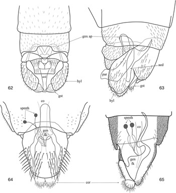
Sternite VIII small. Sternite IX large, fused with gonocoxites to form prominent hypandrium. Hypandrial lobes large, paired, hiding small setaceous gonostyli in lateral view (Figs. 45, 46). Cercus small, obscure, differentiated from proctiger primarily by texture. Aedeagus tube-like (Fig. 46, 62, 63). Parameres broadly flattened laterally, fused medially, flared at apex (Fig. 45). See Wood (Reference Wood1981, Reference Wood1991) for further detailed illustrations. Details of internal structure are presented by Borkent and Sinclair (Reference Borkent and Sinclair2012) in this volume.
Thorax and wing of adult Axymyia furcata. 59, Thorax, dorsal view; 60, thorax, lateral view; 61, wing. Abbreviations: abd I = abdominal segment I, anepm = anepimeron, anepst = anepisternum, aprn = antepronotum; a spr = anterior spiracle, bas = basalare, cerv scl = cervical sclerite, cx 1–cx 3 = coxa 1–3; epm = epimeron, hlt = haltere, kepm = katepimeron, kepst = katepisternum, ltg = laterotergite, mtg = mediotergite, mr = meron, mtanepst = metanepisternum, mtepm = metepimeron, mtkepst = metakatepisternum, presct pit = prescutal pit, presct sut = prescutal suture, pprn lb = postpronotal lobe, presct = prescutum, p spr = posterior spiracle, sctl = scutellum, trn sut = transverse suture.

Terminalia of adult Axymyia furcata. 62, Male, dorsal view; 63, male, lateral view; 64, female, ventral view; 65, female, lateral view. Abbreviations: aed = aedeagus, cer = cercus, co = common oviduct, gen fk = genital fork, gnx ap = gonocoxal apodeme, gst = gonostylus, hyl = hypandrial lobe, par = paramere, spmth = spermatheca.

Female terminalia
Sternite VIII large, curved strongly upward laterally nearly reaching dorsal surface. Sternite VIII with deep posterior furrow, covered in microtrichia and numerous setiform sensilla (Figs. 47, 48). Cercus large, pad-like, two-segmented; proximal segment with abundant setiform sensilla dorsally; distal segment with numerous cuticular projections, each with elongate macrotrichia basally and large, rod-like sensillum apically (Figs. 49, 50). Genital fork Y-shaped in ventral view (Fig. 64), sinuate in lateral view (Fig. 65). Common oviduct unsclerotized, indistinct posteriorly. Accessory glands not apparent. Reproductive tract simple Spermathecae spherical, sclerotized, two in number; spermathecal ducts sclerotized distally, inflated and membranous basally (Figs. 64, 65).
Discussion
Axymyiidae is an enigmatic family of semi-aquatic nematocerous flies whose phylogenetic placement has remained problematic. Although we have not attempted to resolve the phylogenetic relationships of this unusual family, we hope that some of the morphological data presented here may be useful to future investigations of evolutionary relationships. Our data on first instars represents the first SEM study of this life stage in Axymyiidae and uncovers some unexpected results (e.g., lack of egg burster). Other larval characters suggest affinities with certain bibionomorph and psychodomorph families (e.g., larval antenna reduced); however, many other features (e.g., conical labrum, horizontal mandibular rotation, fused postgenae, simple postmentum) are either more generalized or widespread across many larval nematocerans. Still other characters are clearly autapomorphic and presumably related at least partly to their xylophilic lifestyle. For example, the complex structure of respiratory siphon apex appears to function as a grapple, securing the siphon to the gallery opening (Fig. 51 (inset)). Other features (e.g., elongate siphon with elastic qualities, structure of anal papillae) may be adaptations to periodical inundation by water and life in saturated wood.
A study of pupal structure and homology across all nematocerous flies is desperately needed. Detailed studies of pupal structure in other groups (e.g., Arens Reference Arens1995, Reference Arens1997; Borkent Reference Borkent2010) have demonstrated that pupae can provide a rich source of phylogenetic information. Although we expect many features of axymyiid pupae will be unique, again due to xylophyly, we hope that other characters may provide a useful phylogenetic signal.
Adult axymyiids have been the subject of several phylogenetic studies, usually as part of a broader investigation of many nematoceran families (e.g., Wood and Borkent Reference Wood and Borkent1989; Oosterbroek and Courtney Reference Oosterbroek and Courtney1995; Michelsen Reference Michelsen1996). Other studies have focused on particular character systems (e.g., Borkent and Sinclair Reference Borkent and Sinclair2012). For the most part, those studies have demonstrated that axymyiids are fairly generalized in structure (e.g., terminalia), or possess unique features (i.e., autapomorphies) that provide no evidence of phylogenetic relationship.
Acknowledgments
The museums listed in materials and methods generously loaned preserved specimens of Axymyia furcata for this morphological study. We are also grateful to Art Borkent and Brad Sinclair for careful review of an earlier draft of this paper. This project was supported in part by National Science Foundation grants (EF-0334948 and DEB-0933218) to Gregory W. Courtney and by funds from the Dean of Agriculture and Life Sciences International Research Grant and Biosystematics Travel Fund to Matthew W. Wihlm. This journal paper of the Iowa Agriculture and Home Economics Experiment Station, Project no. 6693, was supported by Hatch Act and State of Iowa funds.