Isolated congenital complete atrioventricular block occurs mostly in presence of maternal connective tissues disease, or anti SSA/Ro or anti SSB/La antibodies, with otherwise structurally normal hearts.Reference Taylor, Scott, Gerlis, Esscher and Scott1, Reference Buyon, Hiebert and Copel2 Such congenital complete atrioventricular block is known to develop in 1 to 2% of all pregnancies in mothers that carry anti-Ro/La antibodies.Reference Ho, Esscher and Anderson3 The passive transplacental passage of the maternal autoantibodies occurs since the middle of the second trimester, and is thought to be responsible for inflammation and fibrosis of the conduction system, with documented evidence of binding of the antibodies to the myocytes of the conduction tissues.Reference Ho, Esscher and Anderson3
Signs of myocarditis and endocarditis, as well as endocardial fibroelastosis and dilated cardiomyopathy, have been reported both in necropsy specimens and in clinical studies.Reference Lang, Wilhelm, Gildein, Vaith, Schillinger and Peter4–Reference Moak, Barron and Hougen8 The antibodies may also involve other organs, inducing the clinical and the laboratory manifestations of the neonatal lupus syndrome.Reference Taylor, Scott, Gerlis, Esscher and Scott1, Reference Buyon, Hiebert and Copel2, Reference Cimaz, Catelli, Luzzana, Panzeri and Meroni9 The fetuses with complete atrioventricular block with a low heart rate, less than 50 to 55 beats per minute, can rapidly develop hydrops, often leading to intrauterine or postnatal death.Reference Schmidt, Ulmer, Silverman, Kleinman and Copel10, Reference Groves, Allan and Rosenthal11
In the last decade, it has been suggested that treatment of the fetus by maternal administration of steroids might produce regression of the conduction disorder, or improve the fetal compensation.Reference Bierman, Baxi and Jaffe12–Reference Vignati, Brucato and Pisoni17. As an alternative, others have used sympaticomimetics to increase the fetal heart rate.Reference Groves, Allan and Rosenthal15 The benefits of the treatment in the fetal life with respect to outcomes are still under evaluation. With this in mind, we have analysed retrospectively a series of fetuses diagnosed with isolated congenital complete atrioventricular block, examining their characteristics, their course during fetal life and after birth, and the differences in outcome with respect to the heart rate and cardiac compensation with or without maternal-fetal treatment.
Patients and methods
Between 1992 and 2004, congenital complete atrioventricular block was diagnosed echocardiographically in three centres in 28 fetuses of 27 pregnant women. The mothers were aged from 23 to 35 years, with a median of 28 years, one mother being followed during 2 pregnancies. All were found to carry anti-Ro/La antibodies. The age of the fetuses at presentation ranged from 19 to 32 weeks gestation, with a median of 25 weeks. Of the mothers, 11 had known autoimmune disease, lupus erythematosus in 10, none on specific treatment, and one had autoimmune thyroiditis. There was 1 case of a bichorionic, biamniotic twin pregnancy, and 1 trichorionic, triamniotic, triple pregnancy, after in-vitro fertilization. The mother carrying the twins had previously borne a fetus with complete atrioventricular block who died at 25 weeks’ gestation. We followed this mother during both her pregnancies.
The mothers were usually referred subsequent to the discovery of fetal bradycardia. The mother with the twin pregnancy had been followed regularly since the onset of pregnancy due to the previous history, while the mother with triplets had been referred at 21 weeks gestation for signs of myocarditis in the 2 female fetuses, with a subsequent appearance of complete atrioventricular block in one of them, at 24 weeks, with a rapid progression to hydrops. At presentation, 8 fetuses already showed signs of fetal hydrops, mild in 3, moderate in 3, and severe in 2, while the other 20 fetuses were compensated, with no evidence of hydrops.
Methodology
Echocardiography was performed using Acuson 128 XP/10, Acuson Sequoia 512, Imagegate, Siemens, Erlangen, Germany and Vingmed System Five, General Electrics machines, using transducers 5 or 3.5 megaherz according to the gestational age. A complete ultrasonic scan was performed in all cases.
The fetal heart was analysed by means of M-mode or Colour Doppler M-mode, evaluating the sequences of atrial and ventricular contractions. Complete atrioventricular block was diagnosed when atrial contractions were independent and dissociated from ventricular contractions. We used the cut in the parasternal long axis view showing both the aorta and the left atrial wall so as properly to assess the contractions of the atrial wall. Doppler and Colour Doppler were used to detect any abnormal regurgitant flows across the atrioventricular valves. Since 2000, we have also evaluated the sequences of flow in the superior caval vein, the ascending aorta, and the venous duct. The general state and behaviour of the fetus was analysed by means of Doppler interrogation to provide the pulsatility index of the umbilical artery and median cerebral artery, eventual fluctuation pattern of the flow in the umbilical vein, and by observing fetal movements. We diagnosed fetal hydrops when serous effusions were observed in at least 2 compartments.
Protocol of management
We checked both the clinical state and laboratory findings in all the mothers. All had initially been hospitalized, permitting evaluation of routine laboratory examinations.
Having documented anti-Ro/La antibodies, we started treatment of the fetus by dosing the mother in 21 cases, in 16 with non-hydropic fetuses, and in 5 with hydrops. In 20 cases, we used dexamethasone at 4 mg per day, combined in 2 mothers with salbutamol. In 1 mother seen early in our experience, we used isoprotenerol, the fetus in this case having a very low heart rate, 32 beats per minute, at presentation. In most cases, treatment was started within 2 weeks of presentation, albeit that in 3 of the fetuses without hydrops, and 2 with hydrops, we started treatment after a delay of 4 to 7 weeks, having noted a decrease in the heart rate. Treatment was usually continued until the end of the pregnancy, lasting from for 1 to 14 weeks, with a median of 4 weeks. The dose of dexamethasone was progressively reduced towards the planned time for delivery.
We did not attempt treatment in 7 mothers, 4 of the ones seen earlier in our experience who were carrying non-hydropic fetuses, with heart rates of 50, 54, 80, and 73 beats per minute respectively, and in 3 hydropic fetuses. In the latter group, 1 fetus had a very low heart rate and severe anasarca, and was considered to be preterminal, while the other 2 had only minor degrees of cardiac failure, and showed stable heart rates. We followed all mothers at short time intervals, usually once or twice a week, until the ends of their pregnancies. In all instances, management was made by a multidisciplinary team of a gynaecologist, a paediatric cardiologist, and a neonatologist.
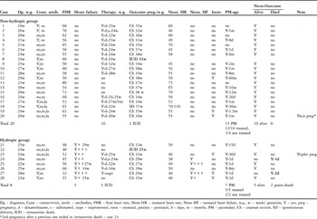
After birth, we have followed the infants for a median of 7 years, with a range from 3 to 14 years. The postnatal follow-up included clinical and echocardiographic evaluation at short time intervals initially, then 2 to 3 times a year, according to the individual situation. When they were around 2 years of age, we also had the neurodevelopmental state assessed by a specialist in our group who used the developmental scale developed by Griffiths. We summarise our data in Table 1.
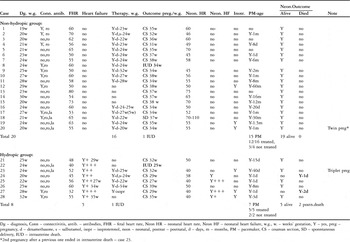
Data of the series.
Dg – diagnosis, Conn – connectivitis, antib. – antibodies, FHR – fetal heart rate, Neon HR – neonatal heart rate, Neon HF – neonatal heart failure, w.g., w. – weeks’ gestation, Y – yes, preg – pregnancy, d – dexamethasone, s – salbutamol, isopr – isoproterenol, neon – neonatal, postnat – postnatal, d – days, m – months, PM – pacemaker, CS – cesarean section, SD – sponntaneous delivery, IUD – intrauterine death.
*2nd pregnancy after a previous one ended in intrauterine death – case 23.
Statistical analysis
Data is expressed as a frequency or percentage for nominal variables, as a median with range for categorical variables, and as a mean ± standard deviation for continuous variables.
We analysed as dependent variables whether the fetuses were alive or had subsequently died, the need for insertion of a pacemaker, and if required, the timing of its implantation. As independent variables we included gestational ages and heart rate of the fetuses at presentation, their age at delivery, the neonatal heart rate, whether the mothers had been treated, and the presence or absence of fetal hydrops. Fetal hydrops, if present, was graded as mild, moderate, or severe. We grouped heart rates for fetuses and neonates as less than 50 beats per minute, from 50 to 60 beats per minute, or more than 60 beats per minute. Implantations of pacemakers were grouped for insertions at 1 to 30 days, 1 to 2 months, 2 to 12 months, or longer than 12 months. We performed univariate analysis using the chi-squared test, Fisher’s exact test, the unpaired Student’s t test, the Wilcoxon rank sum test, the paired t test, and the Mann-Whitney U test. Multivariate analysis was performed by using multiple logistic regression analysis. The diagnostic of the regression model was made obtaining the standardized residuals and plotting them by use of the Wilks-Shapiro/rankit plot test. Independent variables were included in the multivariate model if in the univariate analysis p was less than 0.2. Odds ratios with 95% confidence intervals were calculated for independent variables included in the multivariate model. All tests were two-sided. A probability value of p less than 0.05 was considered as significant.
Results
Characteristics
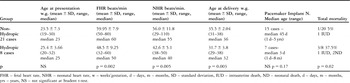
In Table 2, we present the main data of the hydropic as opposed to non-hydropic fetuses. Age at presentation of the fetuses in the 2 groups was not significantly different, but both fetal and neonatal heart rates were lower in those with hydrops (p less than 0.002 and 0.005 respectively). The heart rate reduced throughout pregnancy in fetuses of both groups (p less than 0.05 in those without hydrops, and p less than 0.08 in those with hydrops).
Main data of non-hydropic and hydropic fetuses at statistical analysis.
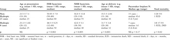
FHR – fetal heart rate, NHR – neonatal heart rate, w – weeks’gestation, d – days, m – months, SD – standard deviation, IUD – intrauterine death, ND – neonatal death, d – days, m – months, yrs – years, NS – not significant at Student t-test.
Outcomes
Non-hydropic fetuses
All 4 fetuses without hydrops that were not treated survived. In 3 of them (cases 12, 15 and 16), pacemakers were implanted at 12, 16, and 60 months, while the fourth child remains stable at 3 years.
Of the 17 fetuses without hydrops that received treatment, 1 (case 34) died at 34 weeks gestation having presented at 24 weeks gestation with a heart rate of 60 beats per minute. There had been a sudden drop in the heart rate at 34 weeks, followed by death. Of the remaining fetuses, 15 were delivered at a median gestational age of 34 weeks, with a range from 31 to 37 weeks, with 4 requiring inotropic treatment after birth, and all 15 having pacemakers implanted at a median age of 45 days, 6 requiring implantation at 1 to 30 days, the remaining fetuses having the devices implanted later, at 1.5 to 60 months.
Hydropic fetuses
Of the mothers with hydropic fetuses, 3 received no treatment. Amongst these, one mother lost a fetus with anasarca at 25 weeks gestation, after presentation at 24 weeks (case 22). The fetal heart rate in this case progressively reduced from 45 to 25 beats per minute. The other 2 fetuses survived (cases 21 and 28), and had pacemakers implanted at 15 and 3 days.
We treated the other 5 mothers, 2 of these were carrying fetuses in which fetal hydrops progressed to severe anasarca and died both after birth, despite implantation of pacemakers on the day of birth (cases 24 and 27).
The other 3 fetuses were delivered at 27, 32, and 38 weeks gestation, respectively, and survived following implantation of pacemakers at 1 day, 40 days, and 8 months.
Age at delivery
The non-hydropic fetuses were delivered at later gestational ages, of 35.5 +− 2.04 weeks as opposed to 31.7 +− 3.8 weeks for the hydropic fetuses (p less than 0.003 – Table 2). Emergency caesarean section was required in 6 cases because of an abrupt decrease of the heart rate in 5 instances, and because of premature rupture of the membranes in the triplet pregnancy. The remaining mothers underwent planned delivery according to the situation, all but one by caesarean section.
Effect of maternal treatment
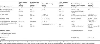
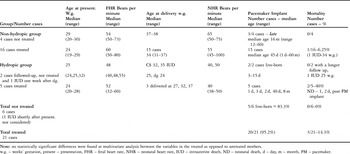
In no case did we observe any regression of the conduction disorder after treatment. There was, however, a rapid improvement in hydrops in 3 of the 5 hydropic fetuses after treatment, with no noted deterioration of cardiac compensation observed in 15 of the 16 treated non-hydropic fetuses, albeit that 3 of these suffered an abrupt decrease of the heart rate and underwent emergency caesarean sections as reported above. In Table 3, we summarise the results for treated as opposed to untreated cases. We failed to detect any statistically significant differences.
Data of treated compared to untreated cases.
Note: no statistically significant differences were found at multivariate analysis between the variables in the treated as opposed to untreated mothers.
w.g. – weeks’ gestation, present – presentation, FHR – fetal heart rate, NHR – neonatal heart rate, IUD – intrauterine death, ND – neonatal death, d – day, m – month, PM – pacemaker.
Adverse effects of dexamethasone treatment
We did observe adverse effects of dexamethasone in 2 fetuses, noting a reduction of amniotic fluid leading to progressive cessation of therapy. No other adverse effects were observed, either in fetuses or mothers. Fetal growth was within the normal limits, between the 10th and 50th centile for the gestational age, with a median of the 25th centile. The weights at birth ranged from 1600 to 3100 grams, with a median of 2400 grams.
Pacemaker implantation
We implanted devices in 22 of the 26 fetuses born alive (84.6%). Of these, 15 had not suffered hydrops and 12 had the devices implanted at a median age of 45 days, with a range from 1 day to 5 years. In the cases not treated in fetal life, devices were implanted at a later median age of 16 months, with a range from 12 to 60 months. In the 7 liveborn infants who had suffered hydrops, 5 of which having been treated, devices were implanted at a median age of 3 days, with a range from 1 day to 8 months (Tables 2 and 3). Epicardial pacemakers were implanted using subxyphoid access, positioning the device in the upper abdomen, and using VVI pacing at a heart rate of 90 beats per minute. At multiple logistic regression analysis, no predictive variables were found.
Mortality
Four of the 28 fetuses died (14.3%), 2 during fetal life and 2 after birth. Of the 21 fetuses treated, 3 died (14.3%), but none of the fetuses not treated died, if we exclude the fetus with severe anasarca who died shortly after fetal presentation (Table 3). Mortality was lower in cases without hydrops, 1 from 20 (5%) than for those with hydrops, of whom 3 of 8 died (37.5% – p less than 0.02).
In all cases, death was due to cardiac failure. At multiple logistic regression analysis, the only predictive variable for outcome was the presence and degree of hydrops (ß coefficient equal to 2.2; p less than 0.018; OR: 9,2(95% CI: 1.5 to 58).
Postnatal and longterm outcomes
Of our initial cohort, 24 infants (82.7%) are alive at a median follow-up of 7 years, with a range from 5 to 14 years. None have developed dilated cardiomyopathy, nor showed any abnormal neurological development as estimated using Griffith’s neurodevelopmental scale. The cases born prematurely suffered from disorders of prematurity, albeit without any significant consequences. One of our twin infants presented after birth moderate pulmonary stenosis, with a transvalvar gradient of 50 mmHg, that subsequently improved without the need for an invasive procedure. The patient is now well at 6 years.
The second female of the triplet pregnancy showed, after birth, the first and episodical second degree atrioventricular block. She is currently stable at the age of 7 years. In her case, a slightly prolonged P-R interval, of 0.14 msec, was seen 2 weeks before the caesarean section at 32 weeks gestation when using the Doppler evaluation of the pattern of flow in the superior caval vein compared to the ascending aorta.
Discussion
The natural history of isolated immune-mediated congenital complete atrioventricular block depends mainly upon the degree of fetal bradycardia, the fetal heart being limited in its ability to produce a valid cardiac output in the setting of very low heart rates. A high morbidity and mortality has been reported for such fetuses, depending usually on the hydropic state.Reference Buyon, Hiebert and Copel2, Reference Schmidt, Ulmer, Silverman, Kleinman and Copel10, Reference Jaeggi, Hamilton and Silverman16, Reference Vignati, Brucato and Pisoni17 This data, however, mostly reflects cases studied in the previous era, when no maternal-fetal treatment was available.
The reasoning for treatment of the mother with steroids so as to influence the fetus is to act against the immune-mediated inflammatory process produced by the passive passage of antibodies, with the dual aims of, first, potentially producing regression in the degree of the conduction disorder, and second, to act against all other sites of the inflammatory process that characterize the neonatal lupus syndrome. Prophylatic use of such transplacental treatment for prevention of congenital heart block is not justified in women with anti-Ro/La antibodies, since there are no known predictive markers for its development, and the incidence of atrioventricular block is low in this population.Reference Buyon, Hiebert and Copel2, Reference Gladman, Silverman and Yuk-Law18–Reference Costedoat-Chalumeau, Georgin-Lavialle, Amoura and Piette20
Also, some manifestations of the interactions of the maternal antibodies in the fetus, such as myocarditis and endocardial fibroelastosis, can develop only during the pregnancy, or after the delivery. Myocarditis preceded the development of the conduction disorder to different degrees in our triplet pregnancy, with complete atrioventricular block seen in one fetus and first and second degree block in the second, but with the third fetus showing only mild signs of neonatal lupus after birth.Reference Fesslova, Mannarino and Salice5 Equally, in our twin pregnancy, only one fetus was affected by complete atrioventricular block, albeit this being a recurrence, with a previous sibling suffering the same disorder, but with the second twin being completely free of symptoms of neonatal lupus syndrome. Our data confirms, therefore, the known discordance in the clinical manifestations of neonatal lupus in multiple pregnancies, mainly in bi- or tri-chorionic ones.Reference Watson, Scheel and Petri21, Reference Cooley, Keech, Melny, Menaheim, Morahan and Kay22 Females appear to be at higher risk for neonatal lupus and complete atrioventricular block, as was also the case in our triplet pregnancy.Reference Fesslova, Mannarino and Salice5, Reference Miyagawa, Dohi, Yoshioka and Shirai23
Several previous retrospective studies have suggested that the use of glucocorticoids might temper immune-mediated fetal cardiac damage, and indirectly improve cardiac contractility in suspected fetal myocarditis and, rarely, lessen the degree of atrioventricular block or even produce its regression.Reference Jaeggi, Hamilton and Silverman16, Reference Buyon, Swersky, Fox, Bierman and Winchester24, Reference Copel, Buyon and Kleinman25 Jaeggi and colleaguesReference Jaeggi, Fouron, Silverman, Ryan, Smallhorn and Hornberger14 stated that the routine use of dexamethasone given to mothers at the time of diagnosis of fetal heart block, in combination with ß-sympathomimetic therapy for persistent fetal bradycardia of less than 55 beats per minute, significantly improved survival compared with untreated fetuses that had a lower postnatal survival due to a higher frequency of immune-mediated complications and endocardiacal fibroelastosis. In contrast, Saleeb and colleaguesReference Saleeb, Copel and Friedman26 reported no regression of complete atrioventricular block, but observed in some cases a resolution of incomplete atrioventricular block, and a resolution of effusions and fetal hydrops, albeit without any benefit in outcome in comparison with untreated fetuses. Equally, Breur and colleagues,Reference Breur, Visser, Kruize, Steoutenbeek and Meijboom27 in a review of 19 studies on 93 fetuses with atrioventricular block, failed to find any cases having regression of complete block, but did document regression of incomplete block in a few cases.
In our study, the treatment was not randomized, but rather depended on the era of the diagnosis of complete atrioventricular block, and on the severity of the fetal compensation. Since the mid 1990s, we have adopted a policy to treat the patients by dexamethasone shortly after the documentation of anti-Ro/La antibodies, both in cases with and without cardiac failure.
The majority of our cases, 20 in all, were treated, with additional use of a beta- sympathomimetic in 2 fetuses having a low heart rate. In one of our first cases, having a very markedly reduced heart rate of 32 beats per minute, we used immediate treatment with isoproterenol, albeit without benefit, as documented for other severely compromised cases.Reference Jaeggi, Fouron, Silverman, Ryan, Smallhorn and Hornberger14, Reference Groves, Allan and Rosenthal15
Treatment with steroids did not reverse the degree of the block in our fetuses. The main effect of treatment was to produce a rapid improvement in the degree of fetal hydrops in 3 of 5 cases. Thus, we have failed to show any statistically significant benefit of treatment on the eventual outcome of our fetuses suffering complete atrioventricular block, possibly due to the small number of cases not treated. We found no statistically significant differences in mortality, age at delivery, or in the need for implantation of permanent pacemakers, between the fetuses whose mothers had or had not been treated. There are limitations in the comparisons to be made between our treated and untreated fetuses, nonetheless, not least because treatment was generally not started until after the 22nd week of gestation, when the conduction disorder was already well established
We found it necessary to insert permanent pacemakers in most of our children, their ventricular rates being less than 50 to 55 beats per minute. We timed the insertion of the device on the basis of the heart rate, but also of the weight of the infants. In a few very premature babies, we preferred to offer initial postnatal support with inotropes, proceeding to implantation of the pacemaker when the neonate weighed around 1900 to 2000 grams. In all, we have inserted pacemakers in 22 of the 26 infants born alive, this being necessary at an earlier age in the hydropic compared to the non-hydropic fetuses. Despite this, 2 of our hydropic fetuses subsequently died soon after birth. It was possible to delay insertion of the pacemakers in the untreated non-hydropic cases because of their better fetal and postnatal heart rates.
It is evident from our results that the main negative prognostic factors in our series were the presence and degree of cardiac failure, and a lower heart rate at presentation, confirming previous data.Reference Jaeggi, Fouron, Silverman, Ryan, Smallhorn and Hornberger14, Reference Breur, Visser, Kruize, Steoutenbeek and Meijboom27, Reference Brucato, Gasparini and Vignati28 We found statistically significant differences between the fetuses with and without cardiac failure at presentation, those with hydrops having a lower mean heart rate at presentation and after birth, being delivered at a younger gestational age, and carrying higher mortality. In all those dying, death was due to the cardiac failure. The presence and degree of fetal hydrops, furthermore, were found to be the only predictive variable for outcome at multiple logistic regression analysis. Hydropic infants needed permanent pacemakers implanted sooner after birth.
Side effects of the maternal steroid therapy have already been reported,Reference Jaeggi, Fouron, Silverman, Ryan, Smallhorn and Hornberger14, Reference Breur, Visser, Kruize, Steoutenbeek and Meijboom27 mainly oligohydramnios, which occurred to mild degree in 2 of our cases, but reversed promptly after reduction and eventual cessation of the therapy. We encountered no instances of fetal retardation of growth, nor did we observe any neurodevelopmental side effects having carried out specific neurodevelpmental assessment around the age of 2 years.Reference Brucato, Li Destri and Astori29, Reference Tincani, Rebaioli and Frassi30
Equally, we did not find any instances of the postnatal late dilated cardiomyopathy reported by some authors.Reference Moak, Barron and Hougen8, Reference Udink ten Cate, Breur and Cohen31 This may be due to progressive myocardial damage, absence of improvement after the pacemaker implant, or the type of pacing, with VVI pacing favouring ventricular desynchronization and possibly predisposing to myocardial damage. In our population, only the hydropic fetuses showed signs of endocardial fibroelastosis, with a higher echogenicity of the ventricular walls and impaired contractility. This was especially the case in 2 of the triplets, who presented with myocarditis, albeit that both improved after implantation of the pacemaker and provision of a better cardiac output.
In conclusion, the outcome of our fetuses was mainly dependent on the presence and degree of hydrops at presentation. Our data does not show any significant differences between fetuses treated or not treated by steroids, recognising that only a small number of our cases were not treated. Treatment did not produce regression of the conduction disorder, but did have a favourable effect on the fetal hydrops. Randomized, and larger scale, prospective studies are needed in order to fully define the efficacy of the treatment with steroids in fetuses with complete atrioventricular block.