Introduction
Episodic memory is a form of declarative memory that consists of the ability to store and recall a seemingly endless series of experiences, many of which occur only once (Tulving, Reference Tulving1983). Converging findings from lesion and electrophysiological studies indicate that structures in the mesial temporal lobe, including the parahippocampal gyrus and the hippocampus, are crucial for encoding and recall of episodic memory in animals and in humans (for a review, see Squire et al. Reference Squire, Wixted and Clark2007). Similar to other brain regions, several studies have also suggested that there is a critical range of dopamine signaling that physiologically regulates activity of the parahippocampal–hippocampal network during episodic memory (Lisman & Grace, Reference Lisman and Grace2005). Functional imaging studies in humans have largely been consistent with these data (Bertolino et al. Reference Bertolino, Rubino, Sambataro, Blasi, Latorre, Fazio, Caforio, Petruzzella, Kolachana, Hariri, Meyer-Lindenberg, Nardini, Weinberger and Scarabino2006b, Reference Bertolino, Di Giorgio, Blasi, Sambataro, Caforio, Sinibaldi, Latorre, Rampino, Taurisano, Fazio, Romano, Douzgou, Popolizio, Kolachana, Nardini, Weinberger and Dallapiccola2008; Schott et al. Reference Schott, Seidenbecher, Fenker, Lauer, Bunzeck, Bernstein, Tischmeyer, Gundelfinger, Heinze and Duzel2006).
Deficits in episodic memory are important features of schizophrenia (Aleman et al. Reference Aleman, Hijman, de Haan and Kahn1999; Lepage et al. Reference Lepage, Sergerie, Pelletier and Harvey2007; Leavitt & Goldberg, Reference Leavitt and Goldberg2009) and they are in part genetically determined, as suggested by studies in unaffected first- and second-degree relatives (Cannon et al. Reference Cannon, Zorrilla, Shtasel, Gur, Gur, Marco, Moberg and Price1994; Glahn et al. Reference Glahn, Almasy, Blangero, Burk, Estrada, Peralta, Meyenberg, Castro, Barrett, Nicolini, Raventos and Escamilla2007; Lefebvre et al. Reference Lefebvre, Cellard, Tremblay, Achim, Rouleau, Maziade and Roy2009). In patients, these deficits seem to be stable over time, largely independent of psychotic symptoms (for a review, see Ranganath et al. Reference Ranganath, Minzenberg and Ragland2008), less responsive to current pharmacological treatment, and they are related to functional outcome (Goldberg et al. Reference Goldberg, Goldman, Burdick, Malhotra, Lencz, Patel, Woerner, Schooler, Kane and Robinson2007). Neuropsychological studies have also suggested that impairments in encoding are central to episodic memory deficits in schizophrenia (Goldberg et al. Reference Goldberg, Egan, Gscheidle, Coppola, Weickert, Kolachana, Goldman and Weinberger2003; Gold, Reference Gold2004; Danion et al. Reference Danion, Huron, Vidailhet and Berna2007), whereas storage and retrieval phases may be relatively unimpaired (Boyer et al. Reference Boyer, Phillips, Rousseau and Ilivitsky2007). Consistent with these behavioral data, functional imaging studies of memory encoding in schizophrenia have documented abnormal hippocampal engagement (for a meta-analysis, see Achim & Lepage, Reference Achim and Lepage2005). Furthermore, other studies with functional imaging using different cognitive paradigms in patients with schizophrenia have also demonstrated abnormal activity of the mesial temporal lobe, including the hippocampus and the parahippocampus (Meyer-Lindenberg et al. Reference Meyer-Lindenberg, Poline, Kohn, Holt, Egan, Weinberger and Berman2001, Reference Meyer-Lindenberg, Olsen, Kohn, Brown, Egan, Weinberger and Berman2005).
Catechol-O-methyltransferase (COMT) plays a key role in the regulation of dopamine signaling in the cortex (Karoum et al. Reference Karoum, Chrapusta and Egan1994) through inactivation of extracellular dopamine. In the brain of mammals, COMT mRNA is expressed abundantly in the prefrontal cortex and also in granular and pyramidal neurons of the dentate gyrus and cornu ammonis regions of the hippocampus (Matsumoto et al. Reference Matsumoto, Weickert, Akil, Lipska, Hyde, Herman, Kleinman and Weinberger2003; Papaleo et al. Reference Papaleo, Crawley, Song, Lipska, Pickel, Weinberger and Chen2008). The activity of COMT is affected by a common single nucleotide polymorphism (SNP) determining a valine to methionine substitution at codon 158 (rs4680, G→A, COMT Val158Met) (Lachman et al. Reference Lachman, Morrow, Shprintzen, Veit, Parsia, Faedda, Goldberg, Kucherlapati and Papolos1996). The Val allele is associated with greater COMT protein levels and enzymatic activity compared to the Met allele, presumably leading to reduced dopamine availability at the synaptic cleft (Chen et al. Reference Chen, Lipska, Halim, Ma, Matsumoto, Melhem, Kolachana, Hyde, Herman, Apud, Egan, Kleinman and Weinberger2004). Consistent with this neurobiology, recent experiments in transgenic and knockout animals have indicated that genetic variation in COMT strongly affects cognitive functions, including episodic memory (Papaleo et al. Reference Papaleo, Crawley, Song, Lipska, Pickel, Weinberger and Chen2008). In humans, evidence for association of this variant with brain activity investigated with functional imaging is robust and consistent (for a meta-analysis, see Mier et al. Reference Mier, Kirsch and Meyer-Lindenberg2009). For example, it has been reported that the COMT Val158Met polymorphism affects hippocampal physiology and hippocampal–prefrontal coupling during encoding and retrieval of recognition memory (Bertolino et al. Reference Bertolino, Rubino, Sambataro, Blasi, Latorre, Fazio, Caforio, Petruzzella, Kolachana, Hariri, Meyer-Lindenberg, Nardini, Weinberger and Scarabino2006b, Reference Bertolino, Di Giorgio, Blasi, Sambataro, Caforio, Sinibaldi, Latorre, Rampino, Taurisano, Fazio, Romano, Douzgou, Popolizio, Kolachana, Nardini, Weinberger and Dallapiccola2008; Krach et al. Reference Krach, Jansen, Krug, Markov, Thimm, Sheldrick, Eggermann, Zerres, Stocker, Shah and Kircher2010), accounting at least in part for individual differences in recognition memory performance. We have also demonstrated that this SNP interacts epistatically with another variant in the dopamine transporter gene (DAT 3′ UTR VNTR) on hippocampal physiology during memory encoding, suggesting a non-linear relationship between the COMT and DAT polymorphisms (Bertolino et al. Reference Bertolino, Fazio, Di Giorgio, Blasi, Romano, Taurisano, Caforio, Sinibaldi, Ursini, Popolizio, Tirotta, Papp, Dallapiccola, Borrelli and Sadee2009).
Several studies in schizophrenia have focused on the COMT Val158Met polymorphism. Nonetheless, evidence supporting association of this coding variant with schizophrenia is still inconclusive (Egan et al. Reference Egan, Goldberg, Kolachana, Callicott, Mazzanti, Straub, Goldman and Weinberger2001; Shifman et al. Reference Shifman, Bronstein, Sternfeld, Pisante-Shalom, Lev-Lehman, Weizman, Reznik, Spivak, Grisaru, Karp, Schiffer, Kotler, Strous, Swartz-Vanetik, Knobler, Shinar, Beckmann, Yakir, Risch, Zak and Darvasi2002; Glatt et al. Reference Glatt, Faraone and Tsuang2003; Owen et al. Reference Owen, Williams and O'Donovan2004; Fan et al. Reference Fan, Zhang, Gu, Li, Sun, Wang, Feng, St Clair and He2005), and the isolated association of this polymorphism with the phenotype of diagnosis is likely to be small. However, some studies have demonstrated statistical epistasis, in that other genetic variants are associated with schizophrenia when the analysis is conditioned on having Val allele (Nicodemus et al. Reference Nicodemus, Kolachana, Vakkalanka, Straub, Giegling, Egan, Rujescu and Weinberger2007). These studies indicate that COMT may interact with other neurobiological mechanisms to determine risk for schizophrenia. Consistent with this idea, and given that dopamine neurobiology may be modified in schizophrenia (Weinberger, Reference Weinberger1987; Carlsson, Reference Carlsson1988, Reference Carlsson2006), it is important to evaluate the effect of functional genetic variants on the functional circuitry of specific cognitive paradigms in patient populations. Although genetic risk can be determined from studies in healthy subjects, the specific direction of the effect on the phenotype may not be immediately derived unless patients are also included, potentially unmasking interaction with other disease mechanisms (Bertolino & Blasi, Reference Bertolino and Blasi2009; Bertolino et al. Reference Bertolino, Fazio, Di Giorgio, Blasi, Romano, Taurisano, Caforio, Sinibaldi, Ursini, Popolizio, Tirotta, Papp, Dallapiccola, Borrelli and Sadee2009). In line with this idea, findings from a recent positron emission tomography study have suggested that resting-state neurophysiology in brain structures strongly involved in the pathophysiology of schizophrenia (i.e. including the hippocampus and the dorsolateral prefrontal cortex) may be differentially predicted by COMT genetic variation in patients with schizophrenia and in controls (Eisenberg et al. Reference Eisenberg, Sarpal, Kohn, Meyer-Lindenberg, Wint, Kolachana, Apud, Weinberger and Berman2009). Other studies with functional imaging during verbal fluency in schizophrenia have demonstrated that this may indeed be the case for COMT Val158Met (Prata et al. Reference Prata, Mechelli, Fu, Picchioni, Kane, Kalidindi, McDonald, Howes, Kravariti, Demjaha, Toulopoulou, Diforti, Murray, Collier and McGuire2009a,b).
The aim of the present study using blood oxygen level-dependent functional magnetic resonance imaging (BOLD fMRI) was to extend earlier studies to investigate the potential association between the COMT Val158Met (rs4680) polymorphism and brain physiology during encoding of recognition memory in schizophrenia. In particular, based on the putative dysregulation of dopamine in the disorder and on the above-mentioned studies, we hypothesized that this genetic variant would differentially affect the parahippocampal–hippocampal physiology in healthy subjects and in patients with schizophrenia.
Method
Subjects
Twenty-eight patients with schizophrenia (25 males; mean age±s.d., 30.35±9.35 years) and 33 healthy subjects (25 males; mean age±s.d., 29.84±7.84 years) entered the study. The participants were unrelated, white Caucasians from the province of Bari, and all provided written informed consent after complete description of the study. Protocols and procedures were approved by the local Institutional Review Board. Diagnosis of schizophrenia was confirmed by using the DSM-IV-TR-based Structured Clinical Interview (SCID). Healthy subjects also underwent the SCID to exclude any DSM-IV-TR Axis I disorder. The severity of schizophrenia symptoms was evaluated by a certified psychiatrist (G.C.) with the Positive and Negative Syndrome Scale (PANSS; Kay et al. Reference Kay, Fiszbein and Opler1987) on the day before scanning. Absence of psychotic disorders in first-degree relatives of healthy subjects was assessed by using the Family Interview for Genetic Studies (Maxwell, Reference Maxwell1992). No patients or controls had a history of significant drug or alcohol abuse (no active drug use in the past 6 months), head trauma with loss of consciousness, or significant medical illness. All patients had been on stable pharmacological treatment with first- and/or second-generation antipsychotics for at least 8 weeks before scanning.
Genotyping
COMT rs4680 was determined as a restriction fragment length polymorphism after polymerase chain reaction amplification and digestion with NlaIII, as described previously (Bertolino et al. Reference Bertolino, Caforio, Blasi, De Candia, Latorre, Petruzzella, Altamura, Nappi, Papa, Callicott, Mattay, Bellomo, Scarabino, Weinberger and Nardini2004). Following genotype determination, patients with schizophrenia and healthy subjects were divided based on COMT genotype. To partially control for stratification within other genetic variants associated with hippocampal and parahippocampal physiology, we also genotyped the entire sample for BDNF Val66Met (rs6265) and DISC1 Ser704Cys (rs821616). The DISC1 genotype was determined by using the TaqMan 5′-exonuclease allelic discrimination assay (Livak, Reference Livak1999), as described elsewhere (Callicott et al. Reference Callicott, Straub, Pezawas, Egan, Mattay, Hariri, Verchinski, Meyer-Lindenberg, Balkissoon, Kolachana, Goldberg and Weinberger2005; Di Giorgio et al. Reference Di Giorgio, Blasi, Sambataro, Rampino, Papazacharias, Gambi, Romano, Caforio, Rizzo, Latorre, Popolizio, Kolachana, Callicott, Nardini, Weinberger and Bertolino2008). The BDNF genotype was determined by direct sequencing. In brief, the rs6265 region was amplified using forward 5′-GGTGCAGCTGGAGTTTATCAC-3′ and reverse 5′-GGTCTCGTAGAAGTATTGCTTC-3′ primers. PCR was performed in a 25-μl reaction volume containing 100 ng genomic DNA, 2.5 μl reaction buffer [100 mm Tris (pH 8.3), 500 mm KCl], 2 mm MgCl2, 100 μm dNTP, 2 pmol of each primer and 1 U Ampli Taq Gold Polymerase (Applied Biosystems, USA) with the following cycling profile: 12 min initial denaturation at 94°C, 35 cycles of denaturation at 94°C for 45 s, annealing at 58°C for 45 s, extension at 72°C for 1 min, followed by 7 min of the final extension step at 72°C. PCR products were sequenced in both directions using BigDye Terminator chemistry and run on the ABI Prism 3100 DNA sequencer (Applied Biosystems).
fMRI experimental paradigm
The fMRI paradigm consisted of the encoding and subsequent retrieval of novel, complex scenes, a task that has consistently been shown to produce activation of the parahippocampal–hippocampal network in human neuroimaging experiments (Hariri et al. Reference Hariri, Goldberg, Mattay, Kolachana, Callicott, Egan and Weinberger2003; Bertolino et al. Reference Bertolino, Rubino, Sambataro, Blasi, Latorre, Fazio, Caforio, Petruzzella, Kolachana, Hariri, Meyer-Lindenberg, Nardini, Weinberger and Scarabino2006b, Reference Bertolino, Di Giorgio, Blasi, Sambataro, Caforio, Sinibaldi, Latorre, Rampino, Taurisano, Fazio, Romano, Douzgou, Popolizio, Kolachana, Nardini, Weinberger and Dallapiccola2008). Four encoding blocks were followed by four retrieval blocks in an interleaved design with a passive rest condition, resulting in a total of 16 blocks. Each block was 20-s long, producing a total scan time of 11.2 min. During the encoding blocks, subjects viewed six images, presented serially for 3 s each, and determined whether each image represented an ‘indoor’ or an ‘outdoor’ scene (Bertolino et al. Reference Bertolino, Rubino, Sambataro, Blasi, Latorre, Fazio, Caforio, Petruzzella, Kolachana, Hariri, Meyer-Lindenberg, Nardini, Weinberger and Scarabino2006b). An equal number of ‘indoor’ and ‘outdoor’ scenes was presented in each encoding block. All scenes were of neutral emotional valence and were derived from the International Affective Picture System (Lang et al. Reference Lang, Bradley and Cuthbert1997). During the subsequent retrieval blocks, subjects again viewed six images, presented serially for 3 s each, and determined whether each scene was ‘new’ or ‘old’. In each retrieval block, half the scenes were ‘old’ (i.e. presented during the encoding blocks) and half were ‘new’ (i.e. not presented during the encoding blocks). The order of ‘indoor’ and ‘outdoor’ scenes as well as ‘new’ and ‘old’ scenes was distributed randomly throughout the encoding and retrieval blocks, respectively. During the interleaved rest blocks, subjects were instructed to fixate on a centrally presented cross-hair. Before the beginning of each block, subjects viewed a brief (2 s) instruction: ‘Indoor or Outdoor?’, ‘New or Old?’ or ‘Rest’.
However, because of the blocked paradigm, the retrieval phase is actually a mixture between encoding and retrieval: subjects in this phase view an equal number of new and old stimuli. The new stimuli are likely to engage encoding mechanisms in the parahippocampus and hippocampus, and this activity would be mixed in with any retrieval-related activity (for review, see Schacter & Wagner, Reference Schacter and Wagner1999; Squire et al. Reference Squire, Stark and Clark2004). Therefore, given the objective of the study and this limitation, we decided not to analyze the imaging data during retrieval. During scanning, all subjects responded by button presses with their right hand, allowing for determination of behavioral accuracy (% of correct responses) and reaction time (ms).
fMRI data acquisition
BOLD fMRI images were acquired on a General Electric (USA) 3T scanner with a gradient echo planar imaging (EPI) pulse sequence [twenty-four 4-mm-thick axial slices, repetition time/echo time (TR/TE) 2000/28 ms, flip angle 90°, field of view 24 cm, matrix 64×64] while subjects were performing the experimental paradigm. For all participants who had visual refractive abnormalities, correction was achieved with prescription contact lenses or MRI-compatible plastic lenses in a plastic frame.
fMRI data analysis
fMRI data were preprocessed and analyzed using SPM5 (Wellcome Department of Cognitive Neurology, UK; www.fil.ion.ucl.ac.uk) (Friston, Reference Friston, Frackowiak, Friston, Frith, Dolan, Friston, Price, Zeki, Ashburner and Penny2003) running under Matlab 7.2. The first four volumes were discarded to allow for T1 equilibration effects. In brief, images for each subject were realigned to the first volume in the time series to correct for head motion, normalized to the standard Montreal Neurological Institute (MNI)-305 template and spatially smoothed with a 10 mm full-width at half-maximum isotropic three-dimensional Gaussian kernel to minimize noise and residual differences in gyral anatomy. After realignment, data were also screened for high quality (scan stability), as demonstrated by small motion correction (<2.5 mm translation, <2° rotation).
fMRI data were then analyzed as time series modeled by a sine wave shifted by an estimate of the hemodynamic response. For each experimental condition, a box car model convolved with the hemodynamic response function at each voxel was used. Subject-specific movement parameters obtained from the realignment procedure were included in the model as covariates, taking into account the effects of subject motion.
In the first level analyses, predetermined condition effects at each voxel were calculated using a t statistic, producing a statistical image for the contrasts of encoding versus rest for each subject. These individual contrast images were then entered into a 2×2 factorial ANCOVA (covarying for encoding accuracy) to allow for inferences at the second level (Penny et al. Reference Penny, Holmes, Friston, Frackowiak, Friston, Frith, Dolan, Friston, Price, Zeki, Ashburner and Penny2003). ANCOVA was used to characterize the main effect of the experimental paradigm on brain activity of the whole sample, the main effect of COMT genotype, the main effect of the diagnostic group and the interaction of COMT genotype by diagnosis.
Because of our strong a priori hypothesis based on our earlier fMRI studies of COMT genotype and memory encoding (Bertolino et al. Reference Bertolino, Rubino, Sambataro, Blasi, Latorre, Fazio, Caforio, Petruzzella, Kolachana, Hariri, Meyer-Lindenberg, Nardini, Weinberger and Scarabino2006b, Reference Bertolino, Di Giorgio, Blasi, Sambataro, Caforio, Sinibaldi, Latorre, Rampino, Taurisano, Fazio, Romano, Douzgou, Popolizio, Kolachana, Nardini, Weinberger and Dallapiccola2008; Krach et al. Reference Krach, Jansen, Krug, Markov, Thimm, Sheldrick, Eggermann, Zerres, Stocker, Shah and Kircher2010) as well as on known projections of dopamine neurons, the ANCOVA results were masked with a region of interest (ROI) identified in the parahippocampal–hippocampal regions by using the Automated Anatomical Labeling Atlas provided in the Wake Forest University PickAtlas 1.04 (http://www.fmri.wfubmc.edu/cms/software#PickAtlas). We report results in regions that survived a Family Wise Error (FWE) correction for multiple comparisons at p=0.05 within the ROI. Furthermore, because we did not have other a priori hypotheses regarding brain activity outside these ROIs, we used a statistical threshold of p=0.05 FWE corrected for multiple comparisons across all voxels for these whole-brain comparisons. Because no effects were detected with this threshold and for the sake of completeness, we report results found at p=0.001 uncorrected k=10. Finally, BOLD responses were extracted from significant clusters using MarsBaR (http://marsbar.sourceforge.net/).
Statistical analysis
ANOVAs and χ2 tests were performed as appropriate in Statistica (StatSoft, USA) to evaluate demographic and behavioral differences related to genotype, diagnosis, and their interaction. Tukey's Honest Significant Difference (HSD) test was used for post-hoc analyses. To test the relationship between behavioral performance, genotype, diagnosis and BOLD fMRI activity, we performed a full factorial multiple regression analysis outside of SPM. Behavioral performance served as the dependent variable, genotype and diagnosis as categorical predictors, and BOLD fMRI activity as a continuous predictor. We also performed a Spearman rank-order correlation analysis to address whether BOLD signal change in patients might be related to antipsychotic treatment.
Results
After genotyping, the groups were as follows: patients Val/Val (n=7), Val/Met (n=13) and Met/Met (n=8); and healthy subjects Val/Val (n=7), Val/Met (n=20) and Met/Met (n=6). Allele frequencies in both diagnostic groups did not deviate from Hardy–Weinberg (patients: χ2=0.13, p=1.42; healthy subjects: χ2=1.5, p=0.44).
There were no significant differences between patients and controls in terms of age, gender, handedness, socio-economic status index (all p>0.48), and also BDNF (χ2=0.22, p=0.64) and DISC1 (χ2=0.35, p=0.55) genotype distribution. Within the overall sample and within each diagnostic group, there were no significant differences between genotype subgroups (Val/Val, Val/Met and Met/Met) in any of the demographic and genetic variables (all p>0.4). No demographic variable demonstrated a significant genotype by diagnosis interaction (all p>0.3).
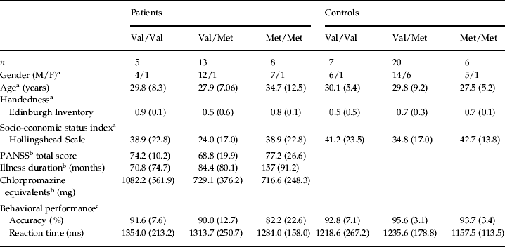
Within patients, genotype subgroups were well matched in terms of clinical variables, including duration of illness (defined as time since the first episode; mean±s.d., 104.92±87.52 months), severity of clinical symptoms (PANSS total scores; mean±s.d., 70.51±20.52) and antipsychotic medication dose (reported in chlorpromazine equivalents; mean±s.d., 825.04±428.38 mg) (all p>0.2). Demographic and clinical data are summarized in Table 1.
Genetics, demographics and behavioral performance

M, Male; F, female; PANSS, Positive and Negative Syndrome Scale.
a Demographics of patients and healthy subjects, bclinical features of patients and cmemory encoding performance of patients and healthy subjects based on the COMT Val158Met genotype.
F and p values for diagnostic group comparisons: age (F=0.15, p=0.69); handedness (F=2.77, p=0.10); socio-economical status index (F=0.04, p=0.82); memory encoding accuracy (F=4.00, p=0.05); memory encoding reaction time (F=3.90, p=0.07).
Values given as mean (standard deviation).
fMRI
Behavioral data
Accuracy data indicated a main effect of diagnosis [patients with schizophrenia performed slightly worse than healthy subjects: F(1, 55)=4.00, p=0.05], no effect of COMT genotype [F(2, 55)=0.98, p=0.38] and no interaction between diagnosis and genotype [F(2, 55)=0.78, p=0.46]. No statistically significant effects were present on reaction time data (all p>0.5).
BOLD response
Main effect of task
Consistent with prior reports (Hariri et al. Reference Hariri, Goldberg, Mattay, Kolachana, Callicott, Egan and Weinberger2003; Bertolino et al. Reference Bertolino, Rubino, Sambataro, Blasi, Latorre, Fazio, Caforio, Petruzzella, Kolachana, Hariri, Meyer-Lindenberg, Nardini, Weinberger and Scarabino2006b, Reference Bertolino, Di Giorgio, Blasi, Sambataro, Caforio, Sinibaldi, Latorre, Rampino, Taurisano, Fazio, Romano, Douzgou, Popolizio, Kolachana, Nardini, Weinberger and Dallapiccola2008), we found a main effect of task in the right (x, y, z coordinates) [(22, −34, −7), k=56, Z=4.42, FWE-corrected p=0.00001; (26, 4, −34), k=22, Z=4.42, p=0.00001] and left [(−30, −37, −15), k=59, Z=5.96, p=0.0001; (−19, 0, −26), k=43, Z=5.46, p=0.0001] parahippocampus, and also in the right [(22, −30, −7), k=32, Z=6.31, p=0.0001] and left [(−34, −30, −11), k=73, Z=5.16, p=0.0001] hippocampus.
Main effect of COMT genotype
No effect of COMT genotype on hippocampal and parahippocampal activity was observed in the overall sample.
Main effect of diagnostic group
ANCOVA demonstrated a main effect of diagnosis in the right [(30, −30, −15), k=53, Z=4.42, FWE-corrected p=0.00001] and left parahippocampus [(−30, −34, −11), k=37, Z=3.97, p=0.002], and also in the right [(30, −34, −7), k=67, Z=4.18, p=0.001] and left [(−34, −30, −11), k=73, Z=4.69, p=0.0001] hippocampus. The specific post-hoc contrast revealed that healthy subjects had greater bilateral hippocampal and parahippocampal activity, whereas no difference resulted in the inverse contrast (patients >healthy subjects).
Group×genotype interaction
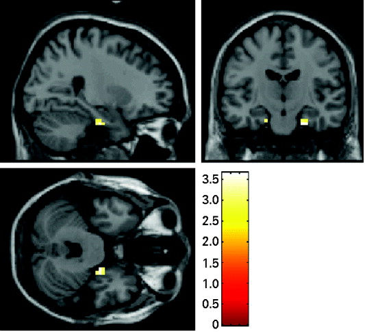
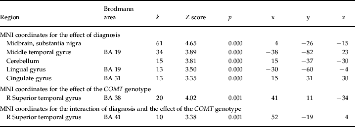
ANCOVA demonstrated an interaction between genotype and diagnosis in the right (x, y, z coordinates) [(22, −15, −30, k=9, Z=3.42, FWE-corrected p=0.02] and left [(−22, −34, −11), k=4, Z=2.88, p=0.05; (−19, −15, −26), k=3, Z=2.86, p=0.06] parahippocampus (Fig. 1a). No significant effects were detected in the hippocampus. The results of the uncorrected exploratory whole-brain analyses are reported in Table 2.
Blood oxygen level-dependent (BOLD) response of the interaction between diagnosis and the catechol-O-methyltransferase (COMT) Val158Met genotype on parahippocampal activity during memory encoding (images thresholded at p=0.005, uncorrected). Color bar represents T score values.

Results of exploratory uncorrected whole-brain statistics (p <0.001, k=10) showing the effect of diagnosis, the effect of the COMT genotype, and their interaction on brain physiology at encoding

COMT, Catechol-O-methyltransferase; MNI, Montreal Neurological Institute; R, right.
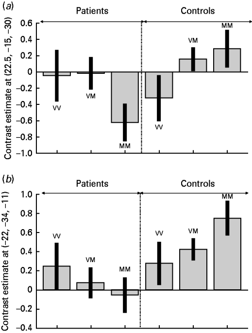
Inspection of BOLD response from these parahippocampal voxels suggested the previously demonstrated Met allele dose effect in healthy subjects whereas the opposite direction was observed in patients with schizophrenia (Fig. 2). Post-hoc Tukey HSD analysis indicated that the Val/Met and Met/Met genotype subgroups in patients had significantly reduced activity in the left parahippocampus compared respectively to Val/Met (p=0.02) and Met/Met (p=0.0003) healthy subjects. The Met/Met genotype subgroup in patients also had reduced BOLD signal in right parahippocampus compared with Met/Met healthy subjects (p=0.01).
Parameter estimates extracted from the cluster of genotype by diagnosis interaction in (a) the right and (b) the left parahippocampus during memory encoding. Error bars show the standard error of the mean. V, Valine; M, methionine.

Finally, to test the relationship between behavioral performance and diagnosis, genotype and activity of the parahippocampus, we performed a multiple regression analysis. This analysis demonstrated a significant relationship between performance and the three-way interaction between genotype, diagnosis and BOLD fMRI activity in the left parahippocampus, suggesting that these three predictors strongly contribute to behavior [whole model: r 2=0.39, F(11, 49)=2.9, p=0.004; three-way interaction: F=7.3, p=0.001]. The same did not hold true when the right parahippocampus was included in the model [r 2=0.20, F(11, 49)=1.1, p=0.3].
Potential correlation between treatment with antipsychotics and activation
Spearman analysis did not demonstrate any significant correlation between dose of antipsychotics (chlorpromazine equivalents) and BOLD signal change in the right (ρ=0.22, p>0.2) and left parahippocampus (ρ=0.31, p>0.1).
Discussion
Extending previous work implicating abnormal activity of mesial temporal lobe structures during memory encoding in schizophrenia, the results of the present study suggest that parahippocampal physiology is differentially associated with COMT Val158Met genotype in healthy subjects and in patients with schizophrenia. This difference is unlikely to be associated with differences in behavioral performance (all analyses were performed with accuracy as covariate) or stratification in the other genetic variants evaluated here with known effects on parahippocampal function, including BDNF Val66Met and DISC1 Ser704Cys. In the hippocampus, however, no effect of COMT Val158Met genotype or interaction with diagnosis was observed.
More specifically, we found an abnormal pattern of hippocampal and parahippocampal activity in patients with schizophrenia during encoding of recognition memory. Extensive literature has demonstrated that these two brain regions are strongly interconnected within the medial temporal lobe (for a review, see van Strien et al. Reference van Strien, Cappaert and Witter2009), and that they are both significantly involved in different subprocesses of encoding and retrieval of recognition memory (for a review, see Squire et al. Reference Squire, Wixted and Clark2007). Evidence from post-mortem (Altshuler et al. Reference Altshuler, Casanova, Goldberg and Kleinman1990; Harrison & Lewis, Reference Harrison, Lewis, Hirsch and Weinberger2003), animal lesion (Lipska & Weinberger, Reference Lipska and Weinberger2000) and imaging studies (for reviews see Wright et al. Reference Wright, Rabe-Hesketh, Woodruff, David, Murray and Bullmore2000; Honea et al. Reference Honea, Crow, Passingham and Mackay2005) has also demonstrated that these regions are crucially involved in the pathophysiology of schizophrenia (Weinberger, Reference Weinberger1999). In vivo studies have indicated small but significant reductions in hippocampal and parahippocampal volumes (Velakoulis et al. Reference Velakoulis, Pantelis, McGorry, Dudgeon, Brewer, Cook, Desmond, Bridle, Tierney, Murrie, Singh and Copolov1999; Witthaus et al. Reference Witthaus, Kaufmann, Bohner, Ozgurdal, Gudlowski, Gallinat, Ruhrmann, Brune, Heinz, Klingebiel and Juckel2009) in first-episode, and also in chronic, schizophrenic patients (Nelson et al. Reference Nelson, Saykin, Flashman and Riordan1998; Honea et al. Reference Honea, Crow, Passingham and Mackay2005) that do not seem to be associated with chronicity of the disorder or with pharmacological treatment. In addition, proton MR spectroscopy studies have indicated a reduced N-acetylaspartate signal in the hippocampus, indicative of neuronal pathology, again present during all phases of the illness (Maier et al. Reference Maier, Ron, Barker and Tofts1995; Bertolino et al. Reference Bertolino, Nawroz, Mattay, Barnett, Duyn, Moonen, Frank, Tedeschi and Weinberger1996, Reference Bertolino, Callicott, Elman, Mattay, Tedeschi, Frank, Breier and Weinberger1998; Deicken et al. Reference Deicken, Pegues and Amend1999). Numerous functional neuroimaging studies, particularly those testing episodic or relational memory, have consistently reported decreased hippocampal activation in patients with schizophrenia (Achim & Lepage, Reference Achim and Lepage2005), whereas only a few studies have demonstrated differences in parahippocampal engagement between patients with schizophrenia and healthy subjects (Ragland et al. Reference Ragland, Gur, Valdez, Turetsky, Elliott, Kohler, Siegel, Kanes and Gur2004). Notably, decreased parahippocampal activation has also been described in genetically at-risk relatives of patients with schizophrenia performing a verbal memory task (Thermenos et al. Reference Thermenos, Seidman, Poldrack, Peace, Koch, Faraone and Tsuang2007). Thus, our results support previous evidence suggesting that parahippocampal regions together with the hippocampus are affected in schizophrenia.
Of note, we report differences in activity of the parahippocampal region between healthy subjects and patients with schizophrenia as a function of the COMT genotype. Val/Met and Met/Met genotype groups in patients had significantly reduced activity in the left parahippocampus compared with the same genotype groups in healthy subjects. The Met/Met genotype group in patients also had reduced right parahippocampal activity compared to the Met/Met group in healthy subjects. A precise understanding of the biological mechanisms explaining this interaction cannot be obtained with the current data and further studies are required. However, some speculations can be proposed based on current knowledge. A series of studies with in vivo recording of single neurons in non-human primates (Zahrt et al. Reference Zahrt, Taylor, Mathew and Arnsten1997; Arnsten & Goldman-Rakic, Reference Arnsten and Goldman-Rakic1998; Lidow et al. Reference Lidow, Koh and Arnsten2003; Vijayraghavan et al. Reference Vijayraghavan, Wang, Birnbaum, Williams and Arnsten2007) and with functional imaging in humans (Egan et al. Reference Egan, Goldberg, Kolachana, Callicott, Mazzanti, Straub, Goldman and Weinberger2001; Meyer-Lindenberg et al. Reference Meyer-Lindenberg, Nichols, Callicott, Ding, Kolachana, Buckholtz, Mattay, Egan and Weinberger2006; Bertolino et al. Reference Bertolino, Di Giorgio, Blasi, Sambataro, Caforio, Sinibaldi, Latorre, Rampino, Taurisano, Fazio, Romano, Douzgou, Popolizio, Kolachana, Nardini, Weinberger and Dallapiccola2008) has demonstrated a non-linear, inverted U-shaped relationship between dopamine signaling within the mesocortical dopamine signaling system (projecting also into the prefrontal cortex) and prefrontal activity. This relationship predicts that too much or too little dopamine is related to disruption of prefrontal activity. More specifically, the Met allele of COMT has been associated with lower (more efficient) prefrontal activity, especially within the context of working memory (Egan et al. Reference Egan, Goldberg, Kolachana, Callicott, Mazzanti, Straub, Goldman and Weinberger2001; Meyer-Lindenberg et al. Reference Meyer-Lindenberg, Nichols, Callicott, Ding, Kolachana, Buckholtz, Mattay, Egan and Weinberger2006). Another set of studies has indicated that a similar nonlinear, inverted U relationship may exist between the mesolimbic dopamine signaling system and activity of the hippocampal region, the amygdala and the nucleus accumbens. In these studies, however, the Met allele of COMT is associated with greater activity of these brain regions (Smolka et al. Reference Smolka, Schumann, Wrase, Grusser, Flor, Mann, Braus, Goldman, Buchel and Heinz2005; Bertolino et al. Reference Bertolino, Caforio, Petruzzella, Latorre, Rubino, Dimalta, Torraco, Blasi, Quartesan, Mattay, Callicott, Weinberger and Scarabino2006a, Reference Bertolino, Di Giorgio, Blasi, Sambataro, Caforio, Sinibaldi, Latorre, Rampino, Taurisano, Fazio, Romano, Douzgou, Popolizio, Kolachana, Nardini, Weinberger and Dallapiccola2008; Drabant et al. Reference Drabant, Hariri, Meyer-Lindenberg, Munoz, Mattay, Kolachana, Egan and Weinberger2006; Yacubian et al. Reference Yacubian, Sommer, Schroeder, Glascher, Kalisch, Leuenberger, Braus and Buchel2007; Krach et al. Reference Krach, Jansen, Krug, Markov, Thimm, Sheldrick, Eggermann, Zerres, Stocker, Shah and Kircher2010). Given this context, the inverted U relationship between dopamine and hippocampal activity may be shifted to the right in schizophrenia because of different levels of dopamine signaling, with the Met allele being associated with lower activity. This speculative interpretation of our present results is consistent with other studies with functional imaging in patients with schizophrenia performing verbal fluency that have demonstrated a similar inversion of the Met allele dose effect in the prefrontal cortex. Therefore, a putative and speculative interpretation of our results is that the inversion of the Met allele load is identified because of different levels of dopamine in schizophrenia (see caveats below). In this sense, the present data might be taken as evidence in humans that parahippocampal physiology and dopamine signaling associated with the COMT Val158Met polymorphism are closely connected in schizophrenia.
Some potential limitations of the present data should be addressed. First, our results are based on a relatively small group size, although the computation of the effect size with Cohen's d supports their statistical robustness. By using the means and standard deviations of the BOLD signal in the right (22, −15, −30) and left (−22, −34, −11) parahippocampus for patients and controls based on COMT genotype, we found a low effect size for Val/Val (right, d=0.49; left, d=0.09), an intermediate effect size for Val/Met (right, d=0.48; left, d=1.08) and a large effect size for Met/Met (right, d=1.9; left, d=2.6). Although replication of our results is necessary, these effect sizes indicate important statistical relationships between the variables examined.
Another caveat of our study is that the behavioral and pathophysiological findings could reflect state- rather than trait-related associations. However, patients had been on stable pharmacological treatment for at least 8 weeks. Moreover, antipsychotics are known to antagonize dopamine transmission whereas our results suggest a shift to the right of the inverted U relationship between dopamine signaling and hippocampal activity, a hypothesis that is consistent with greater levels of dopamine (discussed earlier). Finally, antipsychotic medication may have an effect on the nature of the functional and behavioral abnormalities observed in patients (for a review, see Di Giorgio et al. Reference Di Giorgio, Sambataro and Bertolino2009). However, no significant correlations were found between behavioral data or signal change extracted from the significant cluster showing gene by diagnosis interaction and chlorpromazine equivalents.
In conclusion, these results suggest that genetically determined modulation of dopamine signaling may be important in determining the phenotype of parahippocampal physiology in schizophrenia.
Acknowledgments
We thank R. Masellis, R. Lomuscio and G. De Simeis for data acquisition. We also thank all of the patients who participated in the study and our colleagues (F. Cantalice, G. Cipolla, M. Squeo and G. Abbondanza) who referred them to us.
Declaration of Interest
None.