INTRODUCTION
The Holothuria genus is the only Holothuriidae present in the Mediterranean Sea and north-eastern Atlantic, being found in different marine habitats from coastal shallow waters. They are found in high densities providing important ecosystem services enhancing nutrient cycling and local productivity in oligotrophic carbonate sediments through their bioturbation and deposit feeding activities (Byrne et al., Reference Byrne, Rowe and Uthicke2010).
Holothuria (Roweothuria) arguinensis Koehler & Vaney, Reference Koehler and Vaney1906, belongs to this genus and in recent years it is being considered as a target species in the increasing sea cucumber fishery (Aydin, Reference Aydin2008; Sicuro & Levine, Reference Sicuro and Levine2011). This species had been considered to be a north-eastern Atlantic species that is distributed from Portugal to Morocco and Mauritania, including the Canary Islands (Thandar, Reference Thandar1988; Hanson, Reference Hanson, Costello, Emblow and White2001). It has not been found in other Macaronesian Islands such as Açores, Selvagens or Madeira, or in the Cape Verde Archipelago (Pereira, 1997; Borerro-Pérez et al., 2010; Micael et al., Reference Micael, Alves, Jones and Costa2012). However, its geographical distribution is changing, including the colonization of the Mediterranean Sea, where it was registered for the first time on the Granada coast, southern Spain (Ocaña & Pérez-Ruzafa, Reference Ocaña and Pérez-Ruzafa2004) and its extension to the north Portugal coast in the Berlengas Islands (39°24.56′N 9°30.88′W) (Rodrigues, Reference Rodrigues2012).
Considering this framework, the aims of this paper are: (i) to record the occurrence for the first time of H. arguinensis to the north of the Alborán Sea (south-eastern Spain); (ii) to discuss the colonization process of this species; and (iii) to contribute to the clarification of the taxonomy of Holothuria genus species from the Atlanto-Mediterranean region.
MATERIALS AND METHODS
One specimen was collected in El Mojón (37°50′54.17″N 0°45′39.90″W), south-eastern Spanish coast. It was identified in situ through its external morphology and subsequently this identification was confirmed using traditional taxonomic characters, such as ossicles, on the basis of the original description of this species (Koehler & Vaney, Reference Koehler and Vaney1906) and another relevant taxonomic paper (Thandar, Reference Thandar1988). In addition, cytochrome c oxidase I (COI) gene was amplified according to protocols from Borrero-Pérez et al. (Reference Borrero-Pérez, Gómez-Zurita, González-Wangüemert, Marcos and Pérez-Ruzafa2010) and this sequence was recorded in the GenBank database (accession number JX125700).
RESULTS
SYSTEMATICS
Class HOLOTHUROIDEA
Order ASPIDOCHIROTIDA
Family HOLOTHURIIDAE
Genus Holothuria Linnaeus, 1767
Sugenus Roweothuria Thandar, Reference Thandar1988
Holothuria (Roweothuria) arguinensis Koehler & Vaney, Reference Koehler and Vaney1906
(Figures 1–3)
Holothuria arguinensis Koehler & Vaney, Reference Koehler and Vaney1906, pp. 62–65, pl. 5, figures 5–13, pl. VI, figures 14–21. Cherbonnier, 1950, pp. 106–108, figure 3d.
Holothuria (Lessonothuria) arguinensis Rowe, 1969, pp. 149–150.
Holothuria (Roweothuria) arguinensis Thandar Reference Thandar1988, pp. 48 and 53.
Species length up to 185 mm; dorsal surface quite rounded and more or less wrinkled, cracked and evenly coloured brown on the entire region of bivium; on the sides, this colour is gone gradually to white; ventral surface slightly flattened, whitish and with punctuations that match the pedicels retracted which are light brown. Pedicels evenly distributed throughout the body, more numerous ventrally. Large papillae or warts on the dorsal surface, they are blackish brown to whitish and they are arranged in six longitudinal rows; two of these rows occupy the radial side and they constitute a whole by almost continuous rim; on bivium, each radius has two rows of papillae that alternated more or less irregularly with each other and offer the same dimensions as the lateral papillae. Ossicles: tables few, mostly incomplete with four central openings and without marginal holes; rounded or angular edges; spire with four rods joined to one another by a transverse bar and ending in a cluster of teeth; 60–65 µm. Ventral buttons are oval, very thick, smooth, rounded edges and they vary in size and number of holes, but the most common is 2–3 pairs of small holes; dorsal ones are flatter than the ventral face, and they are not quite the same form as those of the ventral which have rounded edges, while those of the dorsal surface are toothed, very rough, but it is possible to find all the transitions between these two extreme forms; 2–4 pairs of large holes; 50–100 µm. Numerous Polian vesicles; single stone canal (Koehler & Vaney, Reference Koehler and Vaney1906; Thandar, Reference Thandar1988).
SPECIMEN FROM EL MOJÓN (SOUTH-EASTERN SPAIN)
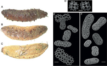
On the basis of H. arguinensis specimens collected and studied previously by us from localities belonging to its original distribution, we must highlight the variability of the coloration on dorsal surface. It ranges from uniform brown to a coloration gradient (brown in the centre and white or yellowish on sides), but always with darker warts (Figure 1A–C). Another important observation is about the rough surface of ventral buttons: Koehler & Vaney (Reference Koehler and Vaney1906) were not clear about this characteristic in H. arguinensis; and Thandar (Reference Thandar1988) described them as smooth buttons. However, they are rough although the dorsal buttons can be toothed too (Figure 1E, F).
Specimens of Holothuria arguinensis from localities belonging to its original geographical distribution. Individuals from Ria Formosa (Portugal) showing coloration variability: (A) dorsal side of individual AFM1; (B, C) dorsal and ventral sides of the individual AFM2. Ossicles of one individual (ACN1) from Canary Islands (Spain); (D) dorsal tables; (E) dorsal buttons; (F) ventral buttons. Scale bar: 20 µm.

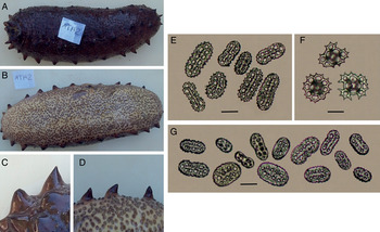
The specimen collected at El Mojón (south-eastern Spain) fits with the characteristics of H. arguinensis presented previously. The length of specimen was 175 mm; dorsal surface is uniform brown with the darker warts alternating more or less irregularly (Figure 2A, B), and with the lateral large papillae arranged all along the continuous rim (Figure 2C, D). Dorsal and ventral ossicles fit with the description presented, observing on this specimen the rough surface of ventral buttons discussed previously (Figure 2E, G). According to the original description, the number of tables is low specially in the ventral side showing incomplete discs, although some complete tables were also found with a marginal ring of holes and pointed edges (Figure 2F).
Specimen of Holothuria arguinensis collected in El Mojón (Spain): (A) complete specimen viewed from dorsal side; (B) complete specimen viewed from ventral side; (C) dorsal view of papilla; (D) ventral view of papilla; (E) dorsal buttons; (F) dorsal tables; (G) ventral buttons. Scale bar: 20 µm.

DISTRIBUTION AND HABITAT
Holothuria arguinensis is distributed in the north-east Atlantic, from the Berlengas Islands (Portugal) to Morocco and Mauritania, including the Canary Islands and the transition zone to the Mediterranean Sea (Alborán Sea). Its range of depth is from 0 to 52 m. Holothuria arguinensis is a species that does not take refuge in crevices, remains visible on sandy or seagrass beds, inhabiting the inter-tidal zone and being able to stand periods of drought during the low tides such as is observed in the Ria Formosa (Portugal) (Figure 3A–D).
Specimens of Holothuria arguinensis: (A, C, D) Ria Formosa (Portugal) during low tide (photographs: M. González-Wangüemert); (B) Arrábida Natural Park (Portugal) (photograph: E. Gonçalves).

TAXONOMIC REMARKS
The taxonomic status of the subgenus Roweothuria, H. arguinensis being the type species, should be revised. Three species H. arguinensis, H. polii and H. vemae belong to this subgenus, considering mainly the spinose, flat rimmed table discs, and smooth to rough or spinose rosette-like buttons (Thandar, Reference Thandar1988). However, recent phylogenetic analysis showed that H.(R.) arguinensis is nearer to H. (Holothuria) tubulosa, H. (Holothuria) mammata and H. (Vaneyothuria) lentiginosa than H.(R.) polii, which is very divergent genetically (Borrero-Pérez et al., Reference Borrero-Pérez, Gómez-Zurita, González-Wangüemert, Marcos and Pérez-Ruzafa2010).
DNA BARCODING
The amplification of the COI mitochondrial gene, a sequence with 531 base pairs in length and the blast in GENBANK (http://www.ncbi.nlm.nih.gov), identified the individual from El Mojón as H. arguinensis with a 99% maximum identity with GQ214754.1 (Canary Islands, Spain) and GQ214755.1 (Algarve, Portugal) but showing a different haplotype which was recorded as JX125700.
DISCUSSION
The current new record of H. arguinensis is supported by the taxonomic information presented in this paper. In addition, this information will be useful to clarify the taxonomy of Holothuria genus species from the Atlanto-Mediterranean region, considering other records of this species in the Mediterranean Sea, which usually have been named as Holothuria tubulosa (Lorenzo-Heller, Reference Lorenzo-Heller2005). Therefore, this species could be under-sighted in the south of the Iberian Peninsula considering its similarity to other species.
The presence of H. arguinensis in El Mojón (south-eastern Spain) is probably related to climate changes and human impacts. The increase of temperatures in the north-eastern Atlantic Ocean (Brander et al., Reference Brander, Blom, Borges, Erzini, Henderson, MacKenzie, Mendes, Ribeiro, Santos and Torensen2003) would allow this species to adapt to warmer waters colonizing the Mediterranean Sea; this ability is reinforced by the high survival of this species to desiccation during the low tides (authors, personal observation in Ria Formosa and Olhos de Agua in South Portugal: Figure 3). Also, the increase of shipping traffic through the Strait of Gibraltar could be favouring the dispersal of this species, mainly during the settled pentacula larvae (Asha & Muthiah, Reference Asha and Muthiah2002; Ivy & Giraspy, Reference Ivy and Giraspy2006).
This finding widens the geographical range of H. arguinensis and demonstrates that its environmental requirements are changing. Future genetic studies should allow us to improve our knowledge about the colonization events of this species from the Atlantic Ocean to the Mediterranean Sea.
ACKNOWLEDGEMENTS
This research was supported by CUMFISH project (PTDC/MAR/119363/2010; Principal Investigator: Dr M. González-Wangüemert) funded by Fundacão para Ciência e Tecnologia (Portugal). M. González-Wangüemert was supported by a FCT postdoctoral fellowship (SFRH/BPD/70689/2010).