



Introduction
Congenitally corrected transposition of the great arteries is a rare cardiac anomaly, representing less than 1% of all congenital heart defects. Reference Marelli, Mackie, Ionescu-Ittu, Rahme and Pilote1 It is characterised by an atrioventricular and ventriculoarterial discordance, which results in physiologically normal blood circulation despite abnormal cardiac anatomy. Namely, the systemic circulation is supported by a morphologically right ventricle. Reference Susheel Kumar2 This unique configuration often delays clinical manifestation until later in life. Dextrocardia, in which the heart is positioned in the right hemithorax, is in up to 20% associated with congenitally corrected transposition of the great arteries and may further complicate the diagnostic process. Reference Tortigue, Nield and Karakachoff3 Many patients with congenitally corrected transposition of the great arteries remain asymptomatic for decades and only present when complications arise. Reference Vasiliu, Seldrum, Dupont, Dormal, Blommaert and De Roy4,Reference Placci, Lovato and Bonvicini5 The most common late complications are conduction disturbances (atrioventricular block), systemic right ventricular failure, and systemic atrioventricular valve dysfunction. Reference Budts, Prokšelj, Lovrić, Kačar, Gatzoulis and Brida6,Reference Kutty, Danford, Diller and Tutarel7 This case underscores the importance of detailed cardiac imaging and high clinical suspicion when evaluating patients with unexplained symptoms and known anatomical variants.
Case presentation
A 71-year-old male with a known history of dextrocardia and stable idiopathic thrombocytopenia was referred for cardiac evaluation due to new-onset exertional dyspnoea that developed during a coronavirus disease 2019 (COVID-19) infection. He was not on any regular cardiac medication and denied other cardiovascular symptoms. Electrocardiogram revealed ectopic atrial rhythm, repolarization abnormalities, and nonspecific intraventricular conduction delay (Figure 1). Pulmonary function tests were within normal limits. A chest X-ray confirmed dextrocardia, with no signs of pulmonary pathology. Transthoracic echocardiography performed by a cardiologist in a regional hospital showed a normal-sized sub-pulmonary (morphologically left) ventricle with preserved systolic and diastolic function (LVEF 73%), but a markedly dilated systemic (morphologically right) ventricle with mildly reduced systolic function (FAC 30%). There was significant interventricular septal shift during systole, consistent with elevated right-sided pressures. A high tricuspid regurgitation gradient (TR max 104 mmHg) was observed, raising suspicion of severe pulmonary hypertension. NT-proBNP was mildly elevated (208 pg/mL).
Electrocardiogram of an adult patient with dextrocardia and congenitally corrected transposition of the great arteries revealing ectopic atrial rhythm, repolarization abnormalities, and nonspecific intraventricular conduction delay.
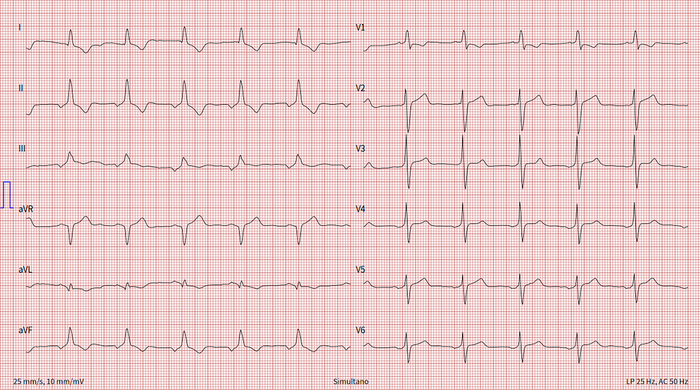
Figure 1 Long description
An electrocardiogram of an adult patient with dextrocardia and congenitally corrected transposition of the great arteries. The ECG reveals ectopic atrial rhythm, repolarization abnormalities, and nonspecific intraventricular conduction delay. The graph consists of multiple leads, each showing distinct patterns of electrical activity in the heart. The leads are labeled with Roman numerals and letters, indicating different views of the heart's electrical activity. The grid background helps in measuring the duration and amplitude of the electrical signals.
Further investigation included cardiac MRI, which revealed situs solitus with dextroversion and findings consistent with congenitally corrected transposition of the great arteries (Figure 2). The systemic right ventricle was enlarged with mild systolic dysfunction.
Cardiac MRI evaluation of anatomy and ventricular function. Panel A: Four-chamber view demonstrating dextrocardia. Panel B: Three-chamber view showing atrioventricular and ventriculoarterial discordance. Panel C: Short-axis view, middle segments, morphological left ventricle on the right, and hypertrabeculated, thickened, morphologically right ventricle on the left. Panel D: Short axis view, basal cut, anteriorly placed aorta arising from the systemic right ventricle. LV= Left Ventricle; SRV= Systemic Right Ventricle; Ao= Aorta; LA= Left Atrium.
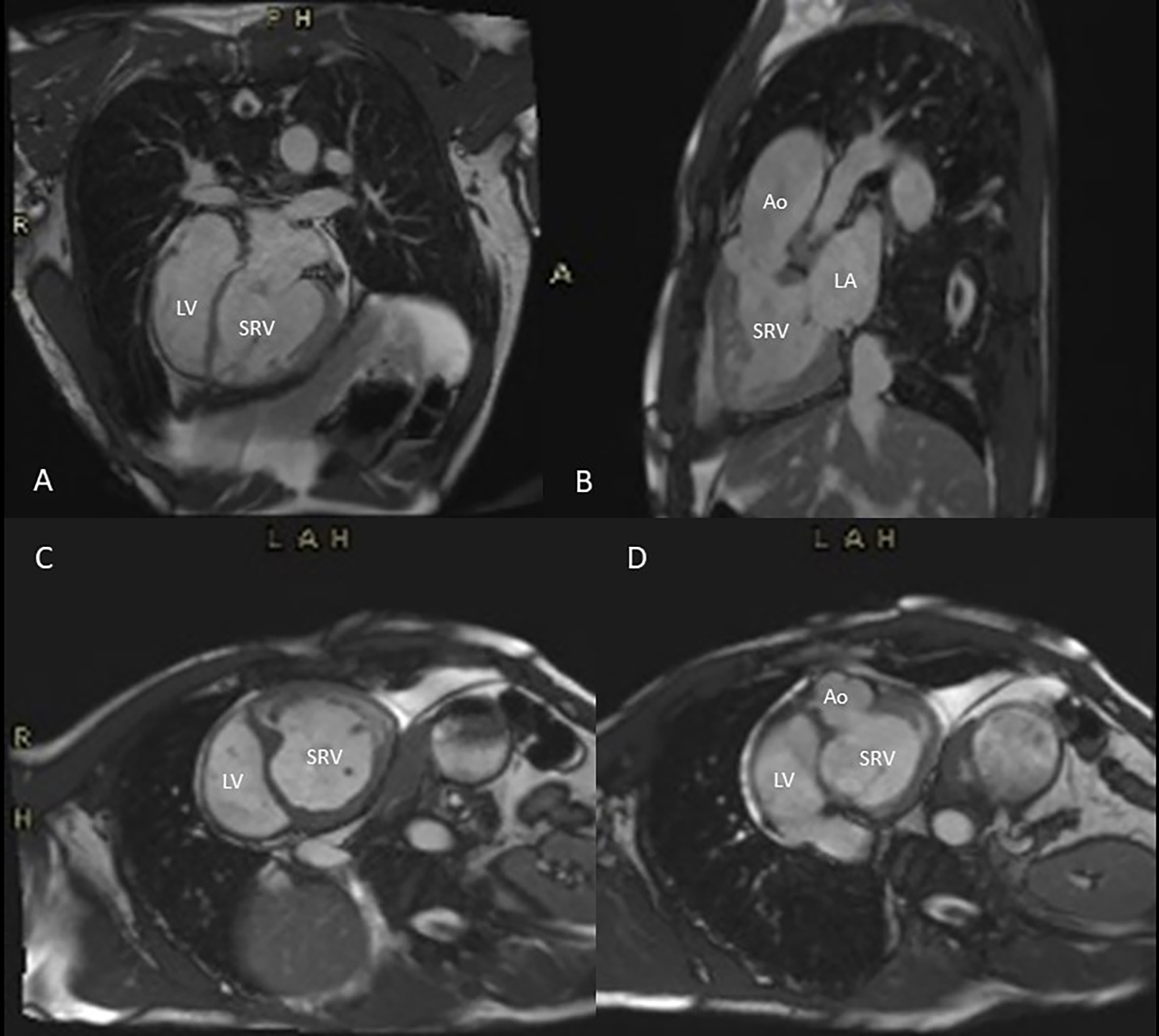
Figure 2 Long description
The image displays a series of cardiac MRI scans. Panel A shows a four-chamber view demonstrating dextrocardia, where the heart is positioned on the right side of the chest. Panel B presents a three-chamber view highlighting atrioventricular and ventriculoarterial discordance, indicating abnormal connections between the heart chambers and arteries. Panel C illustrates a short-axis view at the middle segments, revealing a hypertrabeculated and thickened morphologically right ventricle on the left and the morphological left ventricle on the right. Panel D depicts a short-axis view at the basal cut, showing the aorta arising from the systemic right ventricle. Key structures labeled include the left ventricle (LV), systemic right ventricle (SRV), aorta (Ao), and left atrium (LA).
The patient was referred to our adult CHD centre. Echocardiography confirmed congenitally corrected transposition of the great arteries with dextrocardia, an enlarged systemic right ventricle with mildly to moderately reduced systolic function, and mild systemic atrioventricular (tricuspid) valve regurgitation (Figure 3). Given his stable clinical condition, heart failure therapy was not initiated, but regular follow-up was scheduled.
Transthoracic echocardiography. Panel A: Parasternal short-axis view with both ventriculoarterial valves. Panel B: Parasternal long-axis view with parallel outflow tracts, ventriculoarterial valves, and great arteries. Panel C: Apical four-chamber view with right, systemic ventricle connected to the left atria. Moderator band is seen (asterix). LV= Left Ventricle; LA= Left Atrium; SRV= Systemic Right Ventricle; RA= Right Atrium; Ao= Aorta; PA= Pulmonary Artery.
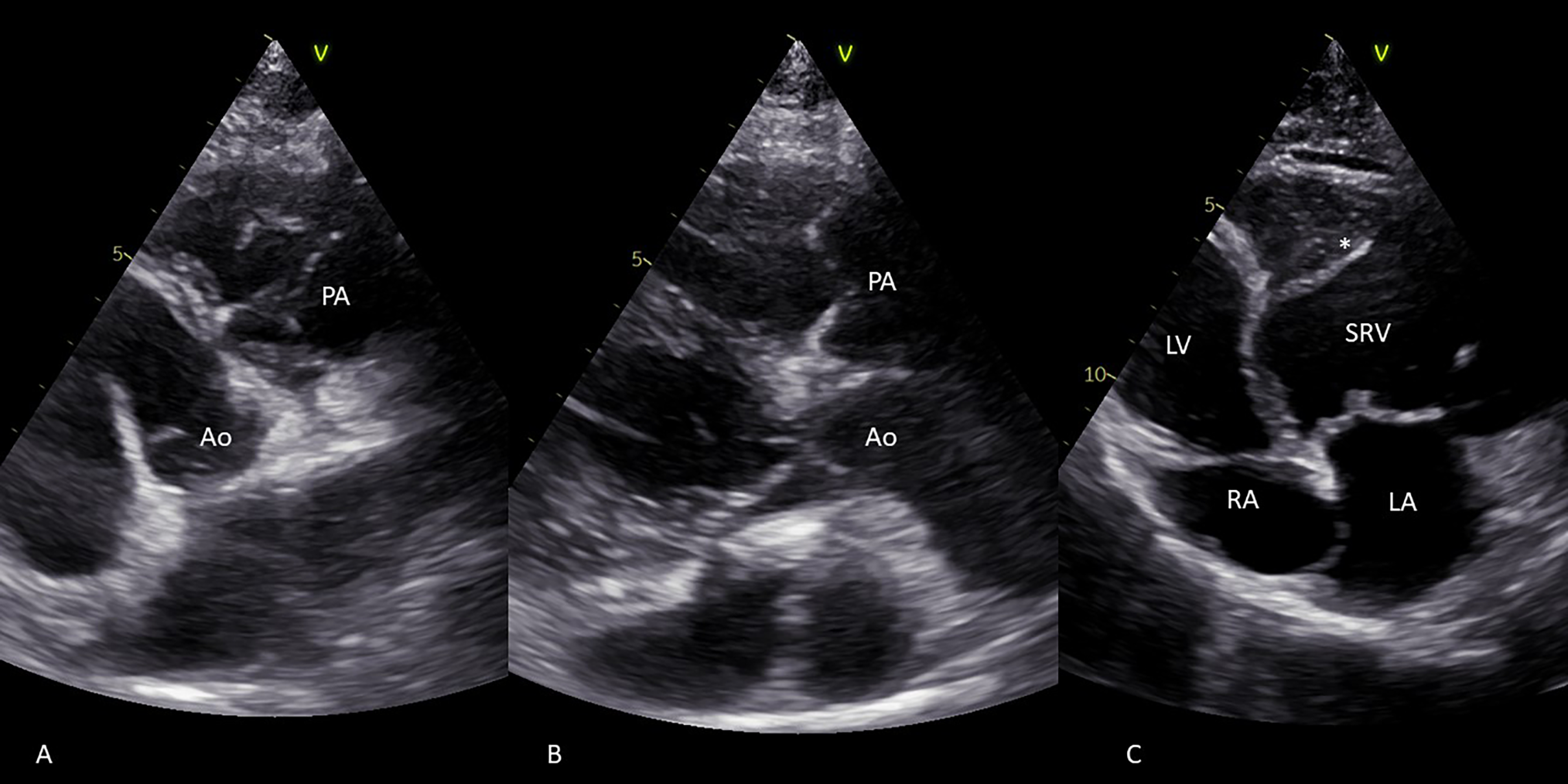
Figure 3 Long description
The image consists of three panels labeled A, B, and C, each showing different views of the heart obtained through transthoracic echocardiography. Panel A presents a parasternal short-axis view, highlighting both ventriculoarterial valves. Panel B shows a parasternal long-axis view, displaying parallel outflow tracts, ventriculoarterial valves, and great arteries. Panel C depicts an apical four-chamber view, where the right, systemic ventricle is connected to the left atrium. A moderator band is visible, marked with an asterisk. The labeled structures include the left ventricle (LV), left atrium (LA), systemic right ventricle (SRV), right atrium (RA), aorta (Ao), and pulmonary artery (PA).
Discussion
Congenitally corrected transposition of the great arteries is a rare and complex congenital defect characterised by a double discordance and a systemic right ventricle. Reference Susheel Kumar2 This specific anatomy ensures physiologic blood flow without cyanosis, which may delay correct diagnosis.
Most patients with congenitally corrected transposition of the great arteries have associated abnormalities such as dextrocardia, ventricular septal defects, pulmonary stenosis, or atrioventricular valve malformations, which may lead to an earlier symptom onset. Reference Kutty, Danford, Diller and Tutarel7 The systemic, morphologically right ventricle, which is not designed to support systemic pressure, is prone to develop heart failure, especially under stress or ageing. Reference Lebherz, Gerhardus and Lammers8 Conduction abnormalities, including complete heart block, are common. Reference Connolly, Miranda, Egbe and Warnes9
In our case, the initial echocardiographic assessment resulted in a misdiagnosis of possible pulmonary hypertension because the systemic right ventricle was not recognised—an oversight that can occur easily in the setting of dextrocardia, where the altered anatomical orientation can make recognition of the systemic ventricle challenging. After referral to a tertiary adult CHD centre, the diagnosis of congenitally corrected transposition of the great arteries was established. The markedly elevated tricuspid regurgitation gradient seen on initial echocardiography reflected systemic pressures in the systemic right ventricle, rather than pulmonary hypertension.
This case highlights the importance of considering congenital heart defects—even in older patients—especially when other abnormalities such as dextrocardia are known. Timely referral for advanced imaging and specialised evaluation is critical for accurate diagnosis and optimal long-term management.
Conclusion
Congenitally corrected transposition of the great arteries, a rare CHD, may remain undiagnosed for many decades. In patients with known dextrocardia and signs of heart failure or right heart strain, additional CHD should be considered. Early referral to a centre with expertise in adult CHD is essential for accurate diagnosis, risk stratification, and management.
Financial support
This research received no specific grant from any funding agency, commercial, or not-for-profit sectors.
Competing interests
The authors declare none.
Ethical standard
This article does not contain any studies with human participants or animals performed by any of the authors.



