Introduction
Saetaspongia Mehl and Reitner in Steiner et al., Reference Steiner, Mehl, Reitner and Erdtmann1993 (type species S. densa Mehl and Reitner in Steiner et al., Reference Steiner, Mehl, Reitner and Erdtmann1993) was first established based on two specimens from the Cambrian (Stage 3) Sansha Fauna (also published as Sansha Sponge Fauna) of Dayong County, Hunan Province, China. The Sansha material is characterized by a small circular body form and has a skeleton made principally of plumose diactines, with the (possible) presence of hexactines. The original diagnosis of Saetaspongia includes two primary characters (Steiner et al., Reference Steiner, Mehl, Reitner and Erdtmann1993): (1) dense accumulations of fine diactines arranged in plumose-appearing bundles, and (2) spicules do not protrude from the outer margin of the sponge body. Some large spicules from particular specimens might be tri-, tetr-, or even hexactines; however, Steiner et al. (Reference Steiner, Mehl, Reitner and Erdtmann1993) suspected that these large spicules might not belong to this sponge because similar spicules were also commonly found in the area located outside the outline of the fossil on the paratype. The possible presence of hexactines suggested that Saetaspongia may have a hexactinellid affinity. However, due to the plumose architecture that is commonly found in certain protomonaxonids, Saetaspongia was assumed to be a protomonaxonid (Steiner et al., Reference Steiner, Mehl, Reitner and Erdtmann1993). Due to limited material and the mode of preservation, the taxonomic position of Saetaspongia was not clarified at that time.
Subsequently, because a specimen (referred to Saetaspongia densa) from the Cambrian (Stage 3) Chengjiang Biota was demonstrated to have hexactine-based spicules (Rigby and Hou, Reference Rigby and Hou1995), the genus was considered a hexactinellid sponge exclusively on the basis of the presence of hexactines. This characteristic was also applied uncritically to other fossil specimens from the Cambrian of China, all of which were assigned to S. densa (Yang and Zhao, Reference Yang and Zhao2000; Zhao et al., Reference Zhao, Yang, Yang and Mao2006; Yang et al., Reference Yang, Zhao, Zhu, Cui and Yang2010; Hou et al., Reference Hou, Siveter, Siveter, Aldridge, Cong, Gabbott, Ma, Purnell and Williams2017). In Finks and Rigby (Reference Finks, Rigby and Kaesler2004), S. densa was classified as a reticulosan and even interpreted as a protospongiid (Chen et al., Reference Chen, Müller, Hou and Xiao2015). Actually, hexactine spicules have been corroborated to be present in many disparate groups of early sponges (Botting and Zhang, Reference Botting and Zhang2013; Botting et al., Reference Botting, Muir and Lin2013; Botting and Muir, Reference Botting and Muir2018), and the presence of hexactines is itself not restricted to hexactinellids (Botting et al., Reference Botting, Muir and Lin2013; Botting and Muir, Reference Botting and Muir2018). So, this spicule type is not diagnostic of hexactinellids in the fossil record (Botting and Muir, Reference Botting and Muir2018). It also should be noted that although the Chengjiang material does not show the plumose structure, it has been referred to Saetaspongia based predominately on the presence of hexactines and parallel hairlike bundles of spicules (Rigby and Hou, Reference Rigby and Hou1995). However, the material from the Cambrian (Stage 3) Sirius Passet Biota of North Greenland does not have hexactines, but has a consistent architecture to the skeleton that contains local plumose arrangements of spicules (Botting and Peel, Reference Botting and Peel2016). Therefore, given the consistent architecture provided by the material from the Sirius Passet Biota, Saetaspongia was assumed to have a protomonaxonid rather than a hexactinellid affinity (Botting and Peel, Reference Botting and Peel2016).
To date, there are different viewpoints on the taxonomic position of Saetaspongia (Wu, Reference Wu2004; Chen et al., Reference Chen, Müller, Hou and Xiao2015; Botting and Peel, Reference Botting and Peel2016). Different specialists studying relevant specimens follow different classification schemes: one based on the plumose architecture (Steiner et al., Reference Steiner, Mehl, Reitner and Erdtmann1993; Botting and Peel, Reference Botting and Peel2016), and the other based on the (possible) presence of hexactinellid-like spicules (Rigby and Hou, Reference Rigby and Hou1995; Yang and Zhao, Reference Yang and Zhao2000; Zhao et al., Reference Zhao, Yang, Yang and Mao2006; Yang et al., Reference Yang, Zhao, Zhu, Cui and Yang2010; Hou et al., Reference Hou, Siveter, Siveter, Aldridge, Cong, Gabbott, Ma, Purnell and Williams2017; Wang et al., Reference Wang, Yang, Zhao and Duan2017). The debate over different classification schemes causes some difficulty in assessing the diversity and biogeography of this genus. Data from the literature (Yang et al., Reference Yang, Zhu, Zhao and Wang2005; Wu et al., Reference Wu, Zhu and Steiner2014; Botting and Peel, Reference Botting and Peel2016) indicated that Saetaspongia sponges had a wide distribution, at least from four assemblages on two paleocontinents (Botting and Muir, Reference Botting and Muir2019). However, due to different specialists following different classification schemes, the identification of some of these specimens is probably unreliable and is strongly biased towards the assessment of the distribution patterns of Saetaspongia. Therefore, fully clarifying these confusions about Saetaspongia fossils requires new relevant specimens that are well preserved and can reveal key taxonomic characters.
Considering the emerging scenario of Saetaspongia, we describe a new taxon and a new record of an existing species here, specifically S. jianhensis n. sp. and S. cf. S. densa. These sponge fossils recovered from the Cambrian (Stage 4) Balang Biota of the Balang Formation can shed light on the confusion surrounding the identification of Saetaspongia fossils and the assessment of the distribution pattern of this genus.
Geological setting
Balang Formation
The Balang Formation is exposed in eastern Guizhou and western Hunan, China, and represents part of the Jiangnan Slope, which represents the transitional slope belt between the Yangtze Platform and the Jiangnan Basin (Fig. 1.1; Peng and Babcock, Reference Peng and Babcock2001; Peng et al., Reference Peng, Zhao, Wu, Yuan and Tai2005; Liu and Lei, Reference Liu and Lei2013; Liu et al., Reference Liu, Wen, Peng, Liang and Wang2017). The lower part of this formation is composed of gray-greenish to yellow-greenish silty mudstone; while the upper part consists mostly of light gray to gray-yellow mudstone interbedded with light gray calcareous mudstone. In the Guizhou area, the Balang Formation is in conformable contact with the overlying Tsinghsutung Formation (also published as Qingxudong Formation) and the underlying Bianmachong Formation (Bureau of Geology and Mineral Resources of Guizhou, 1987; Yin, Reference Yin1996). The Tsinghsutung Formation is dominated by gray-greenish thin-bedded limestone; while the Bianmachong Formation is mainly composed of gray-black mudstone (Fig. 1.2; Bureau of Geology and Mineral Resources of Guizhou, 1987; Peng et al., Reference Peng, Zhao, Wu, Yuan and Tai2005; Peng, Reference Peng2009; Liu, Reference Liu2018). No isotopic dating is available for the Balang Formation, therefore its age is regarded as Stage 4 according to a series of trilobite-based correlations of Cambrian strata between South China and other continents (Zhou et al., Reference Zhou, Yuan, Zhang, Wu and Yin1979, Reference Zhou, Yuan, Zhang, Wu and Yin1980; Zhou and Yuan, Reference Zhou and Yuan1980; Peng and Babcock, Reference Peng and Babcock2001; Yuan et al., Reference Yuan, Zhao and Li2001, Reference Yuan, Zhao and Yang2006, Reference Yuan, Lin, Zhao and Peng2011; Qin et al., Reference Qin, Peng, Fu and Da2010; Yan et al., Reference Yan, Peng, Zhao, Wen and Sun2014; Yuan and Ng, Reference Yuan and Ng2014; Shen et al., Reference Shen, Peng, Wen and Liu2016; Y.F. Wang et al., Reference Wang, Peng, Wang, Wen, Zhang, Du and Shao2020), corresponding to the Qiandongian early Duyunian of the Cambrian chronostratigraphic system of China in slope facies (Peng and Babcock, Reference Peng and Babcock2001).
(1) Map showing the distribution of the Balang Formation (gray areas) in Guizhou Province, the location of the fossil locality (star); and (2) the lithological column of the Lazizhai section.

Biostratigraphy
Zhou and Yuan (Reference Zhou and Yuan1980) first provided a biostratigraphic framework for the Balang Formation and recognized two trilobite zones: the Arthricocephalites-Changaspis Zone and the Arthricocephalus Zone. Subsequently, trilobite biostratigraphy has been revised on several occasions (Zhou et al., Reference Zhou, Yuan, Zhang, Wu and Yin1980; Yuan et al., Reference Yuan, Zhao and Li2001, Reference Yuan, Zhao and Yang2006; Peng, Reference Peng2009; Qin et al., Reference Qin, Peng, Fu and Da2010). Recently, Yan et al (Reference Yan, Peng, Zhao, Wen and Sun2014) included the entire Balang Formation in the Arthricocephalus chauveaui Zone based on trilobite biostratigraphy.
Many fossil assemblages have been discovered from the middle to upper part of the Balang Formation at various localities (Peng et al., Reference Peng, Zhao, Wu, Yuan and Tai2005, Reference Peng, Sun, Zhao and Tai2012a, Reference Peng, Sun, Zhao and Yanb; Peng, Reference Peng2009; Da et al., Reference Da, Peng, Zhao, Ma and Gu2011; Ma et al., Reference Ma, Peng, Zhao, Da and Sun2011; Liu and Lei, Reference Liu and Lei2013; Zhao et al., Reference Zhao, Peng, Wu, Luo, Wen and Liu2015; Y.F. Wang et al., Reference Wang, Peng, Wang, Wen, Zhang, Du and Shao2020), and the Balang Biota comprises at least 80 species of 63 genera (Zhao et al., Reference Zhao, Parsley and Peng2007; Peng et al., Reference Peng, Sun, Zhao and Tai2010a, Reference Peng, Zhao and Sun2012c, Reference Peng, Huang, Zhao and Sun2016; Sun et al., Reference Sun, Babcock, Peng and Zhao2015, Reference Sun, Babcock, Peng and Kastigar2017; Liu, Reference Liu2018; Liu et al., Reference Liu, Peng, Wen and Liang2018a; Wen, Reference Wen2019; Wen et al., Reference Wen, Babcock, Peng, Liu and Liang2019). In Guizhou, collections were conducted mainly in three localities: Xiangshuidong Village in Songtao County; Tongren City and Jiaobang and Lazizhai villages in Jianhe County; and Kaili City. In recent years, abundant fossils have been collected from the middle-upper part of the Lazizhai Section at Lazizhai Village (Fig. 1.1). This fossil-bearing horizon contains a greater diversity of fossils (Liu et al., Reference Liu, Wen, Peng, Liang and Wang2017, Reference Liu, Peng, Wen, Wang and Du2018b), including algae (Morania globosa Walcott, Reference Walcott1919; Sinocylindra sp.; Yuknessia sp.), chancelloriids, cnidarians (Sphenothallus sp.; Byronia sp.), sponges (e.g., Jianhella obconica Wang and Peng in Wang et al., Reference Wang, Peng, Wen, Du, Zhang, Wang and Wang2019; Leptomitus teretiusculus Chen, Hou, and Lu, Reference Chen, Hou and Lu1989; Hamptonia jianhensis Wang and Peng in Q.J. Wang et al., Reference Wang, Peng, Wang, Wen, Zhang, Du and Shao2020), worms (Wronascolex geyiensis Peng et al., Reference Peng, Huang, Zhao and Sun2016), brachiopods (Diandongia cirularis Peng et al., Reference Peng, Zhao, Qin, Yan and Ma2010b; Lingulellotreta malongensis [Rong, Reference Rong1974]), echinoderms (Guizhoueocrinus yui Zhao, Parsley, and Peng, Reference Zhao, Parsley and Peng2007), mollusks, hyoliths (Haplophrentis reesei Babcock and Robison, Reference Babcock and Robison1988; Ambrolinevitus maximus Jiang, Reference Jiang, Luo, Jiang, Wu, Song and Ouyang1982; ?Galicornus seeneus Val'kov, Reference Val'kov1975; Linevitus guizhouensis Sun et al., Reference Sun, Babcock, Peng and Kastigar2017; Meitanovitus guanyindongensis Qian, Reference Qian1978), trilobites (Redlichia [Pteroredlichia] chinensis [Walcott, Reference Walcott1905] Chang, Reference Chang1966; Probowmania [Probowmania] balangensis Yuan and Zhao in Yuan et al., Reference Yuan, Zhao, Wang, Zhou and Chen1997; Arthricocephalus chauveaui Bergeron, Reference Bergeron1899; Arthricocephalites xinzhaiheensis Chien and Lin in Lu et al., Reference Lu, Zhang, Qian, Zhu, Lin, Zhou, Qian, Zhang and Wu1974; A. intermedius Zhou in Lu et al., Reference Lu, Zhang, Qian, Zhu, Lin, Zhou, Qian, Zhang and Wu1974; Duyunnaspis duyunensis Zhang and Qian in Zhou et al., Reference Zhou, Liu, Meng and Sun1977; Balangia balangensis Chien, Reference Chien1961; Changaspis elongata Lee in Chien, Reference Chien1961) and other arthropods (Tuzoia retifera Walcott, Reference Walcott1912; Occacaris lazizhaiensis Wen and Peng in Wen et al., Reference Wen, Peng, Liu and Liang2017; Isoxys acutangulus [Walcott, Reference Walcott1908] Simonetta and Delle Cave, Reference Simonetta and Delle Cave1975; I. auratus [Jiang, Reference Jiang, Luo, Jiang, Wu, Song and Ouyang1982] Hou, Reference Hou1987; I. jianheensis Liu et al., Reference Liu, Peng, Wen and Liang2018a; Comptaluta kailiensis Peng et al., Reference Peng, Sun, Zhao and Tai2010a, C. inflata [Zhang, Reference Zhang1974] Hou et al., Reference Hou, Siveter, Williams and Feng2002; Alutella elongta Peng et al., Reference Peng, Sun, Zhao and Tai2010a; Anomalocaris sp.), as well as many trace fossils.
This fossil assemblage includes benthic, nektobenthic, and nektonic to planktonic organisms. The taphonomic and ecological characteristics of this fossil assemblage indicate an outer-shelf deep setting, and those of the fossil-bearing horizon represent a low-energy depositional environment (Liu et al., Reference Liu, Wen, Peng, Liang and Wang2017, Reference Liu, Peng, Wen, Wang and Du2018b).
Material and methods
Sponges are one of the major components of this biota and may be the second most diverse group within this biota. Sponge specimens include both fully articulated skeletons and partly and completely disarticulated remains (Q.J. Wang et al., Reference Wang, Peng, Wen, Liu and Wang2018, Reference Wang, Peng, Wen, Du, Zhang, Wang and Wang2019, Reference Wang, Peng, Wen, Du, Zhang, Wang and Wang2020) that were collected from the Lazizhai section (Fig. 1.2) at Lazizhai Village, Jianhe County, Kaili City, Guizhou. All specimens are indicated by their accession numbers. The abbreviation JLSHM refers to the sponge fossils discovered from the Lazizhai section at Lazizhai Village, Jianhe County. The Saetaspongia fossils were recovered from the same outcrop of the middle-upper part of the Lazizhai Section (Fig. 1.2).
Photographs were taken using a VHX-100K three-dimensional microscope. Detailed structures were observed and photographed under a Leica M205C stereoscope microscope. The gross morphology and skeletal architecture were described based on standard terminology (Steiner et al., Reference Steiner, Mehl, Reitner and Erdtmann1993; Botting and Peel, Reference Botting and Peel2016).
Repository and institutional abbreviation
Types and figured specimens examined in this study are deposited in the Guizhou Research Centre for Palaeontology (GRCP), Guizhou University, Guiyang, China.
Systematic paleontology
Phylum Porifera Grant, Reference Grant and Todd1836
Family uncertain
Genus Saetaspongia Mehl and Reitner in Steiner et al., Reference Steiner, Mehl, Reitner and Erdtmann1993
Type species
Saetaspongia densa Mehl and Reitner in Steiner et al., Reference Steiner, Mehl, Reitner and Erdtmann1993 from the Niutitang Formation at Sansha, Dayong County, Hunan, China, by original designation.
Other species
Saetaspongia cf. S. densa Mehl and Reitner in Steiner et al., Reference Steiner, Mehl, Reitner and Erdtmann1993; Yang et al., Reference Yang, Zhao, Zhu, Cui and Yang2010, p. 352, pl. 2, figs. 3, 4; Botting and Peel, Reference Botting and Peel2016, p. 473, fig. 6A, B. Saetaspongia procera Botting and Peel, Reference Botting and Peel2016 (p. 475, fig. 6C, E).
Diagnosis
Moderately small to large, thin-walled, circular, prolate ovoid, or elongated to cylindrical sponges. Primary skeleton composed of fine or large spicules (monaxons), and these spicules densely arranged. Hexactines and hexactinellid-like spicules absent. Body wall possessing (at least) two major plumose arrays in the outer part of the body wall. Body surface with irregularly positioned, large or fine spicules (either monaxons or irregular stauractines).
Occurrence
This genus has three previous records reported from the Cambrian (Stages 2–3) of South China (Saetaspongia densa from the Niutitang Formation of Dayong County, Hunan Province, South China and S. cf. S. densa from the Niutitang Formation of Danzhai County, Guizhou Province, South China) and the Cambrian (Stage 3) of North Greenland (S. procera and S. cf. S. densa from the Buen Formation).
Remarks
The presented diagnosis is emended from that of Steiner et al. (Reference Steiner, Mehl, Reitner and Erdtmann1993) and Botting and Peel (Reference Botting and Peel2016), for the reasons discussed below. To date, the accurate phylogenetic position of Saetaspongia is unclear. It is an enigmatic genus that so far cannot be definitely assigned to any class-level crown group (Steiner et al., Reference Steiner, Mehl, Reitner and Erdtmann1993; Chen et al., Reference Chen, Müller, Hou and Xiao2015; Botting and Peel, Reference Botting and Peel2016). Because a competing scenario that involves the diagnostic features of this genus has emerged, the interpretations of Saetaspongia have fallen into two distinct schools: one based on the presence of plumose arrays (Steiner et al., Reference Steiner, Mehl, Reitner and Erdtmann1993; Botting and Peel, Reference Botting and Peel2016), and the other based on the (possible) presence of hexactines (Rigby and Hou, Reference Rigby and Hou1995; Yang and Zhao, Reference Yang and Zhao2000; Zhao et al., Reference Zhao, Yang, Yang and Mao2006; Yang et al., Reference Yang, Zhao, Zhu, Cui and Yang2010; Hou et al., Reference Hou, Siveter, Siveter, Aldridge, Cong, Gabbott, Ma, Purnell and Williams2017; Wang et al., Reference Wang, Yang, Zhao and Duan2017). The diagnosis proposed by Steiner et al. (Reference Steiner, Mehl, Reitner and Erdtmann1993) emphasized only two critical features: (1) the primary skeleton is dominated by dense accumulations of fine diactines arranged in plumose-appearing bundles, and (2) spicules do not protrude beyond the outer surface of the sponge body. Due to rock breakage, the outer margin of some articulated specimens may be damaged and broken. Therefore, diagnostic feature (2) cannot be observed on all relevant specimens and is not demonstrably useful in their classification. However, diagnostic feature (1) is a reliable feature to use in classification and can be effectively applied to fully articulated specimens and even to partly articulated specimens (Botting and Peel, Reference Botting and Peel2016, fig. 6C–E). It should be noted that although some spicules from the specimens described by Steiner et al. (Reference Steiner, Mehl, Reitner and Erdtmann1993) might be tri-, tetr- or even hexactines, these spicules were not considered to belong to those specimens by the authors (Steiner et al., Reference Steiner, Mehl, Reitner and Erdtmann1993). Especially regarding the critical features provided by the material from the Sirius Passet Biota (Botting and Peel, Reference Botting and Peel2016), specimens lacking plumose structures but possessing hexactines should not be assigned to Saetaspongia. In this context, the presence of hexactine spicules is not considered to be a diagnostic feature of the genus, and the assignment of specific specimens to Saetaspongia based on the (possible) presence of hexactines is problematic. It has been clear and unambiguous that Saetaspongia does not have hexactine-based spicules and should not be classified as a hexactinellid.
Saetaspongia jianhensis new species
Figure 2
- Reference Wang, Peng, Wen, Liu and Wang2018
Saetaspongia sp. Wang et al., p. 317, text-fig. 3.l.
Holotype
JLSHM130 (Fig. 2.1), one nearly complete specimen with a fully articulated skeleton and a well-defined outer margin of sponge body; collected from the Balang Formation at Lazizhai Village, Jianhe County, Kaili City, Guizhou, China.
Diagnosis
Moderately small, probably circular sponges. Skeleton consists of loose to dense accumulations of fine spicules arranged in plumose-appearing bundles. These fine spicules of primary arrays are monaxons. There is one major plumose array with an uncertain number (perhaps two) of small plumose domains. The primary skeleton is interspersed with some irregularly oriented individual spicules that probably are monaxons.
Occurrence
The only known occurrence is from the Cambrian (Stage 4) Balang Formation in Lazizhai Village, Jianhe County, Kaili City, Guizhou, China.
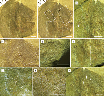
Saetaspongia jianhensis n. sp. (1) the holotype, specimen JLSHM130; (2) an overall image of the sponge showing the positions of the other close-up images (Fig. 2.3–2.6) (dotted and solid boxes); (3) few spicules beyond the margin (arrow); (4) the angle of divergence increasing to 65° at the apical of the large plume (box); (5) a small plumose array; (6) the other small plumose array (box) and this region shown in close-up in Figure 2.7; (7) two minor plumes are superimposed on the primary skeleton (boxes); (8) enlargement of the central region of the fossil specimen, showing that spicules radiate from the center towards the periphery and are in sub-parallel arrangement; (9) the primary skeleton with some irregularly oriented individual spicules (arrows). All scale bars represent 2 mm.

Description
Material consists of a single nearly complete flattened specimen, 13.3 mm long and 9.7 mm wide. The outline of the sponge body is presumed to have been circular to ovoid before compaction. Oscular margin cannot be distinguished, but the outer margin of the sponge body is visible.
Skeletal architecture is distinctive, and spicules are inclined to be densely arranged so that they are in frequent contact (Fig. 2). Specimen shows that these spicules are restricted within the body wall and do not protrude beyond the outer surface of the sponge body (Fig. 2.1). Very few spicules extend up to 1 mm beyond the margin (Fig. 2.3); these slightly projecting spicules may have acted as anchoring spicules or resulted from a taphonomic artefact. Spicules are fine monaxons, probably oxeas. Limitations of preservation prevent clear indications of spicule terminations, and no distinctive or rounded terminations are observed. Majority of visible spicules are 0.28–0.37 mm long and 0.04–0.06 mm wide, but the full length is obscured by adjacent spicules. Many spicules >2 mm long in the outermost part of the specimen (Fig. 2.3), and their maximum observed thickness is ~0.1 mm.
Spicules are inclined to be loosely to densely arranged into plumose arrays; the skeleton is composed primarily of plumose bundles. The plumose region covers approximately two-thirds of the total area of the fossil on the specimen. It seems that there are one major plumose array (Fig. 2.1, 2.2) and at least two small plumose arrays (Fig. 2.5, 2.6). Spicules within the large plume diverge outwards from a well-defined, curved axis; the initial angle of divergence is very shallow, especially in the central area of the plume (Fig. 2.1, 2.4); angle of divergence increases to 65° at the apical part of the large plume (Fig. 2.4). In the other area, there are two additional small plumose spicule arrays (Fig. 2.5, 2.6), the spicules of which are slightly arranged and are parallel to the plumose axis (Fig. 2.5) or parallel to part of the diverging plume spicules (Fig. 2.6). In addition, minor plumose structures also can be found to be superimposed on the primary skeleton and do not affect it (Fig. 2.7). The positioning of these minor plumes relative to body orientation is unknown. Additionally, some spicules generally radiate from the center towards the periphery and are in sub-parallel arrangement (Fig. 2.8). Central region of the fossil specimen is slightly confused, with extremely fine spicules distributed. Dense arrangement of these fine spicules makes full recognition of these individual spicules impossible.
Primary skeleton is occasionally interspersed with some irregularly oriented individual spicules (Fig. 2.9). There is no consistent pattern of ray overlap, which may be only a reflection of the suppression of two separated spicules. Some individual spicules cross the primary skeletal spicules at high angles. These individual spicules are usually 0.1 mm wide, with a few reaching up to 0.75 mm long and 0.5 mm in diameter.
Etymology
The species name jianhensis refers to the fossil discovered from Jianhe County.
Remarks
Saetaspongia fossils have been reported from the Niutitang Formation of South China (Steiner et al., Reference Steiner, Mehl, Reitner and Erdtmann1993; Yang et al., Reference Yang, Zhao, Zhu, Cui and Yang2010, pl. II, fig. 4) and the Buen Formation of North Greenland (Botting and Peel, Reference Botting and Peel2016). These specimens can fully reveal a consistent organization to the skeleton that contains some local plumose arrangements of spicules (Steiner et al., Reference Steiner, Mehl, Reitner and Erdtmann1993, text-fig. 13; Yang et al., Reference Yang, Zhao, Zhu, Cui and Yang2010, pl. II, fig. 3; Botting and Peel, Reference Botting and Peel2016, fig. 6A, B). The plumose architecture is a critical diagnostic feature of Saetaspongia. The fundamental skeletal architecture of the present specimen shows plumose structures and broadly conforms to that of previously reported Saetaspongia fossils (Steiner et al., Reference Steiner, Mehl, Reitner and Erdtmann1993; Yang et al., Reference Yang, Zhao, Zhu, Cui and Yang2010; Botting and Peel, Reference Botting and Peel2016). Therefore, there seems to be little doubt that the material here should be assigned to this genus.
Comparison should be made between the present specimen and the Sansha and Sirius Passet specimens. The Sansha specimens were referred to Saetaspongia densa by Steiner et al. (Reference Steiner, Mehl, Reitner and Erdtmann1993) and somewhat resemble S. jianhensis n. sp. in body form and spicule features, including a circular outline, the plumose structure, and irregular fine spicules. However, the Sansha specimens differ dramatically in the following features (Steiner et al., Reference Steiner, Mehl, Reitner and Erdtmann1993): skeleton composed primarily of a dense, radiating array of fine monaxons; spicules generally radiate towards the periphery; spicules line the outer margins as dense, tangential bundles.
Three specimens from the Sirius Passet Biota have been assigned to Saetaspongia cf. S. densa (Botting and Peel, Reference Botting and Peel2016) and share some similarities in terms of their skeletal architecture with S. jianhensis n. sp., including irregularly oriented individual spicules and several spicule bundles. The Sirius Passet material represents prolate ovoid sponges, with spicules becoming dominantly transverse at the lowest part of the body (Botting and Peel, Reference Botting and Peel2016). However, the irregularly (sub-)transverse to oblique spicules are not dominant in the Balang material.
Saetaspongia cf. S. densa Mehl and Reitner in Steiner et al., Reference Steiner, Mehl, Reitner and Erdtmann1993
Figures 3, 4
- Reference Yang, Zhao, Zhu, Cui and Yang2010
Saetaspongia densa; Yang et al., p. 352, pl. 2, figs. 3, 4.
- Reference Botting and Peel2016
Saetaspongia cf. S. densa; Botting and Peel, p. 473, fig. 6A, B.
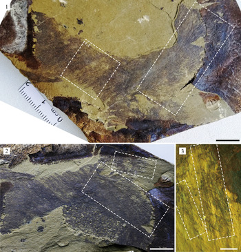
Saetaspongia cf. S. densa. (1, 2) Specimen JLSHM406 and its counterpart, showing a large sponge body, the major plumose array (two large boxes), two small plumose arrays (indicated by a small box and this region shown in close-up in Fig. 3.3) and sub-parallel large spicules frequently crossed or overlapped by many irregularly oriented large spicules (indicated by a small box in Fig. 3.1; this region shown in close-up in Fig. 4.4); (3) two small plumose arrays (boxes). (1, 2) scale bars = 10 mm; (3) scale bar = 2 mm.

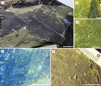
Saetaspongia cf. S. densa. (1) Specimen JLSHM406, showing the positions of the other close-up images (Fig. 4.2, 4.3, 4.5) and many irregularly oriented large spicules (arrows) crossing or overlapping on sub-parallel large spicules; (2) enlargement of Fig. 4.1, showing two probable small plumose arrays; (3) enlargement of Fig. 4.1, showing sub-parallel large spicules crossed by an irregularly oriented large spicule, crossing each other at high angles (indicated by arrow); (4) sub-parallel large spicules are frequently crossed or overlapped by many irregularly oriented large spicules (arrows); this image is the enlargement of a region in Figure 3.1 (indicated by a small box in Fig. 3.1); (5) enlargement of Figure 4.1, showing the plumose array interspersed with some irregularly oriented individual spicules that cross each other at high angles (indicated by arrows). (1, 4, 5) scale bars = 10 mm; (2, 3) scale bars = 2 mm.

Holotype
An articulated specimen (San 102-L) from the Niutitang Formation, Hunan, China (Steiner et al., Reference Steiner, Mehl, Reitner and Erdtmann1993, pl. 3, fig. 1).
Description
A single large specimen (Fig. 3.1, 3.2) represents a partial sponge and shows the critical features that allow identification. Due to irregular rock breakage, no fragment shows the full size of the sponge. The small fragment is partly obscured by rock (Fig. 3.2). The large fragment is >108.46 mm long and 61.46 mm wide (Fig. 3.1). Orientation of these fragments is difficult to determine because the specimen represents a partial sponge.
Spicules are mostly large monaxons, while a few are coarse monaxons. Most spicules are 10.2–15.4 mm long and ~0.05–0.1 mm in diameter. Primary skeleton is dominated by an uncertain number of diverging plumes of large spicules, including one major plumose array (Fig. 3.1, 3.2) and perhaps at least two small plumose arrays (Fig. 3.2, 3.3). Initial angle of divergence is relatively shallow, gradually increasing towards the distal apex of plumose bundles and approaching 15–20° at the apical part (Fig. 3.1–3.3). Central spicules within the plume continue longitudinally towards the apex (Fig. 3.1–3.3). The positioning of these plumose regions relative to the body orientation is unknown. Plumose arrays are interspersed with some irregularly oriented individual spicules (Fig. 4.5). There is no consistent pattern of ray overlap, which may be only a reflection of the suppression of two separated spicules. Some spicules cross each other at high angles (Fig. 4.5). Other areas are composed largely of sub-parallel large spicules (Fig. 4.1, 4.4). These sub-parallel large spicules are frequently crossed or overlapped by many irregularly oriented large spicules (monaxons), with some rays crossing the primary skeletal spicules at variable angles (Fig. 4.3, 4.4).
A few irregularly oriented large spicules are much larger, up to 20.7 mm long and 0.1 mm in diameter. They are superimposed on the primary skeletal layer (Fig. 4.1).
Material
JLSHM406, one partially preserved specimen, including its counterpart; collected from the Balang Formation at Lazizhai Village, Jianhe County, Kaili City, Guizhou, China.
Remarks
According to the specimens described by Steiner et al. (Reference Steiner, Mehl, Reitner and Erdtmann1993) and Botting and Peel (Reference Botting and Peel2016), the current understanding of Saetaspongia is that the primary skeleton consists of some local plumose arrangements of spicules. The plumose architecture is a critical diagnostic feature of Saetaspongia. The fundamental skeletal architecture of the present specimen conforms well with that of S. cf. S. densa described by Yang et al. (Reference Yang, Zhao, Zhu, Cui and Yang2010, pl. II, fig. 4) and Botting and Peel (Reference Botting and Peel2016, fig. 6B), including primary plumose arrays and overlying irregular, coarse monaxons. Additionally, the specimens described here show similar dimensions to those of the largest specimens from the Niutitang Formation of Guizhou (Yang et al., Reference Yang, Zhao, Zhu, Cui and Yang2010) and the Buen Formation (Botting and Peel, Reference Botting and Peel2016). For these reasons, and especially given the typical plumose structure, there seems to be little doubt that the material here should be assigned to the genus Saetaspongia, and therefore is placed tentatively in S. cf. S. densa.
Discussion
Revision of Saetaspongia fossil records.—Since Saetaspongia was erected by Steiner et al. (Reference Steiner, Mehl, Reitner and Erdtmann1993), it has been declared by some specialists that Saetaspongia fossils were widely found in some early and middle Cambrian biotas (Rigby and Hou, Reference Rigby and Hou1995; Yang and Zhao, Reference Yang and Zhao2000; Wu, Reference Wu2004; Zhao et al., Reference Zhao, Yang, Yang and Mao2006; Yang et al., Reference Yang, Zhao, Zhu, Cui and Yang2010; Wu et al., Reference Wu, Zhu and Steiner2014; Botting and Peel, Reference Botting and Peel2016; Hou et al., Reference Hou, Siveter, Siveter, Aldridge, Cong, Gabbott, Ma, Purnell and Williams2017; Q.J. Wang et al., Reference Wang, Yang, Zhao and Duan2017; Y.F. Wang et al., Reference Wang, Peng, Wen, Liu and Wang2018). According to the diagnostic structures provided by the material from the Sansha, Sirius Passet, and Balang biotas (Steiner et al., Reference Steiner, Mehl, Reitner and Erdtmann1993; Botting and Peel, Reference Botting and Peel2016; and references therein), prior assignments of specific specimens to Saetaspongia should be treated with caution and reconsidered critically because certain specimens (Rigby and Hou, Reference Rigby and Hou1995; Zhao et al., Reference Zhao, Yang, Yang and Mao2006; Hou et al., Reference Hou, Siveter, Siveter, Aldridge, Cong, Gabbott, Ma, Purnell and Williams2017; Wang et al., Reference Wang, Yang, Zhao and Duan2017) do not show plumose arrays, but are definitely claimed to have hexactines.
Some specimens assigned to Saetaspongia were reported from the early Cambrian Niutitang Fauna (also published as Niutitang Sponge Fauna) at two localities (Nangao Town, Danzhai County and Songlin Town, Zunyi County) in Guizhou (Yang and Zhao, Reference Yang and Zhao2000; Zhao et al., Reference Zhao, Yang, Yang and Mao2006; Yang et al., Reference Yang, Zhao, Zhu, Cui and Yang2010). Although the two specimens recorded from Danzhai were previously described as Saetaspongia densa (Yang et al., Reference Yang, Zhao, Zhu, Cui and Yang2010, pl. II, figs. 3, 4), they were considered to belong to S. cf. S. densa by other specialists (Botting and Peel, Reference Botting and Peel2016) because one of them showed a dimension and plumose structure similar to those of the largest specimen described in Botting and Peel (Reference Botting and Peel2016, fig. 6B), including the overlying irregular coarse monaxons and localized superimposed plumose arrays. The other specimen recorded in Yang et al. (Reference Yang, Zhu, Zhao and Wang2005, pl. I, fig. 6) was not formally described until 2010, and was identical to specimen NG-73-15 in Yang et al. (Reference Yang, Zhao, Zhu, Cui and Yang2010, pl. II, fig. 4). Although hexactines were claimed to be present in the Niutitang material (Yang et al., Reference Yang, Zhao, Zhu, Cui and Yang2010), this appears to have been largely a result of some spicules crossing each other at high angles. Moreover, there is no trace of additional rays on the exposed surface. Therefore, these specimens do not have hexactines, and assignments of these specimens to Saetaspongia are uncontested.
However, other fossil records are more problematic. The specimens recorded from Zunyi, including two described by Yang and Zhao (Reference Yang and Zhao2000) and three described by Zhao et al. (Reference Zhao, Yang, Yang and Mao2006), were claimed to have tri-, tetr-, and hexactines (Yang and Zhao, Reference Yang and Zhao2000, p. 32), or did not show the typical plumose architecture (Zhao et al., Reference Zhao, Yang, Yang and Mao2006, pl. I, figs. 1–3). Therefore, it may be that these five specimens cannot be assigned to Saetaspongia.
The specimens from the Chengjiang Biota, including one described in Rigby and Hou (Reference Rigby and Hou1995) and two described in Hou et al. (Reference Hou, Siveter, Siveter, Aldridge, Cong, Gabbott, Ma, Purnell and Williams2017), do not show the typical plumose arrangement of spicules, but have prominent hexactines. They were not considered to belong to Saetaspongia (Botting and Peel, Reference Botting and Peel2016).
Many specimens from the Sirius Passet Biota do show well-defined plumose structures and have been assigned to Saetaspongia cf. S. densa Mehl and Reitner in Steiner et al., Reference Steiner, Mehl, Reitner and Erdtmann1993 and S. procera Botting and Peel, Reference Botting and Peel2016 (Botting and Peel, Reference Botting and Peel2016). The fundamental skeletal architecture of S. cf. S. densa is identical to that described in Yang et al. (Reference Yang, Zhao, Zhu, Cui and Yang2010) and our research. Saetaspongia procera is characterized by plumose spicule arrays that extend to produce a tall, thin body form.
Four specimens from the Cambrian (Stage 4) Tsinghsutung Formation were assigned to Saetaspongia densa (Wang et al., Reference Wang, Yang, Zhao and Duan2017). Although they show overlying irregular fine monaxons, it seems that they do not show plumose structures but have tri-, tetr-, or even stauactines (considered hexactines by the authors) (Wang et al., Reference Wang, Yang, Zhao and Duan2017, text-fig. 5A, B). Therefore, these specimens may not belong to Saetaspongia.
Although Saetaspongia was stated to be present in the Kaili Biota in the literature (Wu et al., Reference Wu, Zhu and Steiner2014; Botting and Muir, Reference Botting and Muir2019), the material has never been formally described.
There is a view that paleodepositional environments influenced the preservation potential of plumes, and there was a connection between the paleodepositional settings and the lack of plumes in the Chengjiang and Tsinghsutung specimens. However, the sixteen specimens (two from the Sansha Fauna, 12 from the Sirius Passet Biota, and the present two from the Balang Biota) suggest that Saetaspongia fossils discovered from different paleodepositional settings all have plumose structures, but do not have hexactines. In fact, the Niutitang assemblages suggest relatively deep shelf facies (Yang et al., Reference Yang, Zhao, Zhu, Cui and Yang2010). The paleoenvironment of the Chengjiang Biota has been widely debated and the sponge-dominated assemblages may represent a slope to basin setting (Sun et al., Reference Sun, Zhao, Steiner, Li, Na, Pan, Yin, Zeng, Iten and Zhu2020), whereas the Sirius Passet Biota indicates a relatively deep water slope setting below an escarpment (Ineson and Peel, Reference Ineson and Peel2011). It should be noted that the fossil assemblages from the Lazizhai section of the Balang Formation and the Jianhe section of the Tsinghsutung Formation represent similar shelf environments around storm wave base (Wang et al., Reference Wang, Yang, Zhao and Duan2017; Liu et al., Reference Liu, Peng, Wen, Wang and Du2018b). However, the Sirius Passet and Balang specimens have plumes, but do not have hexactines. The Chengjiang and Tsinghsutung specimens do not have plumes. So, taphonomic differences (e.g., Botting and Muir, Reference Botting and Muir2018) are also insufficient to explain both the absence of hexactinellid-like spicules in the Saetaspongia specimens from the Sirius Passet Biota and the Balang Biota and the absence of plumes in the Chengjiang and Tsinghsutung specimens.
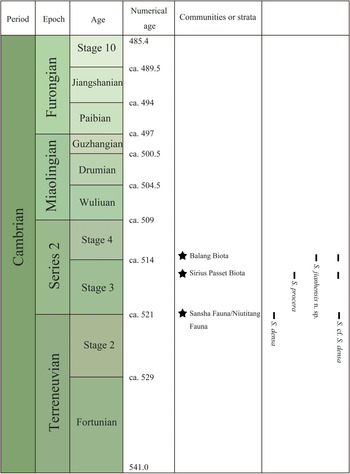
In summary, considering the literature and the new data described herein, the genus Saetaspongia can be identified only in the Sansha Fauna (Steiner et al., Reference Steiner, Mehl, Reitner and Erdtmann1993), Niutitang Fauna (Yang et al., Reference Yang, Zhao, Zhu, Cui and Yang2010), Sirius Passet Biota (Botting and Peel, Reference Botting and Peel2016), and Balang Biota. Based on previous studies, the known stratigraphic range of Saetaspongia has been limited to Cambrian Stage 3, with the oldest representative found in the Niutitang Formation (Steiner et al., Reference Steiner, Mehl, Reitner and Erdtmann1993; Yang et al., Reference Yang, Zhu, Zhao and Wang2005, Reference Yang, Zhao, Zhu, Cui and Yang2010) and the youngest occurrence in the Sirius Passet Biota (Botting and Peel, Reference Botting and Peel2016). Thus, the new discovery of Saetaspongia from the middle-upper part of the Balang Formation further extends the stratigraphic distribution of this genus upwards to Stage 4 (Fig. 5).
Stratigraphic chart showing the distribution of Saetaspongia sponges. Temporal correlations of the occurrences of Cambrian biotas based on Botting and Muir (Reference Botting and Muir2019), Zhu et al. (Reference Zhu, Yang, Yuan, Li, Zhang, Zhao, Ahn and Miao2019) and Q.J. Wang et al. (Reference Wang, Peng, Wen, Du, Zhang, Wang and Wang2020). Numerical ages in Ma.

Biogeography
The oldest clear record of Saetaspongia was recorded from the Niutitang Formation (Steiner et al., Reference Steiner, Mehl, Reitner and Erdtmann1993; Yang et al., Reference Yang, Zhu, Zhao and Wang2005, Reference Yang, Zhao, Zhu, Cui and Yang2010) and indicated that this genus may have been established by the Terreneuvian-Series 2 transition. Saetaspongia was originally considered to be an apparently endemic element because it was only known from a single locality (Muir et al., Reference Muir, Botting, Carrera and Beresi2013). However, together with the reliable fossil record from the Cambrian of North Greenland (Botting and Peel, Reference Botting and Peel2016), the fossil evidence suggests that Saetaspongia occurred across at least two paleocontinents (South China and Laurentia) (Botting and Muir, Reference Botting and Muir2019) and confirms that at least this genus had a broad biogeographical distribution during the early Cambrian, based on four assemblages (i.e., Sansha, Niutitang, Sirius Passet, and Balang) at different paleolatitudes (Fig. 6). Data on Saetaspongia fossil occurrence from the Sirius Passet Biota (Botting and Peel, Reference Botting and Peel2016) suggest that Saetaspongia sponges were present in high numbers in fossil collections (of the 150 sponge specimens from this biota, 12 specimens were Saetaspongia), and they may have been one of the significant components of Cambrian sponge communities.
Paleogeographical distribution of the Cambrian biotas containing Saetaspongia sponges. Paleogeographical reconstruction referred to McKerrow et al. (Reference McKerrow, Scotese and Brasier1992) and Wen et al. (Reference Wen, Babcock, Peng, Liu and Liang2019).

Phylogenetic relationship
Saetaspongia fossils from the Sansha, Niutitang, Sirius Passet, and Balang biotas (Steiner et al., Reference Steiner, Mehl, Reitner and Erdtmann1993; Yang et al., Reference Yang, Zhao, Zhu, Cui and Yang2010; Botting and Peel, Reference Botting and Peel2016; and references therein) could fully reveal a consistent architecture of the skeleton in that it contains some local plumose arrangements of spicules. There seems to be little doubt that plumose structures are a fundamental skeletal feature characteristic of the genus. Fossil evidence shows that the plumose architecture is commonly developed in certain protomonaxonid taxa (e.g., Wapkia Walcott, Reference Walcott1920; Hamptonia bowerbanki Walcott, Reference Walcott1920; Hazelia Walcott, Reference Walcott1920) (Steiner et al., Reference Steiner, Mehl, Reitner and Erdtmann1993; Finks and Rigby, Reference Finks, Rigby and Kaesler2004; Botting et al., Reference Botting, Muir and Lin2013; Botting and Peel, Reference Botting and Peel2016). Although plumose structures have also been found to occur in non-protomonaxonid sponges (Finks and Rigby, Reference Finks, Rigby and Kaesler2004), these sponges have hexactines or differ dramatically from Saetaspongia in skeletal architecture (Finks and Rigby, Reference Finks, Rigby and Kaesler2004). Therefore, Saetaspongia was assumed to have a protomonaxonid rather than a hexactinellid affinity because the plumose architecture has been confirmed to commonly occur in certain protomonaxonid sponges (Botting and Peel, Reference Botting and Peel2016). Although the plumose, radiating spiculation is considered a primitive feature of the Protomonaxonida (Steiner et al., Reference Steiner, Mehl, Reitner and Erdtmann1993; Botting et al., Reference Botting, Muir and Lin2013), it remains unclear whether there is a direct link between Saetaspongia and these protomonaxonids or whether Saetaspongia evolved convergently within a different protomonaxonid lineage (Botting and Peel, Reference Botting and Peel2016). Therefore, the new material described herein provides additional support for the view (Steiner et al., Reference Steiner, Mehl, Reitner and Erdtmann1993; Botting and Peel, Reference Botting and Peel2016) that Saetaspongia has a protomonaxonid affinity and does not have hexactine-based spicules.
Conclusions
Two Saetaspongia sponges, S. jianhensis n. sp. and S. cf. S. densa, are recognized from the Stage 4 Balang Biota of South China based on their skeletal characters and/or body forms. Combined with previous studies, fossil evidence suggests that the plumose architecture is a critical feature diagnostic of Saetaspongia, and that this genus does not have hexactine-based spicules. The new material described herein provides additional support for the view that Saetaspongia has a protomonaxonid rather than a hexactinellid affinity. Data on Saetaspongia fossil occurrences indicate that Saetaspongia had a wide biogeographical distribution during the early Cambrian and that the stratigraphic distribution of this genus further extends upward to Stage 4.
Acknowledgments
We are extremely grateful to L.A. Muir (Amgueddfa Cymru–National Museum Wales, Cathays Park, UK) and L.D. Mouro (Federal University of Santa Catarina, Florianópolis, SC, Brazil) for their constructive and helpful comments that improved the manuscript significantly. We are also particularly grateful to the editors, B. Hunda, J.P. Botting, and J. Kastigar, for their meaningful advice about revising the manuscript and their kind assistance in submitting the revised manuscript. Thanks also to T. Lan (Guizhou University) for his assistance in taking photographs of specimens, and to S. Liu and R.Q. Wen for their assistance in fieldwork. This research was financially supported by the National Natural Science Foundation of China (41672005, 42002012), the Guizhou Science and Technology Department Foundation of China (Gui. Sci. Z. [2014]4003, Gui. Sci. [2017]1057, Gui. Sci. G. [2017]5788, Gui. Sci. [2019]1124), and the Strategic Priority Research Program of Chinese Academy of Sciences (XDB2600000).