Introduction
Our knowledge of pre-Cenozoic land snails is poor. Few findings are known, and their systematic position is often uncertain, especially in the case of Paleozoic and early Mesozoic records (e.g., Solem and Yochelson, Reference Solem and Yochelson1979; Harbeck, Reference Harbeck1996; Bandel, Reference Bandel2002; Ponder and Lindberg, Reference Ponder and Lindberg2008; Bouchet et al., Reference Bouchet, Rocroi, Hausdorf, Kaim, Kano, Nützel, Parkhaev, Schrödl and Strong2017; Nordsieck, Reference Nordsieck2017). However, findings are important to shed light on phylogeny and divergence of higher clades. Many groups of nonmarine gastropods have been claimed to originate in the Mesozoic (e.g., Taylor, Reference Taylor1988; Dayrat et al., Reference Dayrat, Conrad, Balayan, White, Albrecht, Golding, Gomes, Harasewych and de Frias Martins2011), but those dates often rely on the sparse fossil record. A major difficulty in supporting or rejecting previous hypotheses is the apparent gap in the fossil record of land snails in the Triassic and Early/Middle Jurassic (Wade et al., Reference Wade, Mordan and Naggs2006). A prime example for a debate resulting from that gap is the origin of the Stylommatophora. Depending on the systematic classification of the late Paleozoic families Anthracopupidae and Dendropupidae, the supposed origin of the Stylommatophora has been shifted between the Late Jurassic (Tillier et al., Reference Tillier, Masselot, Tillier and Taylor1996; Bandel, Reference Bandel2002; Ponder and Lindberg, Reference Ponder and Lindberg2008) and late Carboniferous (Solem and Yochelson, Reference Solem and Yochelson1979; Solem, Reference Solem, Trueman and Clarke1985; Nordsieck, Reference Nordsieck2017; see also discussion in Dayrat et al., Reference Dayrat, Conrad, Balayan, White, Albrecht, Golding, Gomes, Harasewych and de Frias Martins2011). Such a big discrepancy of more than 150 Myr has major implications for the diversification history of this highly diverse land snail group.
The alleged record of Carychiinae (Ellobiidae) by Pan (Reference Pan1982) from the Lower Jurassic of Hunan, China, is thus a highly welcome source of information that may further elucidate the history of the Stylommatophora. The family Ellobiidae was considered to have originated in the Early Cretaceous (Dayrat et al., Reference Dayrat, Conrad, Balayan, White, Albrecht, Golding, Gomes, Harasewych and de Frias Martins2011) and the subfamily Carychiinae not before the Cenozoic (Strauch, Reference Strauch1977; Villatte, Reference Villatte1979). The earliest and most robust record of Carychiinae is Carychium munieri Briart and Cornet, Reference Briart and Cornet1889 from the early Paleocene of Belgium (Villatte, Reference Villatte1979). Records from older strata have been considered questionable in terms of their identification and in respect to their ecology (Tracey et al., Reference Tracey, Todd, Erwin and Benton1993; Bandel, Reference Bandel1994, Reference Bandel1997).
Our aim is to restudy the two species assigned to the genus Protocarychium Pan, Reference Pan1982 and to discuss its systematic position in light of new data available via CT images. We make comparisons of the shell morphology with extant and fossil representatives of stylommatophoran, ellobioid, and caenogastropod families to determine the most likely systematic position of Protocarychium. Finally, we provide translations of the original Chinese taxon descriptions.
Geological setting
The material studied herein derives from the Tabakou Member (sometimes spelled Dabakou) of the Guanyintan Formation (also known as Guanyingtan) in Hunan Province, China. Apart from the investigated gastropods, the Tabakou Member contains a rich assemblage of mollusks, ostracods, and plants (Zhou, Reference Zhou1984; Chen, Reference Chen, Zhang, Chen and Palmer2003). The Gastropoda were studied by Pan (Reference Pan1982) and include species she assigned to the families Ataphridae, Colloniidae, Craspedostomatidae, Hydrobiidae, Neritidae, Naticidae, Pachychilidae, Procerithiidae, and Pseudomelaniidae. Pan (Reference Pan1982) suggested a brackish-water paleoenvironment with strong marine influence, perhaps a deltaic environment. According to that author, Protocarychium would have dwelled in the intertidal zone. The brackish character is also confirmed by the presence of the isognomoniid bivalve Waagenoperna (Chen and Xu, Reference Chen and Xu1980; Pan et al., Reference Pan, Sha, Wang, Zhang, Yao, Peng and Rao2013) and a diverse fauna of darwinulid and limnocytherid ostracods (Cao, Reference Cao1984) that indicate estuarine conditions (Wang and Cao, Reference Wang, Cao and Westermann1992). The Guanyintan Formation is renowned for its highly diverse floral assemblage dominated by Bennettitales and leptosporangiate ferns, along with common Ginkgophyta, Cycadales, Pteridospermatophyta, Caytoniales, and conifers (Zhou, Reference Zhou1984). Typical for the Tabakou Member is the so-called Marattiopsis-Otozamites Assemblage, also known as ‘Guanyintan Flora’ (Zhou, Reference Zhou1984; Chen, Reference Chen, Zhang, Chen and Palmer2003). Paleobiogeographically, it belongs to the Southern Floristic Province and is indicative of a warm and humid climate (Wang et al., Reference Wang, Nia, Jiang and Tian2008; Escapa et al., Reference Escapa, Bomfleur, Cuneo and Scasso2015).
While originally considered to be of Late Triassic age (Chen et al., Reference Chen, Wen, Zhou, Li, Lin, Zhang, Li, Liu and Li1980; Zhang, Reference Zhang2009), most following authors, variably working on the stratigraphy, flora, and fauna, concluded the Guanyintan Formation was Early Jurassic in age (Liu and Chen, Reference Liu and Chen1981; Pan, Reference Pan1982; Cao, Reference Cao1984; Zhou, Reference Zhou1984; Chen, Reference Chen, Zhang, Chen and Palmer2003; Pan et al., Reference Pan, Sha, Wang, Zhang, Yao, Peng and Rao2013; Sha et al., Reference Sha, Wang, Pan, Yao, Rao, Cai and Zhang2016). According to Chen (Reference Chen, Zhang, Chen and Palmer2003), the Guanyintan Flora is characteristic of a Hettangian to early Pliensbachian age (201.3–187.5 Ma; absolute ages after Gradstein et al., Reference Gradstein, Ogg, Schmitz and Ogg2012; Franceschi et al., Reference Franceschi, Dal Corso, Posenato, Roghi, Masetti and Jenkyns2014).
Materials and methods
We restudied the material published by Pan (Reference Pan1982) that was collected from deposits of the Tabakou Member, Guanyintan Formation, near Taochuan village (Jiangyong-taochuan), Yongzhou City, Hunan Province, China (approximate GPS: 25.11194°N, 111.08306°E). The type material of Protocarychium mirum Pan, Reference Pan1982 and P. arcidentata Pan, Reference Pan1982 includes three specimens, which are preserved as internal casts lacking the outer shell surface. Original genus diagnosis and species descriptions translated from the Chinese text of Pan (Reference Pan1982) are provided. The terminology on apertural dentition follows Solem and Yochelson (Reference Solem and Yochelson1979).
Image acquisition
Protocarychium casts were scanned via the SU-3500 SEM (Hitachi Limited, Japan). All three specimens were imaged at the micro-CT lab of NIGPAS using a three-dimensional X-ray microscope (3D-XRM), Zeiss Xradia 520 Versa (Jena, Germany). Because of the small specimen size, a charge-coupled device (CCD)-based 4× objective was used, providing isotropic voxel sizes of 0.5 mm via geometric magnification at a voltage of 4 w and 0.08 mA. During the scan, the running voltage for the X-ray source was set at 50 kV. A thin filter (LE1) was used to avoid beam-hardening artifacts. To achieve high signal-to-noise ratio, 2,001 projections over 360° were collected. Exposure time for each projection was set as 1 s for P. mirum (NIGP47581, NIGP47582) and 1.5 s for P. arcidentata (NIGP47583). Volume data processing was performed using software Vgstudio Max (version 3.0, Volume Graphics, Heidelberg, Germany).
Repository and institutional abbreviation
The examined and figured type species, Protocarychium mirum Pan Reference Pan1982 (NIGP47581 [holotype] and NIGP47582 [paratype]) and Protocarychium arcidentata Pan, Reference Pan1982 (NIGP47583 [holotype]), are deposited in the Nanjing Institute of Geology and Paleontology, Chinese Academy of Sciences (NIGPAS), Nanjing, China.
Systematic paleontology
Class Gastropoda Cuvier, Reference Cuvier1795
Order Stylommatophora Schmidt, Reference Schmidt1855
?Infraorder Pupilloidei Turton, Reference Turton1831
Superfamily Dendropupoidea Wenz, Reference Wenz and Schindewolf1938
Family Anthracopupidae Wenz, Reference Wenz and Schindewolf1938
Diagnosis
Pupilloid shell with up to 6.5 whorls; shell surface may bear low to distinct axial riblets; early whorl partitions present throughout ontogeny (not resorbed); columella simple, partly hollow; aperture bears one columellar barrier, 1–2 parietal barriers, and 1–2 palatal ridges; dentition does not extend into penultimate whorl.
Remarks
The diagnosis was emended from Solem and Yochelson (Reference Solem and Yochelson1979) on the basis of the inclusion of Protocarychium in the family. See Discussion for more details.
Subfamily Anthracopupinae Wenz, Reference Wenz and Schindewolf1938
Genus Protocarychium Pan, Reference Pan1982
Type species
Protocarychium mirum Pan, Reference Pan1982 (by original designation), China, Hunan, Jiangyong-taochuan; Guanyintan Formation, Tabakou Member.
Original diagnosis
Shell minute in size, dextral, ovate-conical to ovate-pupiform. Apex obtusely round and spire moderately high, having 4–6 roundly convex whorls and body whorl highly large, anomphalous. Aperture subovate in outline, with a strong lamellar parietal tooth and a distinct lamellar columellar tooth, outer side of the whorl near the aperture bearing two lamellar teeth, which do not attain to peristome. Aperture angular above and round below; the surface is ornamented by fine growth lines.
Protocarychium mirum Pan, Reference Pan1982
Figures 2, 3
1982 Protocarychium mirum Pan, p. 105, pl. 4, figs. 1–6, text-fig. 2.
1982 Protocarychium arcidentata Pan, p. 106, pl. 4, figs. 7–9, text-fig. 3.
Geographical overview of the study area north of Taochuan village, Yongzhou City, in southern Hunan Province, China. The red asterisk marks the Lower Jurassic Guanyintan Formation from which the studied material derives. The geology of the area was simplified after the Geological Map of Hunan obtained from http://drr.ikcest.org/map/m02d5. The underlying topographic map derives from https://maps-for-free.com/.

CT and SEM images of the holotype of Protocarychium mirum Pan, Reference Pan1982 (NIGP47581): (1, 2, 6) CT images showing external palatal indentations and the characteristic anthracopupid deep and channeled suture; (3, 4) micro-CT images using maximum intensity projection (MIP) mode; (5) SEM image of abapertural view; (7, 8) cross-sectional images showing the characteristic anthracopupid whorl partitions, the wide basal (and perhaps originally partly hollow) columella, apertural barriers, and structural material (diagenetic or preservational) between new whorl attachment sections.

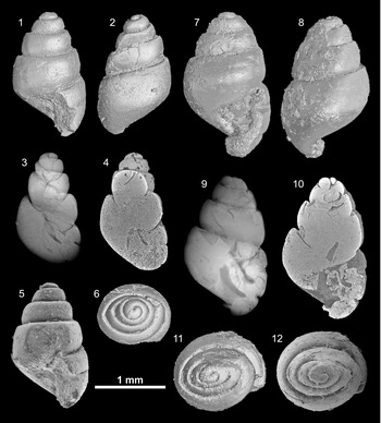
CT and SEM images of Protocarychium mirum Pan, Reference Pan1982: (1, 2, 6) CT images of the subadult paratype shell of Protocarychium mirum (NIGP47582) showing the characteristic anthracopupid deep and channeled suture and the flattened and shouldered whorls; note the apex is laterally compressed; (3) micro-CT image of the same shell using MIP, showing inner whorl partitioning, structural material (diagenetic or preservational) between the whorls, and two apertural barriers; (4) cross-sectional image of the same shell; (5) SEM image of the same shell; (7, 8, 11) CT images of the holotype shell of Protocarychium arcidentata (NIGP47583) showing anthracopupid-like flattened and shouldered whorls, deep suture, and external palatal indentations; shell aperture is filled with sediment; (9) CT image of the same shell showing the well-developed columellar barrier and parietal barrier; (10) cross-sectional perspective of the same shell; (12) SEM image of the same shell in apical view.

Type specimens
The type material includes three specimens: NIGP47581 (holotype of P. mirum), NIGP47582 (paratype of P. mirum), and NIGP47583 (holotype of P. arcidentata), all from Jiangyong-taochuan, Hunan, China; Guanyintan Formation, Tabakou Member (Lower Jurassic).
Occurrence
Known only from the type locality and stratum.
Original (translated) description of Protocarychium mirum
Shell relatively small, ovoid-pupiform, five whorls, apex obtuse, spire half of shell length, conical, whorls gradually increasing with last whorl being larger, approximately three-fifths of height, peristome expanded, suture deep, base without umbilicus, shell surface smooth, aperture oval, upper end pointed, lower end round, outer lip curved, parietal wall oblique, with a long and thin parietal denticle, columellar margin with an oblique columellar tooth, forming a deep impression in the interior mold, extending far inward; two low teeth near the outer lip, the superior one long and the inferior one shorter, neither reaching the peristome.
Original (translated) description of Protocarychium arcidentata
Shell smaller, elongate-oval, with five whorls. Apex obtuse, spire conical, whorls increasing regularly, body whorl taller than spire, about three-fifths of total height. Whorls weakly rounded, suture deep. Base somewhat inflated, without an umbilicus. Aperture oval, angular above, rounded below, outer lip curved, damaged, parietal wall oblique, with a long, narrow parietal tooth leaning to the right, columellar lip with slight impression, with a long and narrow columellar tooth. Two long and thin, low teeth near the palatal lip, the lower being longer and narrower than the upper, neither reaching the peristome.
Revised description using CT data
Casts consist of 4.5 flatly sculpted convex whorls. Suture seemingly channeled, deep, and groove-like; however, this may be due to the nature of preservation as internal casts. Upper whorl partitions present with slight bit of basal growth in cross section (Fig. 2.7, 2.8); columella barely visible in penultimate whorl (Fig. 2.3, 2.4, 2.7, 2.8). Judging from the comparably broad columella at the base, a narrow umbilical chink might have been present (Fig. 2.8). Aperture ovate with thin edge, no sign of parietal callus preserved. Upper parietal barrier (denticle) low and pointed; central parietal barrier very long, wedge-like with a distinct bulbous tip (Figs. 2.4, 2.7, 2.8, 3.3, 3.4, 3.9, 3.10). Palatal dentition consists of two short, narrow, horizontal impressions in the casts, set deep in the aperture (Figs. 2.2, 2.4, 2.7, 3.1, 3.5, 3.7, 3.9, 3.10).
Remarks
Because of the preservation as internal casts, information about shell sculpture, growth line patterns, suture depth, and potential apertural thickening (a feature common for other species assigned to Anthracopupidae) is unavailable.
Pan (Reference Pan1982) differentiated Protocarychium arcidentata from P. mirum on the basis of its smaller size, the more oval shape, the long, thin, and curved parietal barrier, the wider distance between the two palatal ridges, and the lower palatal ridge being thinner and longer than the upper one. After examination of CT and SEM images, we conclude that the singular Protocarychium arcidentata specimen represents an intermediary, subadult growth stage of Protocarychium mirum, and we consider both species as synonyms. The slight variation in dentition does not warrant a taxonomic separation. As first reviser according to ICZN Art. 24.2.2, we choose P. mirum as the accepted name.
Discussion
The systematic position of Protocarychium is uncertain. The initial classification as Carychiinae (Eupulmonata, Ellobiidae), which was followed by Stworzewicz et al. (Reference Stworzewicz, Szulc and Pokryszko2009), can be ruled out on the basis of the apertural barriers not extending into the inner whorls (Figs. 2.3, 2.4, 2.7, 2.8, 3.3, 3.9), a deeply recessed columellar barrier (“extending far inward”; Pan, Reference Pan1982, p. 112; Figs. 2.4, 3.3), the placement of the parietal barrier positioned higher on the parietal region of the shell (Figs. 2.8, 3.3, 3.4), the columellar barrier being flatly lamellar with a bulbous tip and not sinuate in form (Figs. 2.8, 3.10), retention of early whorl partitions (Figs. 2.3, 2.7, 2.8, 3.3), a very thin layer of structure (diagenetic or preservational) of what may have been a periostracal layer between whorls indicating that it was not absorbed before the attachment of the lower whorls (Figs. 2.7, 2.8, 3.3), flatly rounded whorls (Fig. 3.1, 3.2, 3.5, 3.7, 3.8), and a seemingly variable umbilical chink in the columella (Figs. 2.7, 2.8, 3.10), all of which contrast with features observed in the Ellobiidae.
Shells of extant groups of the Stylommatophora such as the Enidae, Subulinidae, Urocoptidae, Achatinidae, and Bulimulidae are considerably bigger, have more whorls, and show different growth patterns compared to Protocarychium. Zonitid and helicoid taxa differ in sculpture, form, whorl count, and size. In whorl number and basic shell shape, Protocarychium is very similar to typical pupillids and tornatellinids with the spire form of Protocarychium being closer to that of the Tornatellinidae. Considering the small size, low whorl number, form and presence of apertural barriers, and tumid shell form, the best match for Protocarychium is the late Paleozoic genus Anthracopupa Whitfield, Reference Whitfield1881.
As for Protocarychium, the systematic position of Anthracopupa is highly uncertain. Several previous authors have discussed potential placements, some of which place the genus even in different gastropod subclasses. Wenz (Reference Wenz and Schindewolf1938) classified it in a new subfamily, Anthracopupinae, within the Ellobiidae. Knight et al. (Reference Knight, Batten, Yochelson and Moore1960) suggested Anthracopupa to belong to the Cyclophoridae (Caenogastropoda), which is unlikely because the aperture of Anthracopupa bears both columellar and parietal barriers, is irregular and more or less auriform in shape, and not round as in Cyclophoridae, and the presence of both columellar and parietal barriers in conjunction with a cyclophorid operculum capable of sealing the shell aperture is improbable. Solem and Yochelson (Reference Solem and Yochelson1979) considered Anthracopupinae a basal stylommatophoran and classified it in the family Tornatellinidae. However, species of Tornatellinidae, with the exception of human-distributed species, are currently restricted to the islands of Polynesia, Micronesia, and Juan Fernandez (Solem and Yochelson, Reference Solem and Yochelson1979). The only other Mesozoic potential Tornatellinidae is Protornatellina isoclina (White, Reference White1895) from the Upper Cretaceous Bear River Formation of Wyoming (Roth, Reference Roth1986). Bandel (Reference Bandel1997), in turn, considered Anthracopupa a Carychiidae. Later, Bandel (Reference Bandel2002) revised his earlier concept and placed the genus in the family Anthracopupidae, which he classified in a separate order, Procyclophorida, which he considered to belong in Architaenioglossa and thus related with recent Cyclophoroidea (Caenogastropoda) (see also Bouchet et al., Reference Bouchet, Rocroi, Hausdorf, Kaim, Kano, Nützel, Parkhaev, Schrödl and Strong2017 for discussion and nomenclatural issues). A relationship of Anthracopupa with Stylommatophora in general was doubted by Wade et al. (Reference Wade, Mordan and Naggs2006) and Dayrat et al. (Reference Dayrat, Conrad, Balayan, White, Albrecht, Golding, Gomes, Harasewych and de Frias Martins2011) because of the lack of clear synapomorphies and the extremely long gap in the fossil record. Nordsieck (Reference Nordsieck2017), in turn, favored assignment to the Achatinellidae (Stylommatophora) or Carychiidae (Ellobiida).
The obvious dilemma of assigning Protocarychium to Anthracopupidae is the long temporal gap between the late Carboniferous and early Permian records of Anthracopupidae (Solem and Yochelson, Reference Solem and Yochelson1979) and the Early Jurassic Protocarychium, as well as the disjunct geographic range. In general, the deficiency of the fossil record complicates any assignment. Only a few land snail assemblages have been reported from the Jurassic, for example, the Upper Jurassic to Lower Cretaceous Purbeck Group of Great Britain and its equivalents in Switzerland and Germany (de Loriol and Jaccard, Reference de Loriol and Jaccard1865; Maillard, Reference Maillard1884; Arkell, Reference Arkell1941; Huckriede, Reference Huckriede1967; Bandel, Reference Bandel1991) and the Morrison Formation in the United States (Yen and Reeside, Reference Yen and Reeside1946; Yen, Reference Yen1952; Evanoff et al., Reference Evanoff, Good and Hanley1998). Nonetheless, striking similarities in several key characters present in Protocarychium and Anthracopupidae, as well as in recent Stylommatophora, suggest a close relationship, leading us to place Protocarychium in the family Anthracopupidae. The morphological concurrence between Anthracopupa and Protocarychium is particularly apparent in the parietal and columellar barriers and the presence of palatal ridges (known in late growth stages; Solem and Yochelson, Reference Solem and Yochelson1979). The two deep indentations on the palatal region of the casts as seen in the CT images (Figs. 2.2, 2.4, 2.7, 3.7, 3.9, 3.10) are indicative of this internal ridge formation as illustrated by Solem and Yochelson (Reference Solem and Yochelson1979, pl. 5, figs. 3–5). As in young Anthracopupa shells (Solem and Yochelson, Reference Solem and Yochelson1979, pl. 5, figs. 1, 2), Protocarychium shows a well-developed columellar barrier and a parietal barrier, both of which continue into the adult growth stage. Moreover, in both genera, dentition does not extend into the penultimate whorl.
Despite these morphological analogies, we tend to keep both genera distinct given the large stratigraphic gap between the Early Jurassic and early Permian taxa as well as the disjunct geographic occurrences. To solve the generic status, additional evidence regarding internal structures of other Anthracopupidae species and their variability is required for a more in-depth assessment of morphological variability present in the group.
In summary, the family Anthracopupidae includes at present the genus Anthracopupa (with the junior synonym Maturipupa; Solem and Yochelson, Reference Solem and Yochelson1979) with five accepted species and the monospecific genus Protocarychium. The inclusion of Protocarychium in Anthracopupidae expands the morphological traits known for the family, and we present here a revised diagnosis of the family (see the precedin). The genus Dendropupa, with four accepted species, is placed in the sister family Dendropupidae, which differs from Anthracopupidae primarily in the abapically expanded columella with raised internal ridges and the more elongated shell with more (up to nine) whorls (Solem and Yochelson, Reference Solem and Yochelson1979). We follow Bouchet et al. (Reference Bouchet, Rocroi, Hausdorf, Kaim, Kano, Nützel, Parkhaev, Schrödl and Strong2017) in assigning both families to the Dendropupoidea Wenz, Reference Wenz and Schindewolf1938, but contrary to their classification in Caenogastropoda, we tentatively consider it to be a basal clade of the Pupilloidei (Stylommatophora, Helicina).
Considering that Protocarychium is indeed a stylommatophoran sheds new light on the age of that group, which is presently considered by many authors to have originated in the Late Jurassic (Tillier et al., Reference Tillier, Masselot, Tillier and Taylor1996; Ponder and Lindberg, Reference Ponder and Lindberg2008). That view is based mostly on the long time gap between the so far known Late Jurassic and Paleozoic records. Nonetheless, already Solem and Yochelson (Reference Solem and Yochelson1979) emphasized that the Stylommatophora are much older than conventionally considered on account of two orders and four families being present in the late Paleozoic. On the basis of the considerations and new evidence presented here, we fully corroborate this view. The Chinese records moreover extend the known geographic range of the Anthracopupidae. Up to now, the family was known from the late Paleozoic of the United States (Illinois, Ohio, Pennsylvania), Canada (Nova Scotia), and Europe (Solem and Yochelson, Reference Solem and Yochelson1979; Stworzewicz et al., Reference Stworzewicz, Szulc and Pokryszko2009). This extension in paleogeographic range of Anthracopupidae and reevaluation of the age of Stylommatophora opens new ground for interpretation of the diversification history of early land snails. We hope future investigations will extend our knowledge of the geographic spectrum of early land snail diversity and help fill the wide gaps in the tenuous fossil record.
Acknowledgments
We are indebted to J. Gerber, Z. Ren, and Y. Chen for their kind help in translating the original Chinese descriptions. We thank H. Pan for her help in providing additional information about the collection site. For his help at the SEM, we acknowledge Y. Mao. We are additionally grateful to the CT operators Z. Yin and S. Wu for imaging Protocarychium. A. Kaim, A. Krapf, and A. Nützel kindly provided rare literature. We thank M. Harzhauser, B. Ruthensteiner, and S. Schneider for fruitful discussions. Finally, we are grateful to M. Harzhauser and A.M. de Frias Martins and the editors, B. Hunda and J. Kastigar, for constructive comments on an earlier draft of the paper. This research was supported by the Strategic Priority Research Program of the Chinese Academy of Sciences (XDB26000000 and XDA19050101) and the National Natural Science Foundation of China (41622201, 41688103).