Introduction
Cetoninae (Coleoptera: Scarabaeidae) are familiar to entomologists for their shiny and brightly coloured bodies, and consist of about 3900 described species that range in size from a few millimetres to over 110 mm (Micó et al., Reference Micó, Morón, Šípek and Galante2008). Most cetonid adults are diurnal and feed on flowers, plant sap and fruits; hence the names fruit and/or flower chaffers (Donaldson, Reference Donaldson1989; Thomas, Reference Thomas1998; Micó et al., Reference Micó, Morón, Šípek and Galante2008). Adults of some cetonid species have been reported to be associated with social insects such as bees (Donaldson, Reference Donaldson1989; Njau et al., Reference Njau, Mpuya and Mturi2009; Torto et al., Reference Torto, Fombong, Mutyambi, Muli, Arbogast and Teal2010) and ants (Alpert, Reference Alpert1994), while their larvae have been reported to feed on decaying plant matter and faeces of herbivores like cattle and donkeys (Donaldson, Reference Donaldson1989; Thomas, Reference Thomas1998; Perissinotto et al., Reference Perissinotto, Smith and Stobbia1999; Micó et al., Reference Micó, Morón, Šípek and Galante2008). A number of cetonid beetles have also been ascribed pest status as a result of their diverse feeding habits (Ritcher, Reference Ritcher1957; Donaldson, Reference Donaldson1989; Thomas, Reference Thomas1998; Johannsmeier, Reference Johannsmeier2001; Torto et al., Reference Torto, Fombong, Mutyambi, Muli, Arbogast and Teal2010).
Identification of insects often relies on morphological characteristics with emphasis on life stages that are commonly encountered in nature. However, among scarab beetles, external larval morphology is increasingly being used to separate species, in addition to adult characters (Ritcher, Reference Ritcher1966; Donaldson, Reference Donaldson1989; Smith et al., Reference Smith, Perissinotto and Villet1998; Perissinotto et al., Reference Perissinotto, Smith and Stobbia1999, Reference Perissinotto, Villet and Stobbia2003; Šípek et al., Reference Šípek, Král and Jahn2008). Besides their identification, an understanding of their life history is imperative to the development of monitoring and management tools for economically important insects (Amarasekare et al., Reference Amarasekare, Mannion, Osborne and Epsky2008) because it reveals their potential for quick reproduction, abundance and their role within their habitats. Behavioural information obtained from life history studies is also useful in separating sibling species (e.g. Alexander and Bigelow, Reference Alexander and Bigelow1960; Craig et al., Reference Craig, Itami, Abrahamson and Horner1993; Boake et al., Reference Boake, DeAngelis and Andreadis1997).
Unlike most adult cetonids, two members of the Oplostomus genus have been reported to be strongly associated with honeybee colonies and are rarely found on nearby flowering plants (Donaldson, Reference Donaldson1989; Johannsmeier, Reference Johannsmeier2001; Njau et al., Reference Njau, Mpuya and Mturi2009; Torto et al., Reference Torto, Fombong, Mutyambi, Muli, Arbogast and Teal2010). Although some information exists on the biology of Oplostomus fuligineus (Olivier) (Donaldson, Reference Donaldson1989), similar information is lacking on O. haroldi (Witte), a closely related species that was recently recorded as a pest of honeybee colonies in Tanzania and Kenya (Njau et al., Reference Njau, Mpuya and Mturi2009; Torto et al., Reference Torto, Fombong, Mutyambi, Muli, Arbogast and Teal2010) where it was observed feeding on bee brood, pollen and honey. Hence, this study was carried out to document the life history of O. haroldi under laboratory conditions and to provide a description of the external morphology of the third instar larva.
Materials and methods
Life history
Collection of insects
Adult specimens of O. haroldi were collected from honeybee colonies in June 2009 during a field survey of various beekeeping sites in the Coast and Eastern provinces of Kenya (Torto et al., Reference Torto, Fombong, Mutyambi, Muli, Arbogast and Teal2010), and brought to the icipe insectary in Nairobi (01°13′25.3″S, 36°53′49.2″E) where they were reared on a substrate of sterilized moistened soil and crushed cow dung mixed in a ratio of 1:1 by volume. The substrate was changed every 3 weeks.
Adult beetles were kept in white rectangular plastic bowls with perforated lids and provided with water by placing moist cotton balls (6.5 cm in diameter) on the rearing substrate with an equal number of males and females placed together. Temperature, relative humidity and photoperiod in the rearing room were maintained at 25 ± 2 °C, 50 ± 5% and 10 h light-14 h dark, respectively.
Eggs
Eggs were recovered by sifting the substrate every 24 h. Each egg was placed on approximately 5 g of the rearing substrate (composed of two parts sterilized soil and one part sterilized and crushed cow dung) placed on moist filter paper in a Petri dish (9 cm in diameter). One to two millilitres of distilled water were added to the filter paper every 2–3 days to keep the eggs moist. Six mated females were kept individually on the rearing substrate and monitored for egg-laying until they stopped. The maximum length and width of each egg was measured every 48 h from day 2 to day 8 using a digital stereomicroscope and the manufacturer's software (EZ4D digital stereomicroscope; Leica Microsystems, Heerbrug, Switzerland) connected to a personal computer. These measurements were used to compute the change in volume of the egg during the incubation period (Donaldson, Reference Donaldson1989). The incubation period and the number of surviving larvae from a subset of eggs (N = 129) were recorded.
Larval development
Larvae were kept on the same rearing substrate as adults, with a substrate change every 2 weeks. Measurements of the maximum head width of each larval instar were made using a digital stereomicroscope. The duration and survival of each larval instar were recorded for a subset of larvae followed through each instar.
Prepupal and pupal development
The arrestment of feeding and construction of a cocoon by the third larval instar marked the prepupal period (and beginning of the pupal stage). Cocoons were transferred into rearing bowls containing moist sterilized soil and monitored daily for adult emergence. The pupal period, as well as the number that emerged as adults, were recorded.
Description of the third instar larva (L3)
Seven third instar larvae (65–70 days old) were prepared for dissection by immersing them first in boiling water (to kill them) and then in 50 and 70% ethanol for 1 and 1.5 h, respectively, to dry them. The dead larvae were decapitated and the head briefly heated in 10% sodium hydroxide (NaOH) at 100 °C to macerate muscle tissues present in the head. Descriptions were based on the head capsule, mouthparts and raster pattern. Voucher specimens were preserved in 70% ethanol and stored in the icipe insect collection. Head parts (which are more conspicuous and easy to visualize in the third instar) were drawn by tracing the images from the digital stereomicroscope. The terminology used in morphological descriptions is similar to that used by Ritcher (Reference Ritcher1966) and Smith et al. (Reference Smith, Perissinotto and Villet1998).
Statistical analysis
The length, width and volume of all eggs followed through incubation were compared across the four different days of measurement by one-way Kruskal–Wallis analysis of variance (ANOVA) and means separated by multiple pairwise Tukey's test (as this test is highly conservative and only separates means with substantial differences). The mean duration of each immature instar was computed from the recorded data for groups of insects followed through each developmental stage. This duration (days) was compared across instars by a one-way Kruskal–Wallis ANOVA and means separated by multiple pairwise comparisons using Dunn's method (which allows mean separation of treatments with unequal replicates). Larval instar head capsule widths were also subjected to similar ANOVA comparisons. The duration from pupa to adult emergence of males and females was compared using Mann–Whitney t test. The lifespan of both male and female beetles under laboratory conditions was compared using Mann–Whitney t test.
Results
Female oviposition behaviour, fecundity and egg development
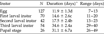
Mated females of O. haroldi started egg-laying 8–10 weeks after collection from the field. Females were observed to lay eggs singly, in pairs or triplets and enclose them within moulded lumps of substrate. Females laid an average of 72.7 ± 22.4 eggs (N = 6) throughout the egg-laying period, which lasted 4–8 weeks. Halting of egg-laying was observed when the rearing substrate became dry. Newly laid eggs were white and oval. As eggs developed, they became spheroid as indicated by their length:width ratio (Table 1). The egg size increased from the day it was deposited up to about day 6 after which it decreased slightly with significant differences in the average length (H = 103.2, df = 3, P < 0.001), width (H = 101.7, df = 3, P < 0.001) and volume (H = 104.5, df = 3, P < 0.001) during development (Table 1). The incubation period was the shortest and lasted 7–13 days (Table 2). A few hours before hatching, the abdominal setae and sclerotized mandibular teeth darkened and became visible under the chorion. Egg survival under the described laboratory conditions was 98.4% (N = 129).
Summary of changes in shape of Oplostomus haroldi egg during incubation period under laboratory conditions

All rearing experiments were carried out under temperature, relative humidity and lighting conditions of 25 ± 2 °C, 50 ± 5% and 10 h light-14 h dark, respectively.
+ Mean values followed by the same letter per column are not statistically different (one-way Kruskal–Wallis ANOVA).
Mean (±SD) of the developmental time (days) of the immature life stages of Oplostomus haroldi
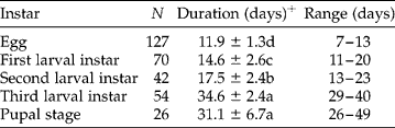
All rearing experiments were carried out under temperature, relative humidity and lighting conditions of 25 ± 2 °C, 50 ± 5% and 10 h light-14 h dark, respectively.
+ Values followed by the same letter per column are not statistically different (one-way Kruskal–Wallis ANOVA).
Larval development
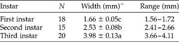
Newly hatched larvae were generally white with a white head capsule that turned brown after a few hours, matching the colour of the setae and spiracles. The width of the head capsule was significantly different between the larval instars (H = 46.03, df = 2, P < 0.001) (Table 3). The mean width of the head capsule was 1.66 ± 0.05 mm for the first instar, 2.53 ± 0.08 mm for the second instar and 3.98 ± 0.13 mm for the third instar (Table 3). The developmental time for the third larval and pupal instars was significantly longer than that for the other instars but not significantly different between the third instar larva and pupa (H = 248.1, df = 4, P < 0.001) (Table 2). The survival rate of the first, second and third instars was 73.7, 93.3 and 96.3%, respectively. Fungal growths observed on some dead larvae were later identified as Metarhizium anisopliae (Metchnikoff) Sorokin (Hypocreales: Clavicipitaceae) (identification carried out by Ouna E., Arthropod Pathology Unit-icipe, Nairobi) strains and may have been the cause of larval death.
Mean (±SD) of head capsule width of the larval instars of Oplostomus haroldi
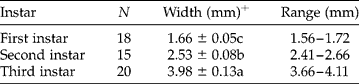
+ Values followed by the same letter per column are not statistically different (based on a one-way Kruskal–Wallis ANOVA).
Pupal development
The pupal stage was recognized by the oval mud cocoon casing. Cocoons were formed within the rearing substrate without any anchoring to the wall of the container. The duration of the pupal stage was shorter compared with total larval developmental time (Table 2). Pupal survival was moderate (54.2%, N = 48), with adults emerging from the surviving pupae with a sex ratio of 1:1 (N = 26 males and females). The developmental time of male and female pupae did not differ significantly (P = 0.979). An examination of cocoons from which adults failed to emerge revealed that all dead insects were in their pupal stage indicating that the larva to pupa transformation was 100%. However, no entomopathogens were observed to have infected the pupae.
Duration of life cycle and adult life span
The total duration of the life cycle from the egg to the newly emerged adult took 86–145 days (about 3–5 months). Adult beetles (both laboratory-bred and field-collected) were observed to live for up to 24 weeks under laboratory conditions, with females living longer than males. Adult males were observed to survive for 29–112 days (N = 55) whereas females survived for 31–168 days (N = 40), these differences being significant under laboratory conditions (U = 767, df = 1, P = 0.012).
Description of the third instar larva (L3)
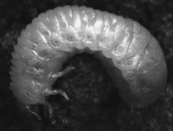
The body of the L3 is yellowish and lightly hirsute, with the posterior abdominal segments greatly enlarged to give a pear-shaped body (Fig. 1). Spiracles are distinctly dark and present on the prothorax and the first eight abdominal segments and enclosed by a c-shaped bulla.
Larva of Oplostomus haroldi (side view) on its rearing substrate composed of cow dung and soil

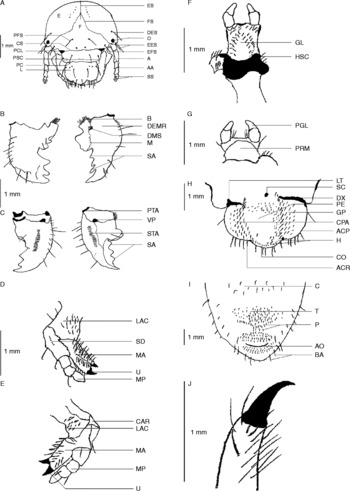
The cranium is reticulate, shiny and yellow to dark brown. Two dark ocelli are present, one above the base of each antenna (Fig. 2A). Epicranial and frontal sutures are present with the former more pronounced than the latter. The epicranial surface has one long seta above each ocellus and two pairs of setae on the lateral edges. The frons has a pair of medially located posterior setae and a pair of exterior setae. The clypeus is trapezoidal in shape (Fig. 2A). There is one long seta present on each lateral margin and a pair of more centrally located setae on the postclypeus (if an imaginary line were drawn to divide the clypeus into two symmetrical parts). Pores are present on the postclypeus and absent on the preclypeus. The preclypeus is less sclerotized than the postclypeus. The labrum is tri-lobed with rounded lateral margins (Fig. 2A). A pair of proximal setae and two pairs of distal setae are present on the lateral lobes, while the apical lobe has a fringe of short setae. The antennae are four-segmented with the first segment twice as long as each of the other three segments (Fig. 2A). The antennae have no setae and the apical segment tapers towards the tip and has two to three round sensory spots on the dorsal surface and two on the ventral surface.
Morphological features of Oplostomus haroldi larva. A, head capsule: A – antenna; AA – anterofrontal angle; CS – clypeofrontal suture; DES – dorsoepicranial seta; E – epicranium; EES – exterior epicranial setae; EFS – exterior frontal seta; ES – epicranial suture; F – frons; FS – frontal suture; L – labrum; O – ocellus; PC – preclypeus; PCL – precoilia; PFS – posterior frontal seta; PSC – postclypeus; SS – sensory spots. B, dorsal surface of mandibles: B – brustia; DEMR – dorsoexterior mandibular region; DMS – dorsomolar setae; M – molar lobe; SA – scissorial area. C, ventral surface of mandibles: PTA – postartis; STA – stridulatory area; VP – ventral process. D, dorsal surface of maxilla: LAC – labacoria; MA – mala; MP – maxillary palp; U – uncus. E, ventral surface of maxilla: CAR – cardo; LAC – labacoria; MA – mala; MP – maxillary palp; U – uncus. F, dorsal surface of labium: GL – glossa; HSC – hypopharyngeal sclerome. G, ventral side of labium: PGL – paraglossa; PRM – prementum. H, epipharynx: ACP – acanthoparia; ACR – acroparia; CO – corypha; CPA – chaetoparia; DX – dexitorma; GP – gymnoparia; H – heli; LT – laeotorma; PE – pedium; SC – sense cone. I, raster: AO – anal opening; BA – barbulae; C – campus; P – palidia; T – tegilla. J, lateral side of mesothoracic claw.

The mandibles are asymmetrical, similar in size and with black, heavily sclerotized scissorial areas (Fig. 2B and C). The scissorial area of the right mandible has four teeth while the left side has three teeth. The outer lateral edge of the mandibles has eight setae, four of which are long and the other four short. The molar part of the mandibles is dark and heavily sclerotized and in the right mandible it is bi-lobed into a large distal lobe and a small proximal lobe. The molar part of the left mandible is tri-lobed with the proximal lobe slightly larger than the other two. A brustia of setae is present on the basal part of each mandible with two to four dorsomolar setae present at the base of the molar teeth. A single seta is present on the ventral surface of the most distal molar tooth of each mandible. A ventral process is also present on each mandible. Areas that might possibly have a stridulatory function are present on the ventral side of each mandible and are lighter in colour than the surrounding area.
A maxillary cardo with fine setae is present on the interior dorsolateral surface (Fig. 2D and E). The exterior dorsolateral surface bears a row of five to seven small denticulate stridulatory teeth. The galea and lacinia are fused to form a mala with two similarly sized unci, one terminal and the other below it. The mala has dorsoventral rows of short sharp setae. The maxillary palp has three segments with a pair of setae present on the middle segment, one located dorsolaterally and the other ventrally.
The labium possesses a pair of short setae on the prementum and the glossa is covered with an ordered pattern of short setae and sensilla (Fig. 2F and G). The hypopharyngeal sclerome is asymmetrical and bears a truncate process on the left side that is absent on the right. The lateral lobe has a group of short setae.
The epipharynx has round margins and the apical margin in corypha region is protuberant (Fig. 2H). Acroparia is prominent and bears long thin setae. The zygum and epizygum are absent. The clithra is also absent. A plegmatium is absent on each lateral margin. The acanthopariae are asetaceous. The gymnopariae have short setae. The chaetopariae have long setae surrounding a slightly indented pedium. The haptomerum has a single row of heli. A sclerotized triangular laeotorma is present on the right side of the epipharynx and an elongated dexitorma on the left side.
The raster lacks a septula (Fig. 2I). The tegilla extends from the barbulae to palidia and is covered with many sharp-pointed short setae. Presepular hamate setae are absent. Barbulae have long slender setae and the campus has sparse short setae. The anal opening is transverse and surrounded by several rows of short setae. The mesothoracic claw is sharp, slightly curved and without setae (Fig. 2J).
Discussion
This study provides for the first time the life history of O. haroldi under laboratory conditions and a description of the third instar larva with emphasis on taxonomically important characters. The third larval stage of O. fuligineus has been described to possess up to 11 maxillary (stridulatory) teeth (Donaldson, Reference Donaldson1989), while this study showed that O. haroldi has up to seven maxillary teeth with similar function, clearly depicting a difference in the number of this structural character. However, lack of pictorial descriptions of the larva of O. fuligineus poses a challenge to the compilation of the morphological differences that exist between larvae of the two species, which could be relevant for taxonomy.
It was evident from our results that the eggs of O. haroldi are smaller than those of O. fuligineus (Donaldson, Reference Donaldson1989). Egg size was used to show changes during incubation because our attempts to weigh them failed due to their highly hygroscopic nature, and, as a consequence, the recorded weight was highly variable. The cessation of egg deposition by females during drier periods implies that females possess the ability to determine moisture levels suitable for egg development and survival of newly hatched larvae. This phenomenon has also been observed in other scarabs (Soltani et al., Reference Soltani, Chaieb and Ben Hamouda2008). The differences in head capsule width of various instars of the larval stage of scarabs have been employed in identifying larval instars (Donaldson, Reference Donaldson1989; Soltani et al., Reference Soltani, Chaieb and Ben Hamouda2008), as well as in taxa other than Scarabaeidae. Data obtained from this study show that differences in head capsule width can be used to delineate the various larval instars.
The results showed that O. haroldi eggs apparently survived better than the other life stages while the pupal stage showed the least survival under laboratory conditions. The enclosure of eggs within lumps of rearing substrate might serve to provide the necessary moisture level for their development and thus explain the relatively high survival rate. Egg enclosure may also offer protection from predators in the natural setting. The high mortality (26.3%) recorded in the first larval instar during the larval stage of the life cycle is an indication of their vulnerability. Although the beetle seemed to survive up to 6 months in the laboratory, it is not known how long beetles can survive inside honeybee colonies. The suitability of the substrate used in this study can only be confirmed by carrying out comparative studies where beetles are kept on different substrates. Initial efforts to rear them on banana in our laboratory, which was reported to be attractive to another member of the Oplostomus genus (Donaldson, Reference Donaldson1989), were not successful, suggesting that this beetle may not be highly polyphagous.
Cocoon formation in O. haroldi was similar to that described by Donaldson (Reference Donaldson1989) for O. fuligineus as they were not attached to the walls of the rearing container. This observation is another behavioural similarity shared by both species in addition to their being pests of honeybee colonies. The cause of the very high pupal mortality observed during the laboratory rearing still remains a mystery and suggests that other factors (both biotic and abiotic) other than faulty larval–pupal transformations and entomopathogens might have been the probable causes of death.
With O. haroldi capable of completing its life cycle within 3–5 months, this species can therefore produce between 2 and 4 generations per year depending on the prevailing climatic conditions. Local climatic conditions can have a direct bearing on the level of honeybee colonies' infestations by shortening or prolonging the life cycle as suggested by Ritcher (Reference Ritcher1957) and Torto et al. (Reference Torto, Fombong, Mutyambi, Muli, Arbogast and Teal2010). Previous studies showed that beetle populations infesting honeybee colonies were male-biased, with female numbers varying significantly between seasons (Torto et al., Reference Torto, Fombong, Mutyambi, Muli, Arbogast and Teal2010). Upon mating inside the hive, female beetles are bound to leave the hives in search of suitable egg-laying sites, and this might explain the difference in sex ratios of beetles inside the hives. No previously reported sex ratio disparities were observed during the laboratory rearing of this beetle.
Initial observations of M. anisopliae infecting larvae of O. haroldi suggest that the entomopathogen could be used to control the pest. However, detailed field studies are required to locate the beetle breeding sites and to assess their ecological importance in nutrient recycling prior to considering using M. anisopliae as a control strategy. This study also demonstrates that it is possible to breed O. haroldi in the laboratory on a diet composed of cow dung and soil.
Acknowledgements
The authors wish to acknowledge D. Munyao, T. Poumo and D. Achinko for their assistance in the rearing of the beetles, J. Gitau for assistance with the preparation of larval samples for dissection and E. Ouna for Metarhizuim isolate identification. The German Academic Exchange Service (DAAD) provided a studentship to A. T. F. to carry out this study.