Introduction
There has been an increasing incidence in head injuries associated with the growing number of accidents involving motor vehicles. Approximately 4 per cent of patients treated for head trauma have skull fractures, and 14–22 per cent of these patients are diagnosed with temporal bone fractures.Reference Brodie and Thompson 1 , Reference Nageris, Hansen, Lavelle and Van Pelt 2 In addition to car accidents, falls and assaults are common causes of temporal bone fractures.Reference Saraiya and Aygun 3 , Reference Ishman and Friedland 4
Temporal bone fractures are clinically significant because the anatomical structure of the temporal bone is complex and any damage to this critical structure may lead to neurological problems. Therefore, it is important to determine a prognosis and to deal with potential complications in advance. The classification of temporal bone fractures serves many important purposes. It assists in the understanding of associated complications, guides treatment and educates.Reference Saraiya and Aygun 3
Temporal bone fractures have traditionally been classified as either longitudinal or transverse, according to the long axis of the fracture line with respect to the long axis of the petrous bone.Reference Cannon and Jahrsdoerfer 5 However, there has been a recent preference for these fractures to be classified as either otic capsule violating or non-violating.Reference Brodie and Thompson 1 , Reference Kennedy, Avey and Gentry 6 The indications and the approach with respect to the surgical repair of cerebrospinal fluid (CSF) fistulae and facial nerve paralysis are also influenced by whether the otic capsule has been violated.Reference Magliulo, Appiani, Iannella and Artico 7 , Reference Dahiya, Keller, Litofsky, Bankey, Bonassar and Megerian 8 Mild symptoms and complications of temporal bone fractures are expected in the otic capsule sparing group, which covers most types of temporal bone fracture. However, more systematic research is required to investigate this category because such fractures can exhibit a variety of neurological symptoms, including facial paralysis.Reference Lim, Jun and Song 9
This study evaluated the clinical aspects of otic capsule sparing temporal bone fractures and analysed them using a new regional radiological evaluation method devised by the authors.
Materials and methods
This retrospective study included 188 temporal bones of 173 patients who were diagnosed with otic capsule sparing temporal bone fractures by high-resolution computed tomography (CT), and who underwent conservative treatment from May 2004 to August 2015. Patients with a history of head trauma, otitis media, neurological disease or skull surgery were excluded. The authors assert that all procedures contributing to this work complied with the ethical standards of the relevant national and institutional guidelines on human experimentation (UC11RISI0100) and with the Helsinki Declaration of 1975, as revised in 2008.
The patients comprised 139 men and 34 women, with a mean age of 39 years. Thirteen men and two women had bilateral fractures of the temporal bone. Right-sided fractures were present in 103 cases and left-sided fractures in 85 cases. High-resolution CT of the temporal bone was obtained in 1 mm sections with a Somatom Definition AS+ 128-slice scanner (Siemens, Seoul, Korea). The temporal bone CT was 800 units long and 2800 units wide. The field of view was 512 × 512 mm2.
Reviewing the medical records of each patient, the otoscopic tympanic membrane findings were evaluated, and symptoms of ear fullness, tinnitus and dizziness were reviewed.
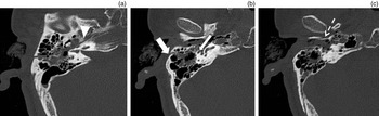
The type of temporal bone fracture was classified using traditional radiological categories (longitudinal, transverse and mixed). The authors also classified fractures, into four types, according to the regional location of the fracture line in the temporal bone: type I, fracture line in the petrous bone; type II, fracture line extended to the middle-ear cavity; type III, fracture line in the mastoid cavity; and type IV, fracture line in the external auditory canal (Figure 1). Cases of otic capsule violating temporal fractures were excluded from the study.
Regional evaluation of the fracture line on temporal bone computed tomography in a single case. Type I = fracture line violates the petrous bone (arrowhead in a); type II = fracture line extends to the middle-ear cavity (thin arrow in b); type III = fracture line is in the mastoid cavity (thick arrow in b); type IV = fracture line violates the external auditory canal (dotted arrow in c).

In addition, the degree of facial paralysis and level of hearing were followed over time. The data collected from the initial and follow-up pure tone audiometry tests were analysed statistically, using the student's t-test, to evaluate the hearing changes during the follow-up period. A value of p < 0.05 was considered significant.
Results
Otological findings and symptoms
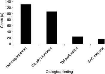
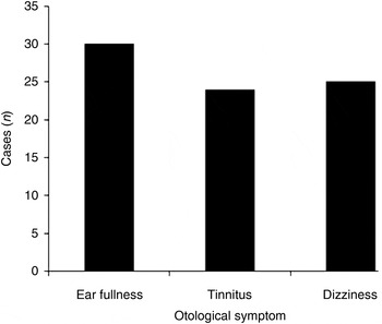
Among the 188 temporal bone fracture cases, there were 131 cases of haemotympanum, 107 of bloody otorrhoea, 24 of tympanic membrane perforation and 17 of external ear canal stenosis (Figure 2). With regard to patients’ symptoms identified, there were 30 cases of ear fullness, 24 of tinnitus and 25 of dizziness (Figure 3).
Otological findings among the 188 cases of temporal bone fracture. TM = tympanic membrane; EAC = external ear canal

Otological symptoms among the 188 cases of temporal bone fracture.

Fracture types
Traditional classification
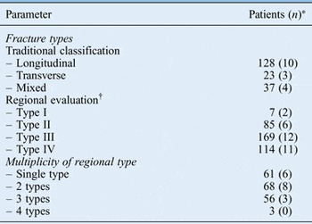
Of the 188 reviewed fractures, classification based on the type of temporal bone fracture revealed that 128 (68 per cent) were longitudinal, 23 (12 per cent) were transverse and 37 (20 per cent) were mixed (Table I).
Classification of Fracture Types and Multiplicity of Regional Evaluation Type

*The numbers in parentheses represent the numbers of patients with facial paralysis. †In type I fractures, the fracture line violates the petrous bone; in type II, the fracture line reaches the middle-ear cavity; in type III, the fracture line is in the mastoid cavity; in type IV, the fracture line violates the external auditory canal.
Regional evaluation
Based on the location of the temporal bone fracture lines in each case, the fracture line was defined (as outlined above) as: type I in 7 cases, type II in 85, type III in 169 and type IV in 114 (Table I). Forty cases of suspicious ossicular injuries were also observed in the type II cases.
There were 61 cases of a single fracture type, 68 involving 2 types, 56 involving 3 types and 3 involving 4 types (Table I). Therefore, more than one regional fracture type may exist simultaneously in a single patient.
Facial paralysis
Among the 188 fracture cases, 17 cases of facial paralysis (9 per cent) were identified. When classified according to the type of fracture line, 10 cases out of 128 (7.8 per cent) were identified as longitudinal, 3 out of 23 (13.0 per cent) were transverse and 4 out of 37 (10.8 per cent) were mixed types (Table I).
When classified according to regional criteria, facial paralysis was revealed in 2 out of 7 (28.6 per cent) type I fracture cases, 6 out of 85 (7.1 per cent) type II cases, 12 out of 169 (7.1 per cent) type III cases, and 11 out of 114 (9.6 per cent) type IV cases (Table I).
When we classified facial palsy according to the presence of a multiplicity of fracture types, facial paralysis was revealed in 6 out of a total of 61 cases (9.9 per cent) involving a single fracture type, 8 out of 68 cases (11.8 per cent) involving 2 types, 3 out of 56 cases (5.4 per cent) involving 3 types, and 0 out of 3 cases (0 per cent) involving 4 types (Table I).
When the 17 patients with facial paralysis were classified according to paralysis severity using the House–Brackmann grading scale, there were 5 House–Brackmann grade II cases, 3 grade III cases, and 9 grade IV cases. Eleven patients had delayed facial paralysis and six had facial paralysis at the initial evaluation. The delayed type was more common in the otic capsule sparing cases. Delayed facial paralysis was seen in five of nine House–Brackmann grade IV cases, in two of three grade III cases and in four of five grade II cases.
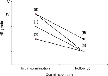
Fourteen patients were identified as having facial paralysis from their medical records after an average follow up of 57.6 days, excluding 2 patients with House–Brackmann grade III and 1 with grade IV paralysis. The degree of facial paralysis improved in nine patients with House–Brackmann grade I paralysis and in five with grade II paralysis (Figure 4).
Fourteen patients with facial paralysis were classified according to severity using the House–Brackmann grading scale. At the initial examination, there were five grade II cases, one grade III case and eight grade IV cases. After an average of 57.6 days, the degree of facial paralysis improved: there were 9 grade I and 5 grade II cases. All five cases with House–Brackmann grade II on follow up had improved from House–Brackmann grade IV at the initial examination. HB = House–Brackmann

Changes in hearing
Thirty-one of the 188 head injury patients with temporal bone fractures underwent initial and follow-up pure tone audiometry. The mean age of these patients was 42.3 years. The initial and follow-up examinations took place at a mean of 21.8 and 79.9 days, respectively, after the temporal bone fracture had occurred.
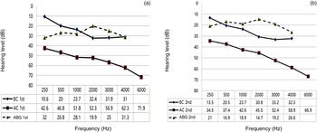
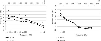
The results of initial and follow-up audiometry are summarised in Figure 5. A frequency-specific analysis of the air–bone gap (ABG) revealed significant closing of the ABG at all frequencies in the follow-up tests (p < 0.05). The closing of the ABG was mainly caused by significant improvement in air conduction (Figure 6a) at all frequencies, except for 3 and 4 kHz. However, bone conduction improved significantly only at 250 Hz (Figure 6b).
Initial (a) and follow-up (b) pure tone audiometry findings for 31 of the 188 temporal bone fracture cases. The examinations took place at a mean of 21.8 and 79.9 days after the injury, respectively. BC = bone conduction; AC = air conduction; ABG = air–bone gap

The change in air (a) and bone (b) conduction at the initial and follow-up examinations. A significant improvement in air conduction (a) was found at all frequencies, except at 3 and 4 kHz. Bone conduction showed a significant improvement only at 250 Hz (b). AC = air conduction; BC = bone conduction

Discussion
Traditionally, temporal bone fractures have been classified as longitudinal, transverse or mixed, depending on their relationship to the petrous pyramid. However, Aguilar et al. studied patterns of temporal bone fractures in 22 patients using high-resolution CT scans, and found that a large number of the fractures could not be easily classified as either longitudinal or transverse.Reference Aguilar, Yeakley, Ghorayeb, Hauser, Cabrera and Jahrsdoerfer 10 Kelly and Tami proposed an alternative nomenclature in 1994, suggesting use of the terms ‘otic capsule sparing’ and ‘otic capsule violating’.Reference Kelly, Tami, Jackler and Brackmann 11 It was suggested that this new terminology could improve surgical planning. Brodie and Thompson included this nomenclature in their 1997 retrospective review of 820 temporal bone fractures, and found that the incidence of facial nerve injury correlated with otic capsule violating injuries.Reference Brodie and Thompson 1 They also found that CSF fistulae were twice as common in otic capsule violating injuries. However, in that study, the vast majority of the temporal bone fractures spared the otic capsule, with only 21 of the injuries fracturing the otic capsule.
Yanagihara et al. suggested another fracture classification scheme in 1997.Reference Yanagihara, Murakami and Nishihara 12 This system was developed to provide an optimal approach to facial nerve surgery. In this system, 81 patients with 97 surgically treated fractures were divided into 4 categories based on the specific anatomy involved in the temporal bone fracture: the mastoid, external auditory canal, tegmen or internal auditory canal. These fracture types were not correlated with complications other than facial nerve paresis, and no comparison was made with the traditional classification system.
In our study, the most common type of fracture identified by the traditional classification system was the longitudinal type, as expected, because severe transverse types of fracture may violate the otic capsule and we excluded such fractures from our study. In our regional analysis, the most common fracture site was the mastoid, which occupies a large part of the temporal bone, with the next most common site being the external auditory canal, which is located superficially relative to the petrous and the middle ear. This feature may explain the vulnerability of the mastoid and external auditory canal.
Ishman and Friedland evaluated 155 temporal bone fractures by analysing the traditional classification system and determining whether the fracture was petrous-violating.Reference Ishman and Friedland 4 They observed a higher incidence of facial paralysis (30.8 per cent) in cases with petrous bone violating fractures.
In our study, a similar incidence of facial paralysis (28.6 per cent) was found in cases of type I fractures that were defined as petrous-violating. However, the specific finding pertaining to the incidence of facial paralysis in the traditionally classified fracture or in cases involving multiple fracture types was not observed in otic capsule sparing temporal bone fractures. All our cases had incomplete facial paralysis below House–Brackmann grade IV. Typically, patients who have delayed-onset or incomplete paralysis are treated with high-dose corticosteroids, with further intervention based on the results of electrodiagnostic testing or imaging.Reference Brodie and Thompson 1 , Reference Ishman and Friedland 4 After medical treatment over a period of approximately 57 days, the facial paralysis was found to have improved to House–Brackmann grade II or lower. Many authors agree that the threshold for surgical intervention is reached when a 90 per cent or greater degeneration of the facial nerve is observed on electroneuronography or a higher facial paralysis grading than House–Brackmann grade V is obtained on physical evaluation.Reference Johnson, Semaan and Megerian 13 – Reference Coker, Kendall, Jenkins and Alford 15
High-resolution CT provides great sensitivity as a diagnostic tool of temporal bone fracture. It offers a superior depiction of the bony anatomy and reconstructions of coronal and sagittal images from the raw axial data.
In our previous study using multiplanar reconstruction images of temporal bone CT, 12 cases of traumatic facial paralysis among those with otic capsule sparing temporal bone fractures were observed with images of the fracture line in which the extension was estimated to approach the otic capsule.Reference Lim, Jun and Song 9
Hearing loss in patients who have a temporal bone fracture may be immediate or delayed. The incidences in all temporal bone fracture cases have been reported as: 26–57 per cent for conductive hearing loss, 14–23 per cent for sensorineural hearing loss (SNHL) (14 per cent for complete SNHL) and 20–55 per cent for mixed hearing loss.Reference Ishman and Friedland 4 , Reference Nosan, Benecke and Murr 14 , Reference Ghorayeb, Yeakley, Hall and Jones 16 , Reference Darrouzet, Duclos, Liguoro, Truilhe, De Bonfils and Bebear 17 Sensorineural hearing loss may occur without gross disruption of the cochlea by concussion.Reference Ohlrogge and Francis 18 Cochlear concussion was reported in 9 per cent of 115 cases of facial nerve paralysis associated with temporal bone fractures.Reference Darrouzet, Duclos, Liguoro, Truilhe, De Bonfils and Bebear 17
Most of the patients visited or were transferred to an emergency room and evaluated carefully by a neurosurgeon or traumatologist to prevent potentially fatal complications involving the brain. The audiological evaluation tends to be delayed or skipped because urgent treatment is required or the patient's condition is unstable. Nevertheless, most of the cases were evaluated using pure tone audiometry at least once. Unfortunately, only 31 cases underwent both initial and follow-up evaluations.
Early audiometric testing is recommended for evaluating the baseline post-injury hearing status, which is used to provide an indication of SNHL or conductive hearing loss. A subsequent full audiological examination should be performed three to six weeks post-injury, to allow sufficient time for the resolution of haemotympanum, because the presence of middle-ear fluid (CSF or blood) results in a conductive loss.
-
• The temporal bone anatomical structure is complex and any damage may cause neurological problems
-
• Mild symptoms and complications of temporal bone fractures are expected in otic capsule sparing cases
-
• There was a high incidence of facial paralysis (28.6 per cent) in petrous-violating cases in the otic capsule sparing patients
-
• Incomplete facial paralysis in otic capsule sparing temporal bone fracture cases was improved to House–Brackmann grade II or lower by two months
-
• Conductive hearing loss in otic capsule sparing temporal bone fracture cases may resolve spontaneously
-
• Conservative treatment with high-dose corticosteroids is suggested for facial paralysis House–Brackmann grade IV or lower
In our study, there was no significant change in bone conduction between the initial and follow-up tests, except at 125 Hz, and this may indicate that there was no occurrence of SNHL by cochlear concussion. In the initial management of conductive hearing loss, most practitioners prefer to wait to determine whether the loss will resolve spontaneously or not. Grant et al. studied 47 cases of traumatic conductive hearing loss with conservative treatment, and reported ABG closure from 24.8 ± 12.1 to 13.2 ± 11.1 dB after the follow-up examination at 9.4 months.Reference Grant, Arganbright and Friedland 19 They recommended that surgical intervention for perforation or conductive hearing loss be undertaken only in rare cases when these conditions persist for longer than six months. Kristensen et al. also suggested that surgical intervention for traumatic tympanic membrane perforation is generally not indicated before three months, because most (88 per cent) tympanic membrane perforations healed spontaneously within this period.Reference Kristensen, Juul, Gammelgaard and Rasmussen 20 In our study, the ABG closed significantly, from 27.2 dB at an average of 21.8 days post-trauma to 19.6 dB at an average of 79.9 days post-trauma, in 31 cases. The difference in follow-up ABG outcomes between the studies was associated with a shorter observation period in our study. Initial conservative treatment for conductive hearing loss is an option in the otic capsule sparing group. However, surgical intervention is recommended for the prevention of chronic otitis media or permanent conductive hearing loss caused by ossicular disruption after the observation period.
Our study was limited in that it was a retrospective study relying on medical records, leading to possible misinterpretations of otological symptoms by reviewers. A systematic evaluation within a prospective study would enhance our understanding of the clinical features of temporal bone fractures.
Conclusion
Mild symptoms and complications of temporal bone fractures were observed in the otic capsule sparing temporal bone fracture group.
Careful radiological evaluation of otic capsule and petrous bone violating temporal bone fractures would help with treatment policy and patient education. Otic capsule sparing temporal bone fractures resulted in incomplete facial paralysis (lower than House–Brackmann grade IV), and most patients had good prognoses; conservative treatment with high-dose corticosteroids is suggested.