Highlights
-
• Movement disorders are frequent but underrecognized in central nervous system tuberculosis.
-
• Common types include ataxia, dystonia, chorea and parkinsonism.
-
• Basal ganglia or thalamic lesions are commonly linked to these disorders.
Introduction
Central nervous system (CNS) tuberculosis (TB) is the most devastating form of extrapulmonary tuberculosis, accounting for approximately 1% of all TB cases and up to 10% of extrapulmonary cases. It remains a significant cause of neurological morbidity and mortality, particularly in endemic regions and among immunocompromised individuals. Reference Garg1,Reference Seddon, Tugume, Solomons, Prasad and Bahr2 CNS TB presents primarily as tuberculous meningitis, intracranial tuberculomas, spinal arachnoiditis or a combination thereof. The pathological processes involved – granulomatous inflammation, vasculitis, infarction, hydrocephalus and immune-mediated injury – can result in a variety of neurological syndromes. Reference Garg1,Reference Seddon, Tugume, Solomons, Prasad and Bahr2
Several case reports and case series have documented a diverse array of movement disorders in patients with CNS TB, particularly tuberculous meningitis, highlighting its complex and often underrecognized neurological manifestations. Reference Alarcón, Dueñas, Cevallos and Lees3–Reference Desai, Yadav, Garg, Schneider and Pal5
Although individual case reports and small series have described diverse movement disorders in CNS TB, the evidence remains fragmented and inconsistent. Cohort studies are limited, and outcome predictors such as lesion location, pathophysiological mechanisms and treatment response have not been systematically synthesized. No prior comprehensive review has pooled available cases and compared them with cohort findings. A systematic review at this time is needed to consolidate dispersed evidence, clarify clinical patterns and identify prognostic factors, thereby addressing a critical gap in the literature and guiding both diagnosis and management.
Methods
This systematic review followed the PRISMA 2020 guidelines for reporting. The review protocol was prospectively registered in the PROSPERO database with the registration number CRD420251075389. Reference Garg, Pandey, Singhal and Mahdi6
Search strategy
A thorough and systematic literature search was conducted across four major databases: PubMed, Embase, Scopus and Google Scholar, to identify all relevant studies reporting movement disorders in patients with CNS TB. We limited Google Scholar screening to the first 50 pages, as results on earlier pages are more relevant, while later ones are largely low-yield or duplicate. The final search was completed on May 31, 2025. Detailed search strategy has been provided in Table 1.
Search strategy

Non-English studies were translated using Google Translate. Critical medical terminology, including diagnoses and outcome measures, was cross-verified against bilingual medical dictionaries, if necessary.
Definitions
CNS tuberculosis was defined as microbiologically, radiologically or histologically confirmed tuberculous infection of the brain or spinal cord, including manifestations such as tuberculous meningitis, intracranial or spinal tuberculomas, abscesses and spinal TB with CNS involvement Reference Dian, Ganiem and van Laarhoven7,Reference Marais, Thwaites and Schoeman8 (Figure 1).
MRI of the brain showing a ring-enhancing lesion in the left thalamus with surrounding edema on post-contrast T1-weighted coronal image (left). Magnetic resonance spectroscopy from the lesion (right) demonstrates a lipid-lactate peak, reduced N-acetylaspartate and elevated choline, suggestive of a tuberculoma. The patient had right hemichorea.

Movement disorders were defined clinically and classified into hyperkinetic (e.g., chorea, dystonia, myoclonus, tremor) and hypokinetic (e.g., parkinsonism, rigidity) types. Cases with insufficient neurological detail to confirm movement disorders were excluded. 9
Eligibility criteria
We included case reports, case series and cohort studies that provided a confirmed diagnosis of CNS TB, documented the presence of one or more clinically observed movement disorders and offered sufficient clinical, radiological or treatment-related details for meaningful analysis. Studies were excluded if they involved non-human subjects, lacked adequate clinical descriptions or were purely radiological or experimental without clinical correlation.
Study selection
Two reviewers (RKG and SS) independently assessed all titles and abstracts for eligibility. Full texts of potentially relevant articles were retrieved and reviewed. Any disagreements were resolved through discussion or, when necessary, consultation with a third reviewer (SP).
EndNote 21 (Clarivate Analytics, Philadelphia, USA) was used for reference management and to remove duplicates. Duplicate verification was independently performed by HSM, with final adjudication by SS. The selection process was documented in a PRISMA flow diagram.
Data extraction
Data were extracted using a predefined structured format by three reviewers (SP, SS and RAM). The following data were extracted from each included study: patient demographics (including age and sex), subtype of CNS TB (such as tuberculous meningitis or tuberculoma), type of movement disorder observed, time to onset of movement-related symptoms, findings from neuroimaging and CSF analysis and treatment approaches including antitubercular therapy, corticosteroids, symptomatic medications and surgical interventions, as well as clinical outcomes encompassing recovery status, persistence of symptoms, complications or death. All discrepancies were resolved through consensus in consultation with a senior reviewer (RKG). Data were compiled in Microsoft Word tables and verified for accuracy before analysis.
Quality assessment
Case reports and case series were evaluated using the criteria proposed by Murad et al., which assess four key domains: selection, ascertainment, causality and reporting. Records meeting all four domains were rated as “good,” those fulfilling three as “fair” and those satisfying only one or two as “poor.” Cohort studies were appraised using the Newcastle–Ottawa Scale, focusing on three domains: selection, comparability and outcome assessment. Disagreements in scoring were resolved by consensus among the reviewers.
Data synthesis
A descriptive synthesis of data was performed. Categorical variables were reported as frequencies and percentages. Results were grouped by type of movement disorder, type of TB, underlying mechanism (vascular, immune-mediated, compressive), diagnostic modalities, treatments given and clinical outcomes. Structured tables and illustrative figures were developed to present a comprehensive summary of findings and clinical patterns in CNS TB-associated movement disorders. Missing data were extracted as “not reported,” and no imputation was performed. Study authors were not contacted for additional clarification. Only descriptive synthesis was undertaken, as the available evidence consisted solely of case reports, case series and a few heterogeneous cohorts, precluding a meaningful meta-analysis.
Results
This review includes 61 patients with CNS TB who developed movement disorders, with data derived from 56 published reports. Reference Valdes and Sibat12–Reference Jackson67 All included cases were assessed for quality, with each rated as either good or fair (Supplementary Item 1). The PRISMA checklist is available in Supplementary Item 2. Detailed patient-level data are summarized in the tables. Figure 2 presents the PRISMA flow diagram outlining the case selection process. Of 4310 records identified, 3514 were screened after removing 796 duplicates, 158 full texts were retrieved, 131 assessed for eligibility and finally, 60 reports (including 4 cohort studies) met inclusion criteria.
PRISMA 2020 flow diagram illustrating the study selection process for inclusion in the systematic review. The diagram shows the number of records identified, screened, excluded and included at each stage of the review, along with reasons for exclusions.

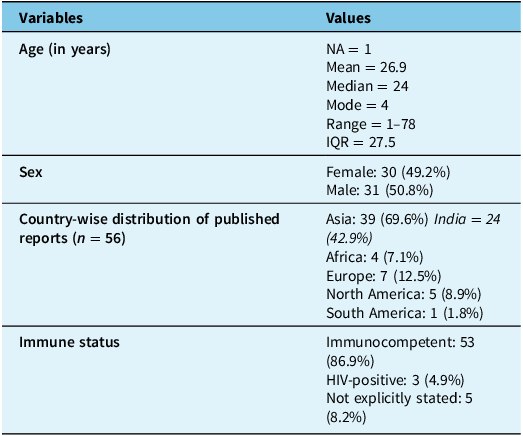
The mean age was 26.9 years, with a median of 24 years and a nearly equal male-to-female distribution. The majority of patients were immunocompetent, and most cases were reported from Asia, predominantly India (Table S1 and Table 2). Tuberculous meningitis was the most frequent type of CNS TB, accounting for nearly two-thirds of the cases, often associated with infarcts, hydrocephalus or tuberculomas. Intracranial tuberculomas without meningitis accounted for about one-fifth, and spinal TB, miliary TB and other forms such as cerebritis and demyelination were less frequent. Neuroimaging showed tuberculomas in about half of the patients, hydrocephalus in 44% and infarcts in 34%, particularly involving the thalamus, basal ganglia and brainstem. Leptomeningeal enhancement, basal exudates and arachnoiditis were also commonly noted. CSF findings typically included lymphocytic pleocytosis with elevated protein and low glucose. Microbiological or histopathological confirmation of tuberculosis was obtained in about two-thirds of the cases, most commonly via CSF GeneXpert, culture or biopsy (Table S1 and Table 3).
Summary of demographic characteristics of CNS tuberculosis cases presenting with movement disorders (n = 61)

CNS = central nervous system; HIV = human immunodeficiency virus.
Summary of clinical, neuroimaging and treatment features of CNS TB cases presenting with movement disorders (n = 61)

AFB = acid-fast bacillus; ART = antiretroviral therapy; ATT = anti-tuberculous therapy; BAL = bronchoalveolar lavage; CBNAAT = cartridge-based nucleic acid amplification test; CNS = central nervous system; EVD = external ventricular drainage; GI = gastrointestinal; H = isoniazid; HIV = human immunodeficiency virus; HRZE = isoniazid, rifampicin, pyrazinamide, ethambutol; ICP = intracranial pressure; INH = isoniazid; IQR = interquartile range; IVIG = intravenous immunoglobulin; NA = not available; OMS/OMAS = opsoclonus-myoclonus syndrome/opsoclonus-myoclonus-ataxia syndrome; PAID = paroxysmal autonomic instability with dystonia; PCR = polymerase chain reaction; PMN = polymorphonuclear; R = rifampicin; RIPE = rifampicin, isoniazid, pyrazinamide, ethambutol; TB = tuberculosis; TBM = tuberculous meningitis; VP = ventriculoperitoneal.
The movement disorders observed were diverse and reflected the underlying pathology and lesion location. Ataxia was the most common manifestation, followed closely by dystonia, chorea or hemiballismus and parkinsonism. Less commonly reported were opsoclonus-myoclonus, segmental myoclonus, postural and resting tremors, torticollis, cervical dystonia, blepharospasm, orofacial dyskinesia, stereotypy, trismus and bobble-head doll syndrome. The onset of movement disorders was subacute in most cases, though some occurred acutely or following initiation of anti-tuberculosis therapy, and a few developed after clinical recovery or months to years later. Associated neurological features included motor weakness, cranial nerve palsies, altered consciousness, raised intracranial pressure, autonomic instability, seizures and behavioral or cognitive abnormalities. Anti-tuberculosis therapy was administered in nearly all patients, often with adjunctive corticosteroids. Symptomatic treatment for movement disorders, including agents such as levodopa, trihexyphenidyl, clonazepam, baclofen and midazolam, was provided in nearly half the cases. Neurosurgical interventions, including ventriculoperitoneal shunting, surgical decompression or lesion excision, were performed when indicated.
Among the patients, 25 (41%) achieved full recovery, while 17 (27.9%) showed marked or significant improvement. Partial improvement was observed in 13 (21.3%) cases, and 11 (18.0%) continued to have persistent or serious problems. One patient (1.6%) died (Table S1 and Table 3).
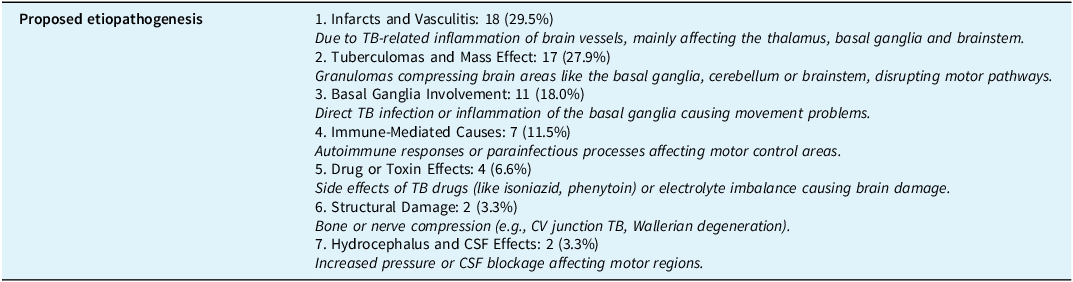
The proposed etiologies of the movement disorders included infarcts and vasculitis affecting the basal ganglia, thalamus or brainstem, compressive effects of tuberculomas, direct basal ganglia involvement, immune-mediated mechanisms, drug-induced neurotoxicity (such as isoniazid toxicity) and complications of hydrocephalus or structural lesions. Outcomes appear less favorable in cases with deep basal ganglia or thalamic infarcts, where persistent parkinsonism, dystonia or hemichorea were commonly reported. Conversely, patients with isolated tuberculomas or cerebellar involvement more often achieved complete or near-complete recovery after anti-TB treatment and adjunctive therapy (Table S1 and Table 4).
Proposed enteropathogenic mechanisms (n = 61)**

TB = tuberculosis; CV = craniovertebral. **Etiopathogenesis was derived from author reports in original case descriptions where explicitly stated; otherwise, categorization was inferred by the reviewers based on clinical, radiological and treatment details.
We also identified four cohort studies that met the PICO criteria. Reference Alarcón, Dueñas, Cevallos and Lees3,Reference Alarcón, Maldonado and Rivera4,Reference Pandey, Kumar, Malhotra, Kohli, Kumar and Verma68,Reference Netravathi, Pal and Devi69 Cohort studies further substantiated these findings. In a pediatric cohort from North India, tuberculous meningitis accounted for 4.3% of all movement disorder cases and typically presented with ataxia or tremors. In a large prospective adult cohort, secondary movement disorders due to CNS TB commonly manifested as dystonia, parkinsonism and tremors, with imaging frequently showing lesions in the thalamus or basal ganglia. Another study of patients with intracranial tuberculomas highlighted the role of focal or unilateral movement disorders caused by mass lesions affecting motor circuits. A separate prospective study of tuberculous meningitis patients found that 87% developed movement disorders, predominantly tremors, chorea, dystonia and parkinsonism, typically within the first three weeks of illness. These cohort findings emphasized that movement disorders are common, often underrecognized consequences of CNS TB, with early imaging and targeted therapy being crucial for improving neurological outcomes (Table 5).
Summary of four cohort studies on movement disorders in central nervous system (CNS) tuberculosis

Discussion
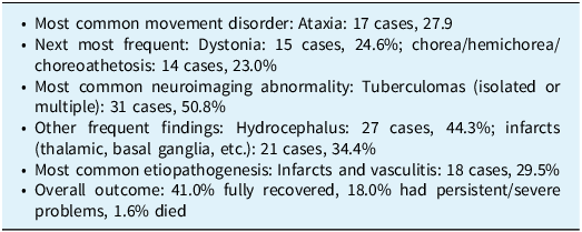
CNS TB, especially tuberculous meningitis, gives rise to a wide variety of movement disorders driven by inflammation, infarction, hydrocephalus or immune-mediated phenomena. Hyperkinetic forms such as chorea, dystonia, tremor, myoclonus, opsoclonus and stereotypies are commonly associated with basal ganglia or brainstem involvement, whereas hypokinetic syndromes like parkinsonism and akinetic mutism are usually linked to thalamic or frontal lobe injury. From a clinical perspective, early recognition of these disorders is crucial since they may precede or persist despite anti-TB therapy and significantly influence long-term outcomes. Active screening, timely neuroimaging and appropriate use of corticosteroids, surgery or symptomatic drugs can substantially improve prognosis (Table 6).
Key findings of our review (n = 61)

Immune-mediated mechanisms
In some patients, movement disorders arise without visible lesions, suggesting an immune basis. Molecular mimicry and cytokine-driven inflammation may lead to parainfectious syndromes such as opsoclonus-myoclonus or dystonia. These cases often respond to corticosteroids or immunotherapy, reinforcing the immune-mediated pathogenesis. Reference Netravathi, Pal and Devi69
Vascular mechanisms
Vasculitis is one of the most common causes of movement disorders in CNS TB. In tuberculous meningitis, infectious vasculitis frequently produces infarcts involving deep brain structures such as the basal ganglia, thalamus, internal capsule and brainstem. These infarcts disrupt motor circuits, leading to manifestations such as chorea, dystonia, tremor or parkinsonism. Reference Misra, Kalita and Maurya70
Structural and compressive lesions
Tuberculomas can exert a mass effect or cause perilesional edema, particularly when located in motor regions like the basal ganglia, thalamus or brainstem. Reference Alarcón, Maldonado and Rivera4 Hydrocephalus, often due to obstruction of CSF pathways by basal exudates, can compress the thalamus or midbrain and disturb motor relays. Reference Rock, Olin, Baker, Molitor and Peterson71 Involvement of the craniovertebral junction or brainstem in spinal or meningeal tuberculosis may also produce abnormal movements through direct compression or inflammation. Reference Synmon, Siangshai, Nongrum and Wezah17,Reference Lee, Oh, Park and Yoon72
Drug-related toxicity
Anti-TB drug neurotoxicity, particularly from isoniazid, may contribute to reversible encephalopathy involving motor pathways, especially in the absence of pyridoxine supplementation. Reference Holla, Netravathi, Kamble, Saini and Pal24 Clinical presentations can mimic vascular or structural complications.
Delayed sequelae
Rarely, movement disorders appear months to years after the initial CNS TB due to sequelae such as gliosis, cortical atrophy or Wallerian degeneration. Reference Alarcón and Giménez-Roldán73 These delayed complications reflect long-term injury and remodeling of motor pathways after the acute infection has subsided.
Taken together, these mechanisms highlight the complex interplay of vascular, inflammatory, immune and drug-related factors underlying movement disorders in CNS TB (Table 3).
This review has several limitations. Most data come from case reports or small series, risking publication bias toward severe or atypical presentations. Variability in clinical descriptions, imaging and diagnostic criteria hindered meta-analysis. Many studies lacked long-term follow-up, consistent treatment details or microbiological confirmation. Immune, drug-induced and infectious causes were often not clearly differentiated. Additionally, geographic bias – mostly from high TB-burden regions like India – limits global generalizability. Future research should focus on prospective cohorts to define the incidence and course of movement disorders in CNS TB. Advanced MRI can clarify mechanisms, while long-term follow-up may reveal prognostic factors. Studies on immune and drug-induced causes are also needed to improve understanding and management.
Conclusions
This review shows that movement disorders are common yet underrecognized in CNS TB. Ataxia, dystonia, chorea and parkinsonism predominate. Causes include infarcts, tuberculomas, hydrocephalus and immune or drug effects. Early diagnosis, neuroimaging, anti-TB treatment and symptomatic therapy improve outcomes. Clinicians should actively screen for these treatable complications to reduce long-term disability.
Supplementary material
The supplementary material for this article can be found at https://doi.org/10.1017/cjn.2025.10436.
Data availability
All data generated or analyzed during this study are contained within the published article and its supplementary materials.
Author contributions
RKG: Conceptualization; Data curation; Formal analysis; Methodology; Project Software; Supervision; Validation; Visualization; Writing – original draft; Writing – review and editing. SP: Methodology; Writing – original draft; Writing – review and editing. RAM: Data curation; Formal analysis; Methodology; Writing – original draft; Writing – review and editing. SS: Project Software; Supervision; Validation; Writing – original draft; Writing – review and editing.
Funding statement
This research received no external funding.
Competing interests
RKG has received honoraria for preparing clinical summaries and articles for MedLink Neurology and UpToDate. The other authors report no conflicts of interest.
Ethical statement
This study did not involve human or animal subjects; therefore, ethical approval was not required.
Ethics approval and consent to participate
As the study did not involve human participants, ethics approval and informed consent were not applicable.