1. Introduction
The pool of cellular RNA is in a state of incessant flux. Even in resting cells, RNA is degraded almost continuously in a steady state, and the nucleotide products of this flux are recycled for fresh rounds of nucleic acid synthesis. RNA turnover is a key aspect of gene regulation and quality control, and in organisms from all domains of life, the regulation of messenger RNA (mRNA) lifetime enables rapid adjustment of protein levels in response to environmental changes or as part of programs of development (Grunberg-Manago, Reference Grunberg-Manago1999; Wilusz & Wilusz, Reference Wilusz and Wilusz2004). Transcript life times are tuned individually in accord with the functional requirements of the encoded protein (Janga & Babu, Reference Janga and Babu2009).
For most bacterial species, transcript turnover is generally rapid, as might be expected for the comparatively short generation times. In Escherichia coli, mRNA half-lives typically range between 2 and 25 minutes (Carpousis, Reference Carpousis2007). Eukaryotic mRNA half-lives are longer, but usually still shorter than a complete cell cycle, and in extreme cases, RNA can be stored in a translationally quiescent state for months or even years as found, for instance, with some maternal RNAs that accumulate in oocytes (Radford et al. Reference Radford, Meijer and De Moor2008). On the other hand, some transcripts are so intrinsically unstable as to undergo only a single round of translation, which ensures that there is a burst of protein expression and a transient signalling event, such as occurs at neuronal synaptic junctions or in cell-cycle regulation (Giorgi et al. Reference Giorgi, Yeo, Stone, Katz, Burge, Turrigiano and Moore2007).
In all domains of life, RNA carries information not only through its passive roles in translation but also by its active capacity for regulation. Non-coding RNA (ncRNA) can affect the lifetime or translation rates of specific transcripts that are recognised through perfect or partial base-pairing complementarity. Like mRNAs, ncRNAs also have well-tuned stability. Given the abundance of cellular RNA, there are likely to be many safeguards against aberrant interactions of ncRNAs with non-specific targets, or even between different transcripts themselves, and these safety checks may include RNA degradation processes (Houseley & Tollervey, Reference Houseley and Tollervey2009).
All organisms have numerous tools for RNA degradation. Included in these complex collections are ribonucleases, helicases, 3′-end nucleotidyltransferases that add tails to transcripts, 5′-end capping and decapping enzymes, and assorted RNA-binding proteins that help to model RNA for presentation as substrate or for recognition. Often these proteins associate into stable complexes in which their activities are coordinated or cooperative. Many of these proteins of RNA metabolism are represented in the components of the multi-enzyme RNA degradosome of E. coli, which contains four canonical components: an endo-ribonuclease, an exo-ribonuclease, an ATP-dependent RNA helicase and a glycolytic enzyme (Carpousis, Reference Carpousis2007). What are the functions of this assembly and how does it work? Why do certain species maintain it throughout evolution while others do not? We address these and other questions about this large assembly. First, to place the RNA degradosome in functional context, a general overview will be provided of RNA turnover and processing in E. coli and other bacteria, and parallels are drawn with analogous and homologous processes and components of the eukaryotes and archaea.
1.1 RNA turnover and processing in E. coli
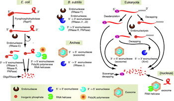
Among the bacteria, mRNA decay is best understood in E. coli, where transcripts are degraded through the sequential actions of well characterised ribonucleases (summarised schematically in Fig. 1, left panel) (Carpousis et al. Reference Carpousis, Luisi and Mcdowall2009; Evguenieva-Hackenberg & Klug, Reference Evguenieva-Hackenberg and Klug2009). A key step committing a transcript to rapid and complete destruction is endoribonucleolytic cleavage by ribonuclease E (RNase E), which has preference for single-stranded substrates, or by RNase III, which cleaves double-stranded RNA. Endonucleolytic cleavage triggers further attack by 3′–5′ exonucleases, such as polynucleotide phosphorylase (PNPase), RNase R or RNase II. Assisting these nucleases in the degradation of structured RNA species are accessory factors such as RNA helicases, which help to denature or remodel substrates. Another accessory enzyme is poly(A) polymerase (PAP), which adds 3′-terminal poly-A tails that become binding sites for processive exoribonucleases such as PNPase (Xu et al. Reference Xu, Lin-Chao and Cohen1993). Processive exonucleolytic degradation generates limit products of 2–4 nucleotides, and these are reduced to single nucleotides by oligoribonuclease, which is an essential enzyme. The general pathway of degradation depicted in Fig. 1 (left panel) is likely to be common even among bacteria that lack RNase E. For instance, in the Gram-positive firmicute Bacillus subtilis, which is a distant species from E. coli, the evolutionarily unrelated ribonuclease RNase Y serves as a functional analogue of RNase E (Fig. 1, top central panel) (Shahbabian et al. Reference Shahbabian, Jamalli, Zig and Putzer2009; Yao & Bechhofer, Reference Yao and Bechhofer2010).
A general schematic of RNA degradation pathway in E. coli and other organisms. RNA degradation requires endo- and exonucleolysis, aided by other enzymatic activities such as decapping, oligoadenylation and RNA unwinding or remodelling. In E. coli and related bacteria (left), nascent transcripts bear a 5′ triphosphate cap and are usually protected by a 3′-terminal stem-loop structure. The 5′-end can be converted to a monophosphate by an RNA pyrophosphohydrolyase (RppH). Degradation of transcripts is initiated by a 5′-monophosphate-dependent endonuclease from the RNase E/G family, and the degradation products that also bear a 5′-monophosphate, undergo subsequent rounds of endonucleolytic cleavage. RNase E also participates in another degradation pathway that bypasses 5′ end sensing (Bouvier and Carpousis, Reference Bouvier and Carpousis2011). RNAs containing deprotected 3′-ends are next digested by 3′-exonucleases, and the 2–5 nucleotide fragments generated are degraded further by oligoribonuclease. 3′-ends of RNA fragments containing secondary structure elements are oligoadenylated by PAP to provide a platform for recruitment of exonucleases. Degradation of stem-loop elements is also facilitated by RNA unwinding or remodelling by RNA helicases. The Gram-positive bacterium B. subtilus (upper middle panel) lacks RNase E, but the endonucleases RNase Y and RNase J can initiate degradation of transcripts. RNase J also provides a 5′ exonuclease activity. In the archaea (lower middle panel), an archael exosome, with 3′→5′ activity, and RNase J homologs, with 5′→3′ activity, have been detected. In eukaryotes (right), mature mRNA is protected by a 5′ 7-methylguanosine cap (m7G) and a long 3′-terminal poly(A) tail that are incorporated co-transcriptionally. RNA is destabilised by removal of these elements, followed by exonucleolytic degradation in the 3′→5′ pathway by the exosome complex or in the 5′→3′ pathway by Xrn1. RNA degradation can also be initiated by an internal cleavage by the endonucleolytic activity of an exosome component Rrp44, or by other endonucleases (such as those involved in the RNA interference pathway). Following 3′→5′ decay, the 5′ cap on the remaining oligomer is metabolised by the scavenger decapping enzyme DcpS. Degradation of RNAs containing secondary structure elements is aided by RNA helicases (e.g. Mtr4 in the nucleus), and also by oligoadenylation (by the TRAMP complex in the nucleus, not shown).

Stable RNA species must also undergo degradation as part of quality control assurance, and this pathway possibly involves the exoribonucleases PNPase and RNase R, since rRNA fragments of defined sizes accumulate in their absence (Cheng & Deutscher, Reference Cheng and Deutscher2003). The degradation of the enormous ribosome particle poses a special challenge, and evidence indicates that the process is initiated when the free 50S and 30S subunits are exposed to endonucleolytic cleavage (Zundel et al. Reference Zundel, Basturea and Deutscher2009), suggesting the possible involvement of RNase E or RNase III in rRNA recycling. The phosphorolytic exoribonuclease RNase PH, which is structurally related to PNPase, plays a key role in ribosome recycling in E. coli during starvation response (Basturea et al. Reference Basturea, Zundel and Deutscher2011). Many nucleases that participate in mRNA decay also play a dual role in the maturation of the more stable, structured RNA molecules (Deutscher, Reference Deutscher2006, Reference Deutscher2009). For example, RNase E processes the precursors of ribosomal RNAs (rRNA), transfer RNAs (tRNA) and other functional RNAs through an internal cleavage, while exoribonucleases can trim 3′-ends from stable RNAs to generate their mature forms (Li & Deutscher, Reference Li and Deutscher2002).
The ability of ribonucleases to gain access to transcripts will be influenced by processes of translation initiation and elongation. While some evidence indicates that transcription and translation are co-compartmentalised and therefore tightly coupled (Montero Llopis et al. Reference Montero Llopis, Jackson, Sliusarenko, Surovtsev, Heinritz, Emonet and Jacobs-Wagner2010), other data indicate that certain transcripts may diffuse far from the site of synthesis (Nevo-Dinur et al. Reference Nevo-Dinur, Nussbaum-Shochat, Ben-Yehuda and Amster-Choder2011). Thus, mRNA decay may follow pathways both on and off ribosomes. As E. coli lacks 5′ exonuclease activity, mRNAs are effectively protected from exonucleolytic degradation if they carry 3′-terminal structured regions, such as the protective stem loop generated by intrinsic (Rho-independent) termination of transcription and if the terminator does not have a 3′ tail longer than 5 nucleotides (so that it is not a favoured substrate for the exoribonuclease RNase R). Another salient feature of bacterial genes is that they are often organised into operons and are transcribed as polycistronic messages. Stem loop structures punctuating the polycistronic transcript play a role in the differential control of expression of the individual genes (Newbury et al. Reference Newbury, Smith, Robinson, Hiles and Higgins1987b). A stable stem loop occurring between the individual open reading frames stabilises the upstream message, resulting in greater expression of the promoter-proximal genes in an operon (e.g. in malEFG operon; Newbury et al. Reference Newbury, Smith and Higgins1987a). Ribonuclease expression may change during growth stage and under stress conditions. For instance, in Caulobacter crescentus, RNase E levels have been found to vary in a cell-cycle specific manner (Hardwick et al. Reference Hardwick, Chan, Broadhurst and Luisi2010). Such changes in patterns of ribonucleases may account for the temporal regulation of certain transcripts, but this hypothesis remains to be validated.
1.2 Key enzymes of RNA metabolism form the E. coli RNA degradosome
In E. coli and many other bacteria, several of the key enzymes of RNA metabolism, mentioned in the previous section, assemble to form the RNA degradosome (Fig. 2). The canonical components of the assembly are the hydrolytic endoribonuclease RNase E, the phosphorolytic exoribonuclease PNPase, the ATP-dependent RNA helicase B (RhlB) and the glycolytic enzyme enolase (Carpousis et al. Reference Carpousis, Van Houwe, Ehretsmann and Krisch1994; Miczak et al. Reference Miczak, Kaberdin, Wei and Lin-Chao1996; Py et al. Reference Py, Causton, Mudd and Higgins1994, Reference Py, Higgins, Krisch and Carpousis1996). Many other proteins are found in cell-extracted degradosome preparations in sub-stoichiometric ratio; these include the RNA chaperone Hfq, the exoribonuclease RNase R, PAP (Carabetta et al. Reference Carabetta, Silhavy and Cristea2010; Ikeda et al. Reference Ikeda, Yagi, Morita and Aiba2011), polyphosphate kinase (Blum et al. Reference Blum, Py, Carpousis and Higgins1997), protein chaperones (GroEL and DnaK) and ribosomal proteins (Kaberdin & Lin-Chao, Reference Kaberdin and Lin-Chao2009; Miczak et al. Reference Miczak, Kaberdin, Wei and Lin-Chao1996; Singh et al. Reference Singh, Chang, Lin, Averina, Kaberdin and Lin-Chao2009). Two inhibitor proteins (RraA and RraB) also interact with RNase E in E. coli and other bacteria (Gao et al. Reference Gao, Lee, Zhao, Qiu, Zhan, Saxena, Moore, Cohen and Georgiou2006; Górna et al. Reference Górna, Pietras, Tsai, Callaghan, Hernandez, Robinson and Luisi2010; Lee et al. Reference Lee, Zhan, Gao, Qiu, Feng, Meganathan, Cohen and Georgiou2003). In stationary phase or in response to cold, alternative helicases may be recruited into the degradosome (RhlE, SrmB and CsdA; Carabetta et al. Reference Carabetta, Silhavy and Cristea2010; Khemici et al. Reference Khemici, Toesca, Poljak, Vanzo and Carpousis2004; Prud'homme-Généreux et al. Reference Prud'Homme-généreux, Beran, Iost, Ramey, Mackie and Simons2004). RNase E-based degradosome-like assemblies occur throughout the gamma-proteobacteria, and we will elaborate upon this later (Ait-Bara & Carpousis, Reference Ait-Bara and Carpousis2010; Erce et al. Reference Erce, Low, March, Wilkins and Takayama2009; Marcaida et al. Reference Marcaida, Depristo, Chandran, Carpousis and Luisi2006; Purusharth et al. Reference Purusharth, Klein, Sulthana, Jäger, Jagannadham, Evguenieva-Hackenberg, Ray and Klug2005).
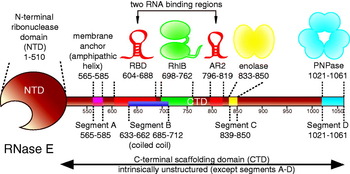
RNase E provides the scaffold for assembly of the E. coli RNA degradosome. A schematic representation of RNase E and the interaction sites with other principle degradosome components. RNase E contains a globular catalytic NTD, and a CTD. Annotated are the RNA-binding sites (RBD, AR2) and the conserved microdomains mediating interactions with cytoplasmic membrane or other degradosome components. The CTD of RNase E is predicted to be disordered. The indicated segments A through D are ‘microdomains’ mediating molecular recognition, and although these segments are likely to be instrinsically unstructured in the free, unbound state, they are likely to bind to their partners through induced fit with an accompanying disorder-to-order transition.

The scaffold for assembling the E. coli degradosome is provided by the non-catalytic C-terminal half of RNase E. In contrast to the N-terminal catalytic domain, the non-catalytic half is not essential for survival (Kido et al. Reference Kido, Yamanaka, Mitani, Niki, Ogura and Hiraga1996; Lopez et al. Reference Lopez, Marchand, Joyce and Dreyfus1999; Vanzo et al. Reference Vanzo, Li, Py, Blum, Higgins, Raynal, Krisch and Carpousis1998). Thus, the degradosome itself is not essential. Nonetheless, the degradosome must be functionally required somehow, because cells lacking the assembly are outcompeted by wild-type cells (Leroy et al. Reference Leroy, Vanzo, Sousa, Dreyfus and Carpousis2002). Perhaps one of the selective advantages conferred by the degradosome is its global impact on gene expression. Not only does the degradosome mediate preferential turnover of different transcripts (Bernstein et al. Reference Bernstein, Lin, Cohen and Lin-Chao2004) but it also affects broadly acting regulatory RNAs, such as the non-coding 6S RNA that sequesters the σ70-containing RNA polymerase holoenzyme (Wassarman, Reference Wassarman2007). The activities of the degradosome are thus embedded in a wide-ranging post-transcriptional regulatory repertoire (Marcaida et al. Reference Marcaida, Depristo, Chandran, Carpousis and Luisi2006).
1.3 Analogous processes, different machinery: RNA degradation in B. subtilis
Although E. coli has been a powerful model for understanding genetic regulation in bacteria and the other domains of life, its post-transcriptional machinery differs in detail, although not effect, among other members of the bacterial kingdom. The model Gram-positive bacterium B. subtilis belongs to an evolutionary lineage that is highly divergent from that of E. coli, and comparison of the RNA metabolism of the two species suggests that at least two distinct pathways have arisen during prokaryotic evolution for 5′-end-dependent mRNA degradation (Condon, Reference Condon2003). Efforts to identify an enzyme with RNase E-like activity in B. subtilis lead to the discovery of the RNase J enzymes (RNase J1 and its paralogue RNase J2) (Condon et al. Reference Condon, Putzer, Luo and Grunberg-Manago1997; de la Sierra-Gallay et al. Reference De La Sierra-Gallay, Zig, Jamalli and Putzer2008; Even et al. Reference Even, Pellegrini, Zig, Labas, Vinh, Brechemmier-Baey and Putzer2005). These RNase J enzymes are clearly unrelated to RNase E in evolution, as they belong to the β-CASP structural family of metallo-β-lactamases (Callebaut et al. Reference Callebaut, Moshous, Mornon and De Villartay2002). In addition to being an endoribonuclease, RNase J has exoribonuclease activity with 5′–3′ directionality, the first such enzyme with this activity identified in a prokaryote (Britton et al. Reference Britton, Wen, Schaefer, Pellegrini, Uicker, Mathy, Tobin, Daou, Szyk and Condon2007; de la Sierra-Gallay et al. Reference De La Sierra-Gallay, Zig, Jamalli and Putzer2008; Mathy et al. Reference Mathy, Benard, Pellegrini, Daou, Wen and Condon2007). In vitro and in vivo evidence suggest that primary transcripts with a 5′ triphosphate group are protected from degradation by RNase J, whereas transcripts with 5′ monophosphate or 5′ hydroxyl-ends are degraded readily (Collins et al. Reference Collins, Irnov, Baker and Winkler2007; Daou-Chabo et al. Reference Daou-Chabo, Mathy, Benard and Condon2009; de la Sierra-Gallay et al. Reference De La Sierra-Gallay, Zig, Jamalli and Putzer2008; Mathy et al. Reference Mathy, Benard, Pellegrini, Daou, Wen and Condon2007).
Assemblies analogous to the E. coli degradosome have been identified in B. subtilis and the pathogen Staphylococcus aureus, but these are based on the endoribonuclease RNase Y (Commichau et al. Reference Commichau, Rothe, Herzberg, Wagner, Hellwig, Lehnik-Habrink, Hammer, Völker and Stülke2009; Lehnik-Habrink et al. Reference Lehnik-Habrink, Pfortner, Rempeters, Pietack, Herzberg and Stulke2010, Reference Lehnik-Habrink, Newman, Rothe, Solovyova, Rodrigues, Herzberg, Commichau, Lewis and Stulke2011; Roux et al. Reference Roux, Demuth and Dunman2011), which also shares no common evolutionary ancestry with RNase E, the scaffold of the E. coli degradosome. The convergent evolution of degradosome-like assemblies in highly divergent bacterial lineages suggests that these machines provide selective benefit. This functional importance is also suggested by the occurrence of analogous assemblies in the archaea and eukarya, to which we now turn.
1.4 RNA turnover and processing in archaea
Because transcription and translation are closely coupled in the archaea (French et al. Reference French, Santangelo, Beyer and Reeve2007), it is anticipated that mRNA degradation in this domain of life might be more closely related to processes in bacteria than in eukaryotes (Evguenieva-Hackenberg & Klug, Reference Evguenieva-Hackenberg and Klug2009). However, the situation is not quite so simple (Fig. 1, lower middle panel). Archaeal proteins with sequence similarity to RNase E have been reported (Lee & Cohen, Reference Lee and Cohen2003), but the only archaeal homologue characterised to date has no detectable ribonuclease activity (Kanai et al. Reference Kanai, Oida, Matsuura and Doi2003). Nor has a homologue of the endoribonuclease RNase Y of B. subtilis, mentioned in the previous section, yet been identified an archaeon genome (Shahbabian et al. Reference Shahbabian, Jamalli, Zig and Putzer2009). On the other hand, most genomes of the euryarchaeal subgroup of the archaea encode a protein identified as a member of the β-CASP metallo-β-lactamase family (Callebaut et al. Reference Callebaut, Moshous, Mornon and De Villartay2002) that is related to B. subtilis RNase J (Even et al. Reference Even, Pellegrini, Zig, Labas, Vinh, Brechemmier-Baey and Putzer2005). These RNase J homologues have an ancient evolutionary origin predating the separation of bacteria and archaea (Clouet-d'Orval et al. Reference Clouet-D'orval, Rinaldi, Quentin and Carpousis2010). The crenarchaeota archaeal branch also has a family of β-CASP proteins that may be related to RNase J (Clouet-d'Orval et al. Reference Clouet-D'orval, Rinaldi, Quentin and Carpousis2010).
As far as known currently, archaeal mRNA is not capped and there are no known archaeal homologues of the eukaryotic enzymes involved in mRNA capping, decapping and 5′–3′ exoribonucleolytic degradation (Anantharaman et al. Reference Anantharaman, Koonin and Aravind2002; Brown & Reeve, Reference Brown and Reeve1985; Evguenieva-Hackenberg & Klug, Reference Evguenieva-Hackenberg and Klug2009). Biochemical evidence has demonstrated that the euryarchaeal RNase J homologues, like their bacterial counterpart, have 5′–3′ exoribonucleolytic activity that is 5′-end-dependent (Clouet-d'Orval et al. Reference Clouet-D'orval, Rinaldi, Quentin and Carpousis2010). In the crenarchaeon Sulfolobus solfataricus, transcripts are protected from a 5′-end-dependent degradation pathway by the translation initiation factor a/eIF2(-g), which binds to RNA 5′-triphosphorylated ends (Hasenohrl et al. Reference Hasenohrl, Lombo, Kaberdin, Londei and Blasi2008). The ribonucleases in this pathway remain to be identified. In another crenarchaeon, Methanococcus jannaschii, mRNA processing at a site 12–16 nucleotides upstream of the AUG translation start codon appears to be performed by an endoribonuclease (Zhang & Olsen, Reference Zhang and Olsen2009). The crenarchaeal β-CASP proteins can potentially account for the ribonuclease activities recently described in S. solfataricus and M. jannaschii, but the enzymatic activity of these proteins has not yet been determined.
As in other domains of life, tRNA processing in the archaea is performed by the ribonucleoprotein RNase P (Frank & Pace, Reference Frank and Pace1998) and the endoribonuclease RNase Z (Redko et al. Reference Redko, Li De Lasierra-Gallay and Condon2007; Schiffer et al. Reference Schiffer, Rosch and Marchfelder2002), indicating an ancient origin for these enzymes. An archaeal RNA-splicing endoribonuclease has been identified that removes introns from archaeal pre-tRNA and processes rRNA (Frank & Pace, Reference Frank and Pace1998; Tang et al. Reference Tang, Rozhdestvensky, D'Orval, Bortolin, Huber, Charpentier, Branlant, Bachellerie, Brosius and Huttenhofer2002). Most archaea have a eukaryotic-like exosome assembly, the core of which shares structural similarity with PNPase, the phosphorolytic exoribonuclease of the degradosome (Koonin et al. Reference Koonin, Wolf and Aravind2001; Lorentzen et al. Reference Lorentzen, Walter, Fribourg, Evguenieva-Hackenberg, Klug and Conti2005). Like PNPase, the archaeal exosome has a 3′–5′ exoribonucleolytic activity. The exception to the exosome rule is the halophilic Haloferax, where this activity is carried out by a homologue of the bacterial hydrolytic exoribonuclease, RNase R (Portnoy & Schuster, Reference Portnoy and Schuster2006).
Much remains to be learned about the maturation of rRNA and the degradation of mRNA in archaea, and it seems likely that more enzymes will be added to the catalogue of archaeal ribonucleases. For example, there is currently a conspicuous lack of an endoribonuclease with RNase E-like or RNase III-like activity in the archaea. Although the archaea have exosomes, whether they have multienzyme complexes analogous to the bacterial RNA degradosomes is an open question.
1.5 RNA turnover and processing in eukarya
In eukaryotes, mRNA degradation can be triggered by removal of the protecting 5′ N7-methylguanosine cap, an irreversible step that often follows deadenylation or uridylation of the 3′-end of the transcript (Rissland & Norbury, Reference Rissland and Norbury2009) (Fig. 1, right panel). Decapping is part of a pathway of constitutive transcript turnover. Decapped transcripts and products of endonucleolytic cleavage with exposed 5′-ends are degraded by 5′→3′ exonucleolysis executed by the exoribonuclease I (Xrn1). Decapping and 5′-end-dependent mRNA degradation by Xrn1 appear to belong to a pathway recently elaborated in evolution (Anantharaman et al. Reference Anantharaman, Koonin and Aravind2002).
mRNA degradation in eukaryotes, like that in the bacteria, can also be initiated by an internal cleavage, but in the eukaryotic cytoplasm the main pathway involves removal of a stabilising poly(A) tail (Garneau et al. Reference Garneau, Wilusz and Wilusz2007). Deadenylated RNA and 5′ fragments of internally cleaved transcripts are next digested by the 3′ exonucleolytic activity of the multi-enzyme exosome assembly. In a remarkable parallel between the bacterial and eukaryotic RNA degradation pathways, polyadenylation at the 3′-end destabilises transcripts in bacteria and in the nucleus of eukaryotes, where 3′-oligoadenylation by the multi-enzyme nuclear TRAMP complex stimulates exosome activity as part of a surveillance mechanism (Lykke-Andersen et al. Reference Lykke-Andersen, Brodersen and Jensen2009). In another functional parallel, the RNA degradative machinery of eukaryotes and prokaryotes can function in both turnover and processing (Houseley & Tollervey, Reference Houseley and Tollervey2009). The mRNA-degrading machinery involved in terminating the synthesis of transcripts with premature termination codons (non-sense mediated decay, NMD), also contributes to global gene expression, and participates in the decay of faulty ribosomal subunits (Cole et al. Reference Cole, Lariviere, Merrikh and Moore2009).
Many essential eukaryotic complexes involved in RNA degradation also contain various nuclease and RNA helicase activities. The active component of exosome, Rrp44, contains both exo- and endonucleolytic domains (Lebreton et al. Reference Lebreton, Tomecki, Dziembowski and Seraphin2008; Schaeffer et al. Reference Schaeffer, Tsanova, Barbas, Reis, Dastidar, Sanchez-Rotunno, Arraiano and Van Hoof2009), and the exosome associates with other components with RNA helicase activity (Mtr4 in TRAMP, or Ski proteins). In the mitochondrial degradosome (mtExo), the RNA helicase Suv3 associates with an RNase II-type enzyme Dss1 in yeast (Dziembowski et al. Reference Dziembowski, Piwowarski, Hoser, Minczuk, Dmochowska, Siep, Van Der Spek, Grivell and Stepien2003; Malecki et al. Reference Malecki, Jedrzejczak, Stepien and Golik2007), and with PNPase in humans (Szczesny et al. Reference Szczesny, Borowski, Brzezniak, Dmochowska, Gewartowski, Bartnik and Stepien2009; Wang et al. Reference Wang, Shu, Lieser, Chen and Lee2009). We will return again to explore in more detail the activities of the exosome and other eukaryotic complexes in the section to follow on the PNPase structure.
The eukaryotic exosome component Rrp44 has preference for 5′-monophosphorylated substrates, as do the Xrn1, Xrn2 and Ago ribonucleases (Bonneau et al. Reference Bonneau, Basquin, Ebert, Lorentzen and Conti2009; Schaeffer et al. Reference Schaeffer, Tsanova, Barbas, Reis, Dastidar, Sanchez-Rotunno, Arraiano and Van Hoof2009). A pyrophosphohydrolase activity has been observed recently for the Rai1 protein that associates with the yeast Xrn2 homologue, Rat1 (Xiang et al. Reference Xiang, Cooper-Morgan, Jiao, Kiledjian, Manley and Tong2009). As noted earlier, 5′ preferences are seen for evolutionarily unrelated ribonucleases, such as bacterial RNase E and archaeal RNase J (Clouet-d'Orval et al. Reference Clouet-D'orval, Rinaldi, Quentin and Carpousis2010; de la Sierra-Gallay et al. Reference De La Sierra-Gallay, Zig, Jamalli and Putzer2008; Even et al. Reference Even, Pellegrini, Zig, Labas, Vinh, Brechemmier-Baey and Putzer2005) and RNase Y (Shahbabian et al. Reference Shahbabian, Jamalli, Zig and Putzer2009). Thus, it is likely that exposure and recognition of the 5′-monophosphate group is an important regulatory step in many RNA degradation pathways in all three domains of life.
2. The modular architecture of the RNA degradosome
2.1 RNase E forms the principal scaffold of the RNA degradosome
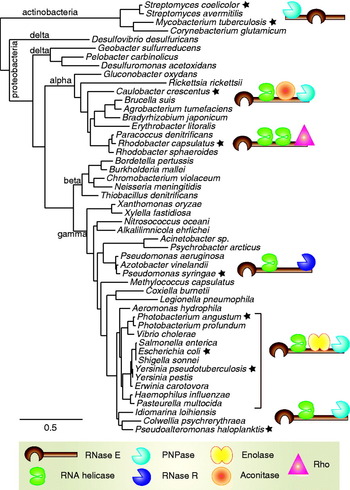
RNase E, the scaffold of the degradosome, belongs to the extensive RNase E/G enzyme family (InterPro family IPR004659; http://www.ebi.ac.uk/interpro). Members of this family occur throughout the proteobacteria (represented by E. coli), actinobacteria (includes Mycobacterium tuberculosis) and firmicutes (includes the orders Listeria and Staphylococcus). RNase E/G homologues are also present in some cyanobacteria and in plant chloroplasts, where it is required for development (Mudd et al. Reference Mudd, Sullivan, Gisby, Mironov, Kwon, Chung and Day2008; Schein et al. Reference Schein, Sheffy-Levin, Glaser and Schuster2008). RNase E and RNase G share in common the conserved ribonuclease domain, but RNase G lacks the degradosome-scaffolding region that characterises E. coli RNase E.
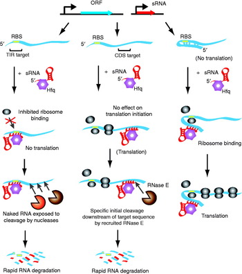
In E. coli, RNase E initiates the turnover of many, if not most, mRNAs by making an internal cleavage of the message (Carpousis, Reference Carpousis2007; Stead et al. Reference Stead, Marshburn, Mohanty, Mitra, Castillo, Ray, Van Bakel, Hughes and Kushner2010) (Fig. 1). RNase E is also involved in mRNA degradation mediated by small regulatory RNA (sRNA), either as a consequence of translational block (Morita et al. Reference Morita, Maki and Aiba2005), or possibly through direct recruitment (Pfeiffer et al. Reference Pfeiffer, Papenfort, Lucchini, Hinton and Vogel2009). RNase E also processes polycistronic transcripts and participates in the maturation of rRNAs, tRNAs, 6S RNA, transfer-mRNA (tmRNA), and the RNase P ribonucleic acid component M1 (Kim & Lee, Reference Kim and Lee2004; Lin-Chao et al. Reference Lin-Chao, Wei and Lin1999; Lundberg & Altman, Reference Lundberg and Altman1995; Ow & Kushner, Reference Ow and Kushner2002). The enzyme is essential for cell viability, possibly due to its multifaceted roles, including regulation of transcripts of cell-division proteins, e.g. FtsZ (Kato & Hashimoto, Reference Kato and Hashimoto2007; Takada et al. Reference Takada, Nagai and Wachi2005).
RNase E can be divided into two domains of nearly equal portions: the N-terminal domain (NTD), which encompasses entirely the endoribonuclease activity, and the C-terminal domain (CTD), which serves as a scaffold for degradosome assembly but has no reported catalytic activity (Leroy et al. Reference Leroy, Vanzo, Sousa, Dreyfus and Carpousis2002; McDowall & Cohen, Reference Mcdowall and Cohen1996) (Fig. 2). These two domains will be described in turn.
2.2 Structure and mechanism of the RNase E catalytic domain
2.2.1 Tertiary structure and quaternary transitions
The crystal structure of the catalytic domain of RNase E, corresponding to amino acids 1–529, is limited to a resolution of 2·9 Å, but nonetheless reveals details of the enzyme and provides testable hypotheses for its catalytic mechanism. Consistent with solution studies (Callaghan et al. Reference Callaghan, Grossmann, Redko, Ilag, Moncrieffe, Symmons, Robinson, Mcdowall and Luisi2003), RNase E is a tetramer in the crystal, and the four subunits are arranged as a dimer-of-dimers (Callaghan et al. Reference Callaghan, Marcaida, Stead, Mcdowall, Scott and Luisi2005a, Reference Callaghan, Redko, Murphy, Grossmann, Yates, Garman, Ilag, Robinson, Symmons, Mcdowall and Luisib; Koslover et al. Reference Koslover, Callaghan, Marcaida, Garman, Martick, Scott and Luisi2008). A tetrameric quaternary structure is likely to be a conserved feature of the extensive RNase E family, and experimental studies show that the plant and mycobacteria homologues are tetramers (Schein et al. Reference Schein, Sheffy-Levin, Glaser and Schuster2008; Zeller et al. Reference Zeller, Csanadi, Miczak, Rose, Bizebard and Kaberdin2007). RNase E oligomerisation is important for catalytic activity (Callaghan et al. Reference Callaghan, Redko, Murphy, Grossmann, Yates, Garman, Ilag, Robinson, Symmons, Mcdowall and Luisi2005b). Disruption of the dimer-to-dimer interface does not abolish activity completely, but in a cellular context it severely compromises organism fitness (Caruthers et al. Reference Caruthers, Feng, Mckay and Cohen2006).
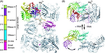
The RNase E protomer is shaped somewhat like a lopsided dumbbell. The fold can be decomposed into sub-domains that belong to widely occurring structural families, and these are indicated in the colour-coded schematic in Fig. 3. The larger sphere of the dumbbell is composed of sub-domains with S1, RNase H and DNase I folds. The smaller sphere has a distinctive fold that will be referred to here as the small domain.
Crystal structure of RNase E catalytic domain. (a) The catalytic domain, corresponding to the NTD shown in Fig. 2, forms a homotetramer. The structural subdomains are indicated by the bar and are coloured for the corresponding segments in the structure, here in complex with a 13-mer RNA (red). (b) Structural basis for 5′-end sensing. The structures of RNA-bound and RNA-free forms of RNase E catalytic domain are shown from the top view on the tetramer (PDB entries 2C0B and 2VMK). The S1 and 5′ sensor domains undergo a large conformational transition upon recognition of the 5′-monophosphate and RNA binding.

The subunits of the principal dimer associate mainly through the self-complementary interactions of DNase I sub-domains. The association is consolidated through the shared coordination of a zinc ion, where cysteine pairs from each of the two protomers form a thiol coordination shell around the metal with tetrahedral geometry (Callaghan et al. Reference Callaghan, Marcaida, Stead, Mcdowall, Scott and Luisi2005a, Reference Callaghan, Redko, Murphy, Grossmann, Yates, Garman, Ilag, Robinson, Symmons, Mcdowall and Luisib). Through this ‘zinc-link’, the principle dimers are tethered by organometallic bonds. The metal-binding CxCxGxG motif occurs throughout the RNase E/G family, and the metal coordination is likely to be conserved. It is interesting to note that the same motif, CxCxGxG, is also used for zinc binding in the DnaJ protein and the B. subtilis trp attenuation system regulator (Shevtsov et al. Reference Shevtsov, Chen, Gollnick and Antson2005), except that in those proteins the two motifs form an intra-molecular pair, whereas in RNase E they form an inter-molecular pair. The elongated protomers of the principal dimer intersect at the zinc-binding site, giving a distinctive scissors-like organisation. RNase G forms dimers in solution (Briant et al. Reference Briant, Hankins, Cook and Mackie2003) and is likely to involve interfaces similar to those seen in the principal dimer of RNase E, including the zinc-link.
The second dimer interface of RNase E, which marries the two principal dimers, is made through self-complementary intermeshing of the small domains. Like the homotypic interface between the DNase I subdomains in the principal dimer, the small domain interface has self-complementarity as a consequence of its two-fold rotational symmetry. Both symmetrical interfaces – large-to-large and small-to-small – are invariant under the large quaternary structural transformations between apo and RNA-bound forms (Koslover et al. Reference Koslover, Callaghan, Marcaida, Garman, Martick, Scott and Luisi2008). How then are such transformations accommodated? In this regard, it is important to note that the scissor-like organisation of the subunits means that there is also a locally asymmetric interface, and this is formed between the small domain of one protomer and the large domain of its partner subunit of the principal dimer. It is this asymmetric interface that can accommodate the quaternary structural change. Such change may permit the tetramer to mould its shape to accommodate larger, folded RNA substrates. In effect, the large-to-small subunit interface resembles a heterotypic protein–protein interaction, and the consequences of local asymmetry in quaternary structural change in RNase E may represent a general mechanism of cooperative behaviour in other homo-oligomeric assemblies.
2.2.2 Substrate recognition
RNase E is single-strand-specific endoribonuclease, with cutting preference for A/U-rich regions (Ehretsmann et al. Reference Ehretsmann, Carpousis and Krisch1992; Lin-Chao et al. Reference Lin-Chao, Wong, Mcdowall and Cohen1994; McDowall et al. Reference Mcdowall, Lin-Chao and Cohen1994) and 5′-monophosphorylated RNAs (Callaghan et al. Reference Callaghan, Marcaida, Stead, Mcdowall, Scott and Luisi2005a, Reference Callaghan, Redko, Murphy, Grossmann, Yates, Garman, Ilag, Robinson, Symmons, Mcdowall and Luisib; Mackie, Reference Mackie1998, Reference Mackie2000). The crystal structure reveals that the RNA (O2′-methyl modified to prevent cleavage) interacts with the surface of the DNase I domain and the S1 domain. The interactions of the RNA with the S1 domain are consistent with NMR studies of the isolated domain (Schubert et al. Reference Schubert, Edge, Lario, Cook, Strynadka, Mackie and Mcintosh2004). The contacts made by the RNA with the S1 and DNase I domains present the scissile phosphate for hydrolytic attack by a water molecule that is activated by a magnesium ion bound to the carboxylates of conserved aspartates from the DNase I domain (Callaghan et al. Reference Callaghan, Marcaida, Stead, Mcdowall, Scott and Luisi2005a, Reference Callaghan, Redko, Murphy, Grossmann, Yates, Garman, Ilag, Robinson, Symmons, Mcdowall and Luisib). Only one magnesium ion could be seen in the crystal structure of the RNA complex of the RNase E catalytic domain (inferred from substitution with the more electron dense manganese ion, and corroborated by anomalous scatter due to X-ray absorption). However, it is possible that a second catalytic magnesium ion also participates in the reaction, based on the mechanism of other Mg2+ catalysed reactions of nucleic acids (Yang et al. Reference Yang, Lee and Nowotny2006). The second metal might be recruited in the Michaelis–Menton complex of RNase E with RNA.
Recognition of the substrate's 5′ monophosphate group boosts the catalytic power of RNase E (Garrey and Mackie, Reference Garrey and Mackie2011; Garrey et al. Reference Garrey, Blech, Riffell, Hankins, Stickney, Diver, Hsu, Kunanithy and Mackie2009; Jiang & Belasco, Reference Jiang and Belasco2004; Jourdan & McDowall, Reference Jourdan and Mcdowall2008). It is still debated whether the increased activity originates through a change in affinity for the substrate or an increase in the catalytic rate constant, k cat. Perhaps this issue will be resolved through better understanding of the effects of RNA fold on enzyme activity, since this seems to be important for certain cases (Kime et al. Reference Kime, Jourdan, Stead, Hidalgo-Sastre and Mcdowall2010). Recognition of the substrate 5′-terminal group requires that it is accessible, since cleavage is hindered by the presence of a stem-loop structure 2–4 nucleotides from the 5′-end (McDowall et al. Reference Mcdowall, Kaberdin, Wu, Cohen and Lin-Chao1995). 5′-monophosphorylated substrates can be generated from nascent triphosphorylated transcripts by the activity of the pyrophosphohydrolase, RppH (Fig. 1, left panel) (Celesnik et al. Reference Celesnik, Deana and Belasco2007; Deana et al. Reference Deana, Celesnik and Belasco2008). RNase E cleavage itself produces a 5′-monophosphorylated product, and therefore the initial cleavage promotes a cascade of downstream cleavages. As noted earlier, exposure and recognition of the 5′-monophosphate group is an important regulatory step in many RNA degradation pathways (Fig. 1).
The basis for the 5′-end sensing by RNase E can be rationalised by the crystal structures of the catalytic domain of RNase E in the RNA-bound form (Callaghan et al. Reference Callaghan, Marcaida, Stead, Mcdowall, Scott and Luisi2005a, Reference Callaghan, Redko, Murphy, Grossmann, Yates, Garman, Ilag, Robinson, Symmons, Mcdowall and Luisib; Koslover et al. Reference Koslover, Callaghan, Marcaida, Garman, Martick, Scott and Luisi2008). This reveals the 5′ phosphate engaged in the pocket of a domain associated with the RNA-binding S1 motif, referred to as the 5′ sensor domain (Fig. 3). The interactions of the 5′ sensor with the RNA help to orient the S1 domain such that it encloses the catalytic site and presents the scissile phosphate for hydrolytic attack. Comparing this structure with the RNA-free form reveals a large conformational switch of the S1 domain with the 5′ sensor domain, which move together as a single-structural unit (Koslover et al. Reference Koslover, Callaghan, Marcaida, Garman, Martick, Scott and Luisi2008) (Fig. 3B). The apo-form structure also reveals that the RNase E catalytic domain can undergo a marked change in the conformation of the whole tetrameric structure through changes in the interfacial contacts between the small and large domains. As noted in the previous section, the changes at this asymmetric interface may permit the tetramer to adjust its quaternary structure to accommodate large substrates.
For some substrates, RNase E can bypass the 5′-end and cleave internally regardless of the terminal group (Kime et al. Reference Kime, Jourdan, Stead, Hidalgo-Sastre and Mcdowall2010; Schuck et al. Reference Schuck, Diwa and Belasco2009), and this operational mode may be the more important activity in vivo (Garrey et al. Reference Garrey, Blech, Riffell, Hankins, Stickney, Diver, Hsu, Kunanithy and Mackie2009; Bouvier & Carpousis, Reference Bouvier and Carpousis2011). One example is the rne transcript encoding RNase E itself, which contains a hairpin structure that is proposed to be the recognition site for the enzyme as part of an autoregulation mechanism (Schuck et al. Reference Schuck, Diwa and Belasco2009). By inference from the available crystal structures, the mechanism of 5′-end bypass might involve recognition of the tertiary fold of the RNA, which mediates the canyon shape of the active site through interactions with the S1 domain.
Two important residues in the 5′-phosphate sensor are R169 and T170, which form hydrogen bonds to the terminal phosphate (Callaghan et al. Reference Callaghan, Marcaida, Stead, Mcdowall, Scott and Luisi2005a, Reference Callaghan, Redko, Murphy, Grossmann, Yates, Garman, Ilag, Robinson, Symmons, Mcdowall and Luisib). The mutations R169K and T170V are predicted to disrupt these hydrogen bonds, and they result in loss of 5′-activation effects for small substrates. Strains expressing RNase E with mutation of the 5′-phosphate sensor (R169Q or T170V) are viable, suggesting that 5′-end sensing is not an essential activity; however, the mutations are lethal when combined with deletions of the CTD (Garrey & Mackie, Reference Garrey and Mackie2011). This finding has lead to the suggestion that RNase E has a ranking of efficiencies for natural substrates that depends on 5′-end access. It also suggests that there is a functionally important interplay between the N-terminal catalytic domain and the C-terminal degradosome-scaffolding domain, to which we now turn.
2.3 A ‘natively unstructured’ scaffold for the degradosome
Inspecting the amino acid sequence of E. coli RNase E, it becomes apparent that the C-terminal half that serves as the degradosome-scaffold has ‘low complexity’, meaning that there is over-representation of only a few residue types. Other salient features of this domain include the under-representation of bulky hydrophobic amino acids, the enrichment of polar and charged residue clusters, the proline-rich stretches and the scarcity of predicted secondary structure. These features are hallmarks of natively unstructured proteins that lack the globular character of a stable fold (Gunasekaran et al. Reference Gunasekaran, Tsai, Kumar, Zanuy and Nussinov2003; Wright & Dyson, Reference Wright and Dyson2009). The absence of secondary structure and lack of globular compaction predicted for the isolated RNase E CTD has been corroborated by circular dichroism spectroscopy and small angle X-ray solution scattering (Callaghan et al. Reference Callaghan, Aurikko, Ilag, Grossmann, Chandran, Kühnel, Poljak, Carpousis, Robinson, Symmons and Luisi2004). Three proline-rich clusters (residues 526–568, 743–796, 819–857; Leroy et al. Reference Leroy, Vanzo, Sousa, Dreyfus and Carpousis2002) may contribute to a more rigid, extended conformation that is inferred from the solution data.
Alignment of CTD sequences from the RNase E family reveals little apparent conservation, with the important exception of a few key sites. These conserved segments, corresponding in size roughly to between 20 and 70 residues, have been experimentally demonstrated to be binding sites for cognate proteins, RNA substrates and the cytoplasmic membrane (Callaghan et al. Reference Callaghan, Aurikko, Ilag, Grossmann, Chandran, Kühnel, Poljak, Carpousis, Robinson, Symmons and Luisi2004; Chandran et al. Reference Chandran, Poljak, Vanzo, Leroy, Miguel, Fernandez-Recio, Parkinson, Burns, Carpousis and Luisi2007; Khemici et al. Reference Khemici, Poljak, Luisi and Carpousis2008; Vanzo et al. Reference Vanzo, Li, Py, Blum, Higgins, Raynal, Krisch and Carpousis1998). Compared with the flanking sequences, some of the conserved segments have markedly greater predicted structural propensity, and owing to their small size, may be described as ‘microdomains’ (Callaghan et al. Reference Callaghan, Aurikko, Ilag, Grossmann, Chandran, Kühnel, Poljak, Carpousis, Robinson, Symmons and Luisi2004; Marcaida et al. Reference Marcaida, Depristo, Chandran, Carpousis and Luisi2006). Part of the RNA-binding segment 632–712 is predicted to form a coiled-coil, but the isolated peptide has no helical structure in isolation, and may require association with RNA (Callaghan et al. Reference Callaghan, Aurikko, Ilag, Grossmann, Chandran, Kühnel, Poljak, Carpousis, Robinson, Symmons and Luisi2004). In general, microdomain recognition is likely to include a degree of folding; for example, chain compaction that clusters the non-polar side chains to form a densely packed interior consolidated by hydrogen bonding interactions (Gunasekaran et al. Reference Gunasekaran, Tsai, Kumar, Zanuy and Nussinov2003; Wright & Dyson, Reference Wright and Dyson2009). Binding of microdomains involves changes of buried surface area that are smaller than those associated with complexes composed of globular proteins, and the binding energies are correspondingly smaller (Nurmohamed et al. Reference Nurmohamed, Vaidialingam, Callaghan and Luisi2009); nonetheless, such weak binding is likely to be highly specific, since the accompanying folding demands precise register of side chains at the interface and within the newly -created compacted core. Crystal structures are available for complexes of enolase and PNPase with their respective RNase E microdomains, and a structural model has been proposed for the association of a helical microdomain with the cytoplasmic membrane; these will be described in further detail in the subsection that follows.
2.4 Interaction with the cytoplasmic membrane is mediated through an RNase E microdomain
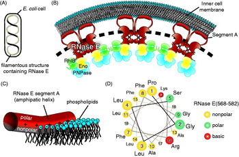
Nearly two decades ago, cell fractionation studies suggested that RNase E is membrane-associated (Miczak et al. Reference Miczak, Srivastava and Apirion1991). This finding has been refined by microscopy studies that locate RNase E and the degradosome complex to the cytoplasmic membrane (Liou et al. Reference Liou, Jane, Cohen, Lin and Lin-Chao2001) where it forms filaments that spiral along the interior surface (Fig. 4) (Taghbalout & Rothfield, Reference Taghbalout and Rothfield2007, Reference Taghbalout and Rothfield2008). The observation that the RNA degradosome is part of a filament has been controversial in part due to the claim that it is associated with a bacterial cytoskeleton (Taghbalout & Rothfield, Reference Taghbalout and Rothfield2007, Reference Taghbalout and Rothfield2008). Nevertheless, a filament was observed by two different techniques: immunofluorescence and direct fluorescence using RNase E or other components of the RNA degradosome fused to yellow fluorescent protein (YFP). It therefore seems unlikely that these images are an artefact of the technique used to label the proteins. Furthermore, the fusion proteins were expressed at normal levels and chemical fixation of the cells before microscopy did not affect the results.
E. coli RNase E can associate with the cytoplasmic membrane. (a) RNase E and the other components of the RNA degradosome are located in a filament-like assembly in E. coli. The assembly follows a helical path that spans the length of the bacterium. (b) A proposed model of the association of the RNA degradosome with the inner leaflet of the cytoplasmic membrane. The dashed lines indicate interactions between adjacent degradosomes in the filament-like assembly. Note that RNase E is a tetramer, but only two segment A regions are shown for clarity. The molecular dimensions and stoichiometry of the assembly were arbitrarily chosen to simplify the figure. (c) A schematic showing how the amphipathic helical segment A of RNase E interacts with the surface of the inner-leaflet of the membrane. The hydrophobic residues are submerged in the acyl interior of the membrane. Favourable electrostatic interactions form between the side chains of the basic amino acids and the negatively charged phosphate head groups. (d) Helical wheel representation for the RNase E segment A. The circle radii correspond to the proximity to the N-terminus, and are colour coded according to physical properties of the side chains.

Molecular genetics and biophysical studies pinpoint the membrane localisation region to a small motif in RNase E, consisting of a 21-residue amphipathic helix and corresponding to one of the predicted microdomains (residues 565–585) (Khemici et al. Reference Khemici, Poljak, Luisi and Carpousis2008). The membrane-anchoring helix has a striking pattern of large non-polar residues on the hydrophobic side, of small polar residues and glycines on the hydrophilic side, and basic amino acids such as arginine or lysine at the boundary where the hydrophobic and hydrophilic surfaces meet. The isolated peptide forms a stable α-helix that binds E. coli lipid vesicles. This in vitro interaction can be disrupted if the conserved hydrophobic residues are replaced with hydrophilic amino acids. The same substitutions when made in the full-length RNase E cause it to dissociate from the cytoplasmic membrane in vivo. Membrane association of RNase E is required for normal growth, suggesting that it impacts on RNase E function (Khemici et al. Reference Khemici, Poljak, Luisi and Carpousis2008), perhaps due to compartmentalisation of degradosome activity, or by helping to support structural organisation of the assembly. The Bacillus subtilus ribonuclease RNase Y, which is analogous but not homologous to RNase E as mentioned earlier, is also localised to the cell periphery (Hunt et al. Reference Hunt, Rawlins, Thomaides and Errington2006) and is predicted to have a N-terminal transmembrane domain (Shahbabian et al. Reference Shahbabian, Jamalli, Zig and Putzer2009).
A potential model for the interaction of the amphipathic helix with a membrane is presented in Figs 4c and 4d. The basic residues at the hydrophobic/hydrophilic boundary form electrostatic interactions with the lipid phosphates, and the non-polar portion of their side chains may be immersed in the membrane acyl layer. Another key sequence signature of the membrane-anchor motif is a proline at the N-terminus, which may play a role in capping of the helix, as found for soluble globular proteins. While glycines are normally destabilising in helices of globular proteins, they appear to be stably accommodated in the amphipathic helix of RNase E, where they create a shallow depression on the hydrophilic surface. A similar sequence pattern is observed in other membrane-associated proteins in E. coli, such as the bacterial cytoskeletal protein MinD, FtsA and the receptor for the signal recognition particle, FtsY (Khemici et al. Reference Khemici, Poljak, Luisi and Carpousis2008). It is likely that these helices interact with the membrane in a similar way to the model proposed for the RNase E amphipathic helix.
Given that RNase E is a tetramer and that the amphipathic α-helix is the membrane-localisation segment, we can return to the filament to ask how it might be organised. Given the dimensions of an E. coli cell, the length of the filament-like structure measured from the published fluorescence images is on average 7500 nm and the width is the limit of resolution of light microscopy (200 nm) (A. J. Carpousis, unpublished results). Given that there are approximately 250 tetramers of RNase E in the cell (Kido et al. Reference Kido, Yamanaka, Mitani, Niki, Ogura and Hiraga1996) and assuming that the tetramer forms the repeat unit of the filament-like structure, the repeat length is 30 nm. The apparent 200 nm width of the filament may therefore be attributed to the resolution limit of conventional light microscopy. We know of no evidence that RNase E actually polymerizes to form a filament, and the possibility that individual RNase E tetramers are localized to a spiral track on the inner cytoplasmic membrane is equally plausible based on the light microscopy work. Recent work involving total internal reflection fluorescence microscopy has shown that MreB in B. subtilis forms discrete patches that move processively along peripheral tracks perpendicular to the cell axis (Dominguez-Escobar et al. Reference Dominguez-Escobar, Chastanet, Crevenna, Fromion, Wedlich-Soldner and Carballido-Lopez2011). It should be interesting to perform similar analyses with fluorescently labeled RNase E in E. coli.
2.5 RNA unwinding and remodelling of protein–RNA complexes by the RhlB DEAD-box helicase
The canonical degradosome component RhlB is a member of the extensive family of DEAD-box RNA helicases, whose members are found in all domains of life (Jankowsky, Reference Jankowsky2011; Pyle, Reference Pyle2008). DEAD-box helicases have ATP-dependent RNA unwinding or translocation activities. They are non-processive, meaning that they act locally to unwind only a few base pairs of duplex RNA, or to remodel RNA–RNA and RNA–protein interactions. The DEAD-box helicases characteristically contain two domains that resemble the RecA recombination protein, a DNA-dependent ATPase. Within the RecA-like domains are numerous conserved sequence signatures, of which motif II (or Walker B motif, required for ATP binding and hydrolysis) has the hallmark D-E-A-D residues for which the family is named (Cordin et al. Reference Cordin, Banroques, Tanner and Linder2006). Crystal structures of DEAD-box helicases reveal that the two RecA-like domains together form a catalytic site for ATP at the buried inter-subunit interface, and an RNA-binding site in which the nucleic acid straddles an exposed surface of both subunits. Interactions with the nucleic acid are mediated through exposed loops within a structural module common to both the RecA-like domains (Milner-White et al. Reference Milner-White, Pietras and Luisi2010). The processes of ATP-binding and hydrolysis trigger relative movements of the two RecA-like domains that change the interactions with the RNA, and in this way power the displacement or unwinding of the bound nucleic acid.
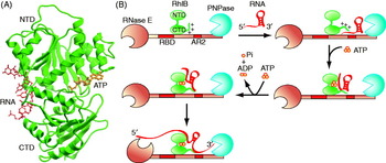
A crystallographic structure for the degradosome RhlB is not available, but a comparative model (Fig. 5a) suggests that the helicase will be very close structurally to the Drosophila Vasa protein (Chandran et al. Reference Chandran, Poljak, Vanzo, Leroy, Miguel, Fernandez-Recio, Parkinson, Burns, Carpousis and Luisi2007). In addition to the two conventional RecA-like domains, RhlB has a C-terminal tail enriched in basic residues. Although this tail is predicted to be natively unstructured, it boosts affinity for RNA and is likely to make extensive electrostatic interactions with the phosphate backbone of the nucleic acid (Chandran et al. Reference Chandran, Poljak, Vanzo, Leroy, Miguel, Fernandez-Recio, Parkinson, Burns, Carpousis and Luisi2007).
Structure and function of the DEAD-box RNA helicase from the E. coli degradosome, RhlB. (a) Comparative model of E. coli RhlB based on the crystal structure of the Drosophila DEAD box helicase Vasa, in complex with RNA and ATP. RhlB has two RecA-like domains and a C-terminal flexible tail that contributes to RNA binding. (b) A model for cooperation of RhlB with the other degradosome components. In the cartoon, PNPase is also engaged with RNase E (enolase is not included for clarity). RNA may bind to RNase E at RBD or AR2 (as indicated here by interaction with the stem–loop RNA structure), or with both RNA-binding sites (not shown). Cycles of ATP binding and hydrolysis may expose the surfaces of the RecA-like domains of RhlB to interact with the RNA during unwinding and (5′→3′) translocation. The exposed terminal regions of the RNA are subject to phosphorolytic cleavage by the adjacent PNPase or hydrolytic attack by the catalytic domain of RNase E.

A distinctive feature of RhlB is that its ATPase activity requires association with RNase E. The site of interaction has been mapped within a 69-residue segment to which the C-terminal RecA-like domain of RhlB binds with a 1:1 stoichiometry with a dissociation constant in the nanomolar range (Worrall et al. Reference Worrall, Howe, Mckay, Robinson and Luisi2008b). The helicase-binding site is not one of the four segments of the CTD predicted to have structural propensity, but it is adjacent to the predicted microdomain that is involved in RNA binding and with which it functions cooperatively (described further in the next section).
In E. coli, RhlB is likely to be exclusively associated with RNase E under normal physiological conditions, where it assists degradosome-mediated processes (Khemici et al. Reference Khemici, Toesca, Poljak, Vanzo and Carpousis2004, Reference Khemici, Poljak, Toesca and Carpousis2005). The helicase may coordinate activity of the degradosome machinery by preparing single-stranded substrate for either or both PNPase and RNase E, and by remodelling protein–RNA interactions (Fig. 5b). Microarray studies have determined that RhlB is necessary for normal mRNA decay and especially affects transcripts of proteins involved in iron uptake, possibly in connection with the action of the sRNA RyhB (Bernstein et al. Reference Bernstein, Lin, Cohen and Lin-Chao2004).
Apart from RhlB, there are four other DEAD-box helicases in E. coli – namely SrmB, RhlE, CsdA (or DeaD) and DbpA (Iost & Dreyfus, Reference Iost and Dreyfus2006) – all of which participate to some extent in ribosome assembly (Charollais et al. Reference Charollais, Dreyfus and Iost2004; Jain, Reference Jain2008; Peil et al. Reference Peil, Virumae and Remme2008; Sharpe Elles et al. Reference Sharpe Elles, Sykes, Williamson and Uhlenbeck2009; Trubetskoy et al. Reference Trubetskoy, Proux, Allemand, Dreyfus and Iost2009). With cold shock stress, CsdA is recruited to the degradosome and may be functionally important for acclimation in cold shock (Iost & Dreyfus, Reference Iost and Dreyfus2006; Prud'homme-Généreux et al. Reference Prud'Homme-généreux, Beran, Iost, Ramey, Mackie and Simons2004). SrmB, RhlE and CsdA have been shown to bind in vitro to RNase E (Khemici et al. Reference Khemici, Toesca, Poljak, Vanzo and Carpousis2004), and SrmB has recently been shown to be associated in vivo in a form of the degradosome that contains PAP (Carabetta et al. Reference Carabetta, Silhavy and Cristea2010). Although the current view is that RhlB is a dedicated component of the canonical RNA degradosome, the other DEAD-box helicases associate under conditions of environmental stress that interfere with the biogenesis of the ribosome.
2.6 RNA-binding domains within the degradosome, and their cooperation with the RNA helicase
The C-terminal half of RNase E contains two arginine-rich (AR) RNA-binding regions, namely the primary RNA-binding domain (RBD, residues 608–688) and a second arginine-rich region (AR2, residues 796–819) (shown schematically in Fig. 2). The RBD coincides with one of the CTD regions with predicted structural propensity region, and although it has little structure in isolation, the microdomain may form a coiled-coil when engaged to RNA (Callaghan et al. Reference Callaghan, Aurikko, Ilag, Grossmann, Chandran, Kühnel, Poljak, Carpousis, Robinson, Symmons and Luisi2004). The RBD and AR2 regions are required for efficient cleavage by RNase E in processing in vitro the precursor of 5S rRNA (Kaberdin et al. Reference Kaberdin, Walsh, Jakobsen, Mcdowall and Von Gabain2000), and their deletion stabilises some transcripts in vivo (Leroy et al. Reference Leroy, Vanzo, Sousa, Dreyfus and Carpousis2002; Lopez et al. Reference Lopez, Marchand, Joyce and Dreyfus1999; Ow et al. Reference Ow, Liu and Kushner2000).
Situated between the RBD and AR2 RNA-binding sites is the RhlB-binding site. The spatial co-localisation of RBD, AR2 and the RNA-binding basic tail of RhlB brings together flexible elements that can cooperate to bind and manipulate RNA. The recent characterisation of the RNA degradosome of Pseudoalteromonas haloplanktis, a bacterium distantly related to E. coli, demonstrates a similar organisation with the RhlB-binding site flanked by the RBD and AR2, and suggests that this ‘module’ within the RNA degradosome is widely conserved among the gamma-proteobacteria (Aït-Bara & Carpousis, Reference Ait-Bara and Carpousis2010). Recruitment of PNPase to the degradosome brings an additional six RNA-binding domains to the degradosome (three S1 and three K-homology (KH) domains in the trimeric PNPase; Fig. 6), with expanded opportunity for complex substrate recognition. Like other multi-dentate interactions (Hunter & Anderson, Reference Hunter and Anderson2009; Whitty, Reference Whitty2008), the clustering of RNA-binding sites is expected to yield cooperative ligand-binding behaviour through chelate cooperativity. Indeed, the interplay of RhlB with the two adjacent RNA-binding regions is likely to be the key to the degradation of structured RNAs and the processing of precursor species (Chandran et al. Reference Chandran, Poljak, Vanzo, Leroy, Miguel, Fernandez-Recio, Parkinson, Burns, Carpousis and Luisi2007). As a speculation, RNA surveillance could be one of the potential functions of the cooperative interplay between RhlB and the clustered RNA-binding sites. In the envisaged proofreading mode, defective RNA or ribonucleoprotein complexes with an unstable structure would be unwound or remodelled by the helicase and subsequently degraded by the ribonucleases, while stably folded RNA would withstand RhlB activity and be released. Partial unwinding or remodelling mediated by RhlB may also be coupled with the processing of structured precursors (Chandran et al. Reference Chandran, Poljak, Vanzo, Leroy, Miguel, Fernandez-Recio, Parkinson, Burns, Carpousis and Luisi2007).
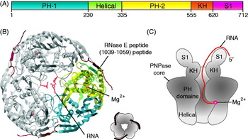
PNPase structure and mechanism. (a) Schematic of structural domain organisation of E. coli PNPase. (b) The crystal structure of PNPase PH1/PH2 core, in complex with an RNase E-derived peptide and ribonucleotides (PDB entry 3GCM). The view is along the molecular three-fold axis. (c) Schematic of the path of the RNA substrate. The KH and S1 domains are flexibly tethered on the entrance surface of the PNPase core ring, and they help thread the substrate through a narrow aperture into the central channel, where the 3′-end is fed into the active site.

2.7 An exoribonuclease within the RNA degradosome: structure and function of PNPase, and its relationship to the archaeal and eukaryotic exosomes
PNPase is a conserved exoribonuclease found not only in bacteria but also in mitochondria and chloroplasts, where it plays key roles in RNA turnover and quality control. Human PNPase mediates the translocation of RNAs into the organelle via the mitochondrial intermembrane space (Wang et al. Reference Wang, Chen, Oktay, Zhang, Allen, Smith, Fan, Hong, French, Mccaffery, Lightowlers, Morse, Koehler and Teitell2010). In bacteria, the recruitment of PNPase into the degradosome provides the multi-functional machinery with a second and mechanistically distinct ribonuclease activity with different substrate preferences. PNPase is processive, meaning that it continues to cut along the same substrate once it has been engaged. The enzyme uses metal-assisted catalysis and cleaves through a phosphorolytic mechanism involving activation of inorganic phosphate to attack the 3′ phophodiester bond of single-stranded RNA. The reaction releases a single nucleoside diphosphate with each successive cleavage. Operating in reverse, PNPase is a 3′-terminal oligonucleotide polymerase and can add 3′ heteropolymeric tails to RNA templates. The enzyme accounts for residual polyadenylation in the absence of PAP (Mohanty & Kushner, Reference Mohanty and Kushner2000). Since phosphorolysis is an energy conserving, reversible reaction, the balance between degradation versus synthesis by PNPase depends on the concentrations of the respective substrates, namely free phosphate and nucleoside diphosphate.
As phosphorolysis has little free energy change, there is little potential for PNPase to couple catalysis with mechanical work, and the enzyme often stalls on structured substrates. Consequently, it would be expected to require assistance from an RNA helicase to degrade structured RNAs. About 10–20% of the total cellular PNPase is bound in the RNA degradosome complex (Liou et al. Reference Liou, Jane, Cohen, Lin and Lin-Chao2001), and this association enables the exoribonuclease to co-operate with degradosome-bound RNA helicases, and is necessary for degradation of certain substrates that are stabilised by extensive stem-loop structures (Coburn et al. Reference Coburn, Miao, Briant and Mackie1999; Khemici & Carpousis, Reference Khemici and Carpousis2004). The coordinated action of both RhlB and PNPase bound to RNase E CTD is required for degradation of resilient RNA structures, such as repetitive extragenic palindrome (REP) elements (Coburn et al. Reference Coburn, Miao, Briant and Mackie1999). In the absence of RNase E CTD, RhlB is reported to still bind to PNPase and aid in the disruption of stem-loop structures that are resistant to exoribonuclease activity (Liou et al. Reference Liou, Chang, Lin and Lin-Chao2002).
E. coli encodes two other 3′→5′ exoribonucleases, RNase R and RNase II, and like PNPase they are metal-assisted and processive, but differ in that they use a hydrolytic mechanism to cleave RNA substrates (Andrade et al. Reference Andrade, Pobre, Silva, Domingues and Arraiano2009). A deletion strain of either PNPase, RNase R or RNase II is viable, and so is the double mutant lacking RNase R and RNase II; however, a double mutant lacking PNPase and either of the other two exonucleases is unviable (Cheng & Deutscher, Reference Cheng and Deutscher2003). This indicates that the loss of PNPase cannot be complemented by any other exonuclease alone, while PNPase is sufficient to perform functions of both RNase R and RNase II. Although PNPase acts mostly in mRNA turnover, it participates also in RNA processing, for instance by trimming 3′ ends from tRNAs (Li & Deutscher, Reference Li and Deutscher2002). PNPase functions also in the decay and quality control of stable RNAs such as rRNA (Cheng & Deutscher, Reference Cheng and Deutscher2003).
PNPase can be divided into modular structural and functional domains, which are illustrated schematically in Fig. 6. The main core of the enzyme is formed from two domains that are both structurally congruent to the phosphorolytic exoribonuclease RNase PH that participates in tRNA processing in prokaryotes. Both domains, like RNase PH, have a left-handed cross-over in a β-α-β element, and the rarity of this local fold in other protein structures makes it clear that PNPase must have originated through the duplication and fusion of an ancestral RNase PH-like module (Symmons et al. Reference Symmons, Jones and Luisi2000, Reference Symmons, Williams, Luisi, Jones and Carpousis2002). A helical domain is situated between the two RNase PH-like domains, and two RBDs, the S1 and KH are at the C-terminus. PNPase has a homotrimeric structure and forms a ring with a central channel. RNase PH forms a very similar ring-like structure, except that it is a hexamer and therefore has six equivalent active sites. In contrast, the pseudo-hexameric PNPase has only three active sites, since only one of the duplicated domains has retained catalytic activity (Symmons et al. Reference Symmons, Jones and Luisi2000). Access to these sites is through the central channel, and while the channel is open at both ends, substrate enters through the slightly constricted end where the bases stack on phenylalanines in highly conserved loops (Nurmohamed et al. Reference Nurmohamed, Vaidialingam, Callaghan and Luisi2009).
The KH and S1 domains are flexibly tethered to one side of the RNase PH-like ring, proximal to a constricted aperture, and an all-helical domain is flexibly tethered in the distal surface (Nurmohamed et al. Reference Nurmohamed, Vaidialingam, Callaghan and Luisi2009; Shi et al. Reference Shi, Yang, Lin-Chao, Chak and Yuan2008; Symmons et al. Reference Symmons, Jones and Luisi2000). In the case of the human PNPase, it seems likely that these same domains stably engage RNA during the transport process in the mitochondria. Exactly how substrates destined for processing are bound by KH domains and thread through the proximal aperture has now been explained by the crystal structures of Caulobacter crescentus PNPase in complex with single stranded RNA (Hardwick & Luisi, in preparation). Here, the three KH domains make non-equivalent interactions with the RNA and each has a different orientation with respect to the catalytic core. A rotary-like movement of the KH domains as a body is associated with transitions between these non-equivalent binding states, and this accompanies quaternary structural changes in the catalytic core. These findings indicate that PNPase responds with allosteric change to ligand binding, with the implication that cycles of threading of substrate may be linked with substrate binding at the successive active sites. Mutagenesis data suggest that the vestigial pocket of the catalytically inactive RNase PH-like repeat may bind to allosteric ligands that may modulate PNPase activity (Nurmohamed et al. Reference Nurmohamed, Vincent, Titman, Chandran, Pears, Du, Griffin, Callaghan and Luisi2011). Further evidence that PNPase may be regulated by allosteric effectors comes from observations that the enzyme responds to the signalling molecule cyclic di-GMP (Tuckerman et al. Reference Tuckerman, Gonzalez and Gilles-Gonzalez2011).
Crystallographic data provide clues of how PNPase and RNase E interact within the degradosome. The co-crystal structure of E. coli PNPase in complex with a segment of RNase E, corresponding to predicted microdomain (residues 1021–1061), reveals that the peptide is engaged to the exposed edge of a β-strand from PNPase to form a pseudo-continuous β-sheet (Fig. 6b; Nurmohamed et al. Reference Nurmohamed, Vaidialingam, Callaghan and Luisi2009). This mode of protein–protein interaction by strand addition occurs frequently in other cases of protein–protein interactions (Remaut & Waksman, Reference Remaut and Waksman2006).
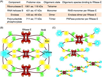
The crystallographic data indicate that a PNPase trimer can bind simultaneously three RNase E-derived recognition peptides. In the context of the degradosome assembly, all the potential binding surfaces can be satisfied if three RNase E tetramers interact with four PNPase trimers. The co-localisation of the PNPase within the degradosome might favour formation of the dimers that are seen in some PNPase crystal forms (Symmons et al. Reference Symmons, Jones and Luisi2000; Hardwick & Luisi, submitted). The dimers are formed by the inter-meshing of all-helical domains, and this could form a degradative chamber (Hardwick & Luisi, submitted).
PNPase and RNase PH share homology with the archaeal and eukaryotic exosomes, which contain a hetero–hexameric RNase PH-like core and a cap of the KH and S1 domains. However, the eukaryotic exosome has no phosphorolytic activity, and mediates RNA degradation through recruitment of the hydrolytic Rrp6 and Rrp44 (Lykke-Andersen et al. Reference Lykke-Andersen, Brodersen and Jensen2009). The central channel may serve to thread single-stranded substrate through the exosome, as it does in PNPase, except that in the exosome the substrate meets ribonucleases on the distal exit site (Bonneau et al. Reference Bonneau, Basquin, Ebert, Lorentzen and Conti2009).
2.8 Structure and function of degradosome-associated enolase
Enolase (phosphopyruvate dehydratase; EC 4.2.1.11) is a universally conserved enzyme that catalyses the metal-assisted dehydration of 2-phospho-D-glycerate (2PG) to form phosphoenolpyruvate (PEP) in the penultimate step of the glycolytic pathway. PEP is a substrate not only for the final step of glycolysis but also for the phosphotransfer systems used for sugar transport in bacteria. In E. coli, roughly 5–10% of enolase is sequestered in the RNA degradosome (Py et al. Reference Py, Higgins, Krisch and Carpousis1996). The precise function of enolase in the degradosome is not established, but it somehow effects the degradation of specific transcripts. Microarray studies have shown that disruption of the RNase E–enolase complex affects the turnover of many mRNAs, especially those encoding enzymes involved in energy-generating pathways, and increases their median half-lives three-fold (Bernstein et al. Reference Bernstein, Lin, Cohen and Lin-Chao2004). The importance of the enolase/RNase E interaction is indicated by the conservation of the cognate microdomain in RNase E of the gamma-proteobacteria, including pathogens (Chandran & Luisi, Reference Chandran and Luisi2006; Nurmohamed et al. Reference Nurmohamed, Mckay, Robinson and Luisi2010). The interaction of enolase with the recognition motif in RNase E of the Gram-negative Vibrio angustum has been experimentally corroborated (Erce et al. Reference Erce, Low, March, Wilkins and Takayama2009) although this interaction is lost in Pseudoaltermonas haloplanktis, a member of the Altermonadales order of bacteria, which are more separated from E. coli than the Vibrionales (Ait-Bara & Carpousis, Reference Ait-Bara and Carpousis2010). The interaction between enolase and RNase E may be restricted to Enterobacteriales, Pasteurellales and Vibrionales.
Through an unknown mechanism, the association of enolase with RNase E affects the stability of ptsG mRNA that encodes the glucose transporter (Morita et al. Reference Morita, Kawamoto, Mizota, Inada and Aiba2004). Under phosphosugar stress, the ptsG transcript is rapidly degraded in an sRNA-mediated, RNase E-dependent manner, but this activated turnover is suppressed in the absence of enolase. The glucose transport system uses PEP, the catalytic product of enolase, to generate phosphoglucose. The interaction with RNase E does not appreciably affect the activity of enolase, so the degradosome is expected to be able to interconvert PEP and 2PG in vivo. An earlier section described how RNase E is associated with the cytoplasmic membrane; perhaps the co-recruitment of enolase to the membrane via RNase E might provide PEP for neighbouring transmembrane sugar transporter or regulate its local concentration by conversion of any excess PEP to 2PG.
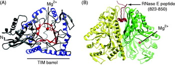
Enolase has a TIM barrel fold – an evolutionary ancient structural motif that is widespread throughout nature and is used as the scaffold for a broad range of enzymes encompassing diverse chemical transformations. The TIM barrel fold comprises a β-sheet that curves and twists into a tight cylinder and is decorated with a circumferential layer of tightly packed helices whose axes are inclined with respect to the central barrel axis (Fig. 7a). Like many other enzymes in this extensive structural family, the active site of enolase is nestled in the cusp of the toroidal surface of the TIM barrel. Enolase is a homo-dimer, and the protomers pack through a conserved interface formed by a subset of helices on the surface of the TIM barrel. Each protomer has additionally an extra helical bundle that strikingly resembles a helical domain of PNPase, although the functional significance of this structural similarity is not clear (Symmons et al. Reference Symmons, Jones and Luisi2000). There are several crystal structures available of enolase in complex with its recognition site from RNase E, and these show that one enolase dimer binds a single RNase E peptide (Fig. 7b; Chandran & Luisi, Reference Chandran and Luisi2006; Nurmohamed et al. Reference Nurmohamed, Mckay, Robinson and Luisi2010). The 2:1 stoichiometry means that the complex is intrinsically asymmetric.
Enolase structure and recognition. (a) Structure of a protomer of E. coli enolase showing the TIM barrel fold. The barrel strands are coloured red and the enveloping helices are in blue. Magnesium is required for catalysis and binds at the active site. (b) The enolase dimer with the RNase E-derived recognition peptide, corresponding to segment C indicated in Fig. 2 (PDB entry 3H8A).

One potential consequence of the interaction between RNase E and enolase is the organisation and presentation of the RNA-binding site (AR2 in Fig. 1) that is physically adjacent to the enolase-binding site in RNase E (Nurmohamed et al. Reference Nurmohamed, Mckay, Robinson and Luisi2010). Alteration of the chromosomal rne gene to express a variant of RNase E lacking the enolase-binding site impacts transcript levels of several enzymes involved in sugar metabolism and growth rates in minimal media with different carbon sources; it also affects recruitment of some sRNAs (Du et al. in preparation). As noted in an earlier section, the Gram-positive bacteria B. subtilis and S. aureus lack an RNase E homologue; however, the functionally analogous ribonuclease RNase Y is observed to interact with glycolytic enzymes in both species (Commichau et al. Reference Commichau, Rothe, Herzberg, Wagner, Hellwig, Lehnik-Habrink, Hammer, Völker and Stülke2009; Roux et al. Reference Roux, Demuth and Dunman2011). Interestingly, the Caulobacter crescentus RNase E associates with aconitase, a Krebs cycle enzyme, in stoichiometric amounts (Hardwick et al. Reference Hardwick, Chan, Broadhurst and Luisi2010). The convergent evolution of the stable interactions highlights a potential important functional role to link ribonuclease with metabolic activities, but this potential relationship remains to be elucidated.
2.9 A model for the organisation and variation of the E. coli RNA degradosome
The structure of the degradosome is currently unknown, but the assembly is likely to be conformationally dynamic. Overall, the CTD may be flexible and loosely packed, at least in the absence of cognate partners. Interactions between the degradosome components and cytoplasmic membrane might support a more compact organisation of the degradosome, and electron micrograph images of degradosomes (either cell-extracted or recombinant) indicate that the assembly is heterogeneous but not extended, with particles of 200–250 Å diameter observed (Górna, Reference Górna2010). In contrast, a truncated version of RNase E encompassing residues 1–762 and which has helicase bound in a stoichiometric ratio, suggested a more elongated structure using small angle X-ray solution scattering and atomic force microscopy (Górna, Reference Górna2010). Based on these observations, the degradosome may be consolidated through interactions of PNPase and enolase with other components. The natively unfolded character of the CTD is envisaged to act as a flexible tether that maintains the globular components in proximity and allows association with the cytoplasmic membrane, while permitting conformational adjustments to accommodate RNA substrates and modulating partners.
The composition of the degradosome changes in response to stress conditions. The expression of PNPase is elevated with cold shock (Gualerzi et al. Reference Gualerzi, Giuliodori and Pon2003), and the PNPase content of the degradosome is affected by cold temperature and also by phosphosugar stress (Worrall et al. Reference Worrall, Górna, Crump, Phillips, Tuck, Price, Bavro and Luisi2008a). PAP (poly A polymerase) associates with RNase E in a growth stage-dependent manner (Carabetta et al. Reference Carabetta, Silhavy and Cristea2010), and this has interesting implications for shunting along different degradative pathways. RNase E is also believed to form an alternative ribonucleoprotein complex with Hfq and sRNAs, for coupled degradation of mRNAs and sRNAs (Morita et al. Reference Morita, Maki and Aiba2005). Recent evidence suggests that Hfq may displace helicase from the assembly (Ikeda et al. Reference Ikeda, Yagi, Morita and Aiba2011). The recently discovered RraA and RraB (regulator of ribonuclease activity A and B, respectively) are proteins that inhibit RNase E activity and may remodel the composition of the degradosome (Gao et al. Reference Gao, Lee, Zhao, Qiu, Zhan, Saxena, Moore, Cohen and Georgiou2006; Lee et al. Reference Lee, Zhan, Gao, Qiu, Feng, Meganathan, Cohen and Georgiou2003). These multiple partners suggest that the degradosome interacts functionally with other cellular machineries and pathways, and that its composition, and thus activity and possibly structure, is modulated according to environmental conditions. The interactions with partners may be ‘dynamic’, in the sense that the components associate and dissociate on second time scale or may be displaced by competing components.
Under normal growth conditions, RNase E co-purifies with the other canonical degradosome components – PNPase, RhlB and enolase – in roughly stoichiometric amounts (Blum et al. Reference Blum, Py, Carpousis and Higgins1997; Carpousis et al. Reference Carpousis, Van Houwe, Ehretsmann and Krisch1994; Py et al. Reference Py, Higgins, Krisch and Carpousis1996), and in vitro the purified components assemble spontaneously into a complex showing degradosome activity (Coburn et al. Reference Coburn, Miao, Briant and Mackie1999; Worrall et al. Reference Worrall, Górna, Crump, Phillips, Tuck, Price, Bavro and Luisi2008a). The hypothetical stoichiometry of the canonical degradosome, based on in vitro biophysical studies and the crystal structures of components, suggest a protomer ratio of 1:1:2:1 for RNase E:PNPase:enolase:RhlB (Chandran & Luisi, Reference Chandran and Luisi2006; Chandran et al. Reference Chandran, Poljak, Vanzo, Leroy, Miguel, Fernandez-Recio, Parkinson, Burns, Carpousis and Luisi2007; Nurmohamed et al. Reference Nurmohamed, Vaidialingam, Callaghan and Luisi2009). A minimal complex that would satisfy this stoichiometry and take into account the oligomeric state of all components has a calculated mass in excess of 4 MDa (Marcaida et al. Reference Marcaida, Depristo, Chandran, Carpousis and Luisi2006), which is greater than that of a ribosome. However, formation of this ‘closed’ complex would depend on the mechanical properties of RNase E CTD, and also would have to allow for the direct association of RNase E with the cytoplasmic membrane.
An alternative structure for degradosome is an ‘open’ network of the RNase E tetramers (degradosome ‘building blocks’), bridged by PNPase trimers interacting with RNase E protomers belonging to different tetramers (Fig. 8). However, in the cell there must exists a means of controlling formation of such indefinite oligomers. This might be achieved by limiting copy number of RNase E, estimated to be ~960 protomers (or 240 tetramers) (Liou et al. Reference Liou, Jane, Cohen, Lin and Lin-Chao2001), or constraints imposed by membrane association (Taghbalout & Rothfield, Reference Taghbalout and Rothfield2007). The excess of total cellular PNPase over available RNase E would also promote saturation with one trimer of PNPase per each RNase E protomer and disfavour the joining of building blocks. It may be that the size of degradosome is not tightly controlled, and instead it exists as a heterogeneous complex, with assemblies of different size co-existing in equilibrium; this could explain the contradictory reports on the size of the degradosome (Carpousis, Reference Carpousis2007). The ‘closed’ model could provide an adjustable chamber like an accordion for limiting diffusion of RNA products, while the ‘open’ model seems to allow for more conformational flexibility.
Models for structure of the degradosome assembly. (a) Panel summarising the canonical degradosome components and their physical properties. (b) A ‘closed’ model for the degradosome complex, in which all the potential binding sites are satisfied. The smallest such complex contains three RNase E tetramers and four PNPase trimers. (c) An ‘open’ model for the degradosome assembly. RNase E tetramers are linked through interactions with shared PNPase trimers into an extended network (here shown arbitrarily for several RNase E tetramers). Brown, green, yellow and blue are RNase E tetramers, RhlB monomers, enolase dimers and PNPase trimers, respectively.

Considering that the flexible RNase E scaffold connects globular, active components, the degradosome could function similarly to the eukaryotic fatty acid synthases (Leibundgut et al. Reference Leibundgut, Maier, Jenni and Ban2008). These multi-enzyme complexes form a chamber with internally displayed active sites, which are visited by a mobile, substrate-bearing acyl carrier protein (ACP) domain. The active sites in fatty acid synthase are spatially arrayed in the order of the reaction cycle, which minimises the distances that the ACP domain must traverse between consecutive catalytic steps. Also, the length of the flexible, unstructured linkers tethering ACP domain is strictly controlled, allowing for substrate shuttling between all active sites, but favouring access to those which catalyze more frequent reaction steps. Similarly, it can be envisaged that the RNA-binding sites within RNase E could present RNA substrate for the catalytic activity of RNase E, RhlB and PNPase. In this case, the substantial length of flexible linker sequence, or the more rigid proline-rich regions that occur between the microdomains in RNase E CTD could serve to control the distances between degradosome components and to organise spatially the active sites for the shuttling of RNA substrate. The interaction of RhlB with the central region of RNase E CTD would also suggest the need for cooperation of the helicase with RNase E as well as with PNPase.
2.10 The puzzle of degradosome assembly
The mechanism of degradosome assembly and localisation on the membrane are not understood. It is not clear whether there is an order of components binding to RNase E. RNase E is the only component of the complex that has clearly been identified as interacting with lipid bilayers. Does the localisation of the degradosome to the membrane occur after the complex has fully assembled, or are the protein components recruited only to the membrane-bound RNase E? In the former case, the complex must undergo a large conformational change, and it is also likely to lose some of its potential geometric symmetry due to the one-sided interaction with the lipid sheet. In the latter case, the recognition sites in RNase E, which have high affinity for the protein partners must be made unavailable until RNase E has been safely deposited on the membrane. It is also not clear whether the assembly of the degradosome or the localisation of RNase E occurs spontaneously, or whether it requires additional factors, such as the chaperones that have been identified as sub-stoichiometric components. Another question is the cooperativity of binding of various RNase E partners to the degradosome scaffold. In the context of the degradosome, binding of one protein to its cognate microdomain may help in recruitment of other components, for instance by favouring weak protein–protein interactions between RNase E partners. Cooperativity originating from multi-dentate interactions has been proposed for multi-component assemblies that involve recognition of natively disordered peptides (Gibson, Reference Gibson2009).
Another unsolved issue is the localisation of the membrane-associated degradosome into the helical filaments that span the length of an E. coli cell (Fig. 4a). Is it a higher-order assembly or does it represent some underlying helical structure punctuated by the occasional degradosome? Both RNase E and RhlB have been reported to localise to these filaments in the absence of each other, and to mediate PNPase association with the filaments; enolase is observed to localise to the filaments but only in the presence of RNase E (Taghbalout & Rothfield, Reference Taghbalout and Rothfield2007, Reference Taghbalout and Rothfield2008). Although RhlB has been found to dimerize (Górna et al. Reference Górna2010), or even form filaments in vitro (Taghbalout & Yang, Reference Taghbalout and Yang2010), this seems driven by factors such as high protein concentration or the addition of calcium ions or ATP. Unlike RNase E, RhlB has also no reported affinity for lipid membrane. Given the small number of RhlB in the cell, and its presumed exclusive association with RNase E, it is unlikely that the helical filaments are composed of tightly packed RhlB. On the other hand, RNase E has been reported to self-interact (Callaghan et al. Reference Callaghan, Aurikko, Ilag, Grossmann, Chandran, Kühnel, Poljak, Carpousis, Robinson, Symmons and Luisi2004; Vanzo et al. Reference Vanzo, Li, Py, Blum, Higgins, Raynal, Krisch and Carpousis1998) and the coiled-coil regions are attractive candidates for the additional oligomerisation sites. It remains to be seen whether the degradosome can oligomerise, or if there exists a factor recruiting degradosome to the filamentous structures. Such a factor could be a protein partner, a specific composition of the lipids (e.g. anionic phospholipid spirals as seen in B. subtilis by Barak et al. Reference Barak, Muchova, Wilkinson, O'Toole and Pavlendova2008), or perhaps even an RNA scaffold.
3. Guided activities of the RNA degradosome
3.1 Non-coding RNAs and their interactions with the RNase E and the degradosome
In prokaryotes as well as the eukaryotes, regulation mediated through RNA is the basis for complex cellular behaviour. While the machinery differs in much of the details in diverse phyla, the convergence of RNA-mediated regulation highlights its importance in the evolution of all domains of life. We turn to a description of RNA-mediated regulation of gene expression in bacteria. RNase E is emerging as a key component of sRNA-mediated regulation in E. coli and Salmonella (Afonyushkin et al. Reference Afonyushkin, Vecerek, Moll, Bläsi and Kaberdin2005; Massé et al. Reference Massé, Escorcia and Gottesman2003; Morita et al. Reference Morita, Maki and Aiba2005; Pfeiffer et al. Reference Pfeiffer, Papenfort, Lucchini, Hinton and Vogel2009).
A variety of ncRNAs in bacteria has been described, from the antisense regulators of plasmid replication, first reported nearly 30 years ago, to the more recently studied, chromosome-encoded transcripts that act in trans to control initiation of translation (Waters & Storz, Reference Waters and Storz2009). The latter are generally referred to as sRNAs, and are best characterised for E. coli and Salmonella (Gottesman, Reference Gottesman2005; Gottesman & Storz, Reference Gottesman and Storz2010; Vogel, Reference Vogel2009). Similarly to eukaryotic miRNAs, bacterial sRNAs are usually transcribed from their own promoter, and act through complementary base-pairing with target mRNA (Fig. 9). Trans-encoded sRNAs base-pair with their targets through short regions of imperfect complementarity, and the interaction often involves the 5′-end of sRNA. Albeit smaller than usual protein-coding transcripts, bacterial sRNAs typically range from 50 to 300 nucleotides in length, and as such are larger than the mature forms of their eukaryotic counterparts. Some sRNAs undergo processing, as is found for ArcZ sRNA (previously named RyhA or SraH), which is cleaved internally to yield ~50 nucleotide mature sRNA with a conserved 5′-end (Argaman et al. Reference Argaman, Hershberg, Vogel, Bejerano, Wagner, Margalit and Altuvia2001; Papenfort et al. Reference Papenfort, Said, Welsink, Lucchini, Hinton and Vogel2009).
Regulation of gene expression by trans-encoded bacterial sRNA. Genes encoding sRNAs (red) are usually located separately from the genes encoding their target RNAs (blue) and only have limited base-pairing complementarity. sRNAs can repress translation by base-pairing with the ribosome-binding site (RBS) and blocking 30S ribosome binding, which also leads to rapid degradation of the sRNA–mRNA duplex by RNases (left panel) (Massé et al. Reference Massé, Escorcia and Gottesman2003; Morita et al. Reference Morita, Maki and Aiba2005). Some sRNAs can target mRNA for degradation by interaction far downstream of the RBS and without inhibition of translation initiation (middle panel) (Pfeiffer et al. Reference Pfeiffer, Papenfort, Lucchini, Hinton and Vogel2009). In this case, RNase E is recruited to the sRNA–mRNA complex and performs the rate-limiting cleavage in the coding region. sRNA can act positively on translation regulation by preventing the formation of an inhibitory structure, which sequesters the RBS (right panel). In all cases, Hfq facilitates sRNA interaction with the mRNA.

The sequence targeted by sRNA usually lies within the translation–initiation region (TIR) of mRNA, which encompasses the Shine–Dalgarno element (the ribosome-binding site) and the translation start codon. Occlusion of the TIR or up to five downstream codons interferes with ribosome binding (Bouvier et al. Reference Bouvier, Sharma, Mika, Nierhaus and Vogel2008) and prevents initiation of translation (Fig. 9). This translational block exposes mRNA to ribonucleolytic attack, and results in rapid coordinated degradation of sRNA and mRNA, involving RNase E or RNase III (Massé et al. Reference Massé, Escorcia and Gottesman2003; Morita et al. Reference Morita, Maki and Aiba2005; Vogel et al. Reference Vogel, Argaman, Wagner and Altuvia2004). Interaction of sRNA with the coding sequence of mRNA, outside of the mentioned five-codon window, does not interfere with initiation of translation; instead, it seems to directly cause RNA degradation. Examples of such downstream-binding sRNAs are few so far and only recently discovered (Papenfort et al. Reference Papenfort, Said, Welsink, Lucchini, Hinton and Vogel2009; Pfeiffer et al. Reference Pfeiffer, Papenfort, Lucchini, Hinton and Vogel2009), but they are likely to be more widespread. Through binding upstream of TIR, sRNAs can also promote translation, by disrupting inhibitory RNA folds that sequester the TIR. This dual regulation of TIR availability resembles the action of 5′-UTR-encoded riboswitches that upon binding of small molecules undergo conformational changes (Roth & Breaker, Reference Roth and Breaker2009), exposing or sequestering the TIR. In this way, translation of the downstream message can be inhibited or activated in response to changes in the concentration of nutrients. Transcription of sRNAs is often activated in response to change in environmental conditions, and can play a similar role to riboswitches in regulation of metabolism-related genes. An important aspect of trans-acting sRNAs is that they are able to target multiple messages, as is seen for RyhB sRNA, which controls levels of a multitude of transcripts in response to iron deficiency (Massé et al. Reference Massé, Salvail, Desnoyers and Arguin2007). Moreover, the same sRNA can use alternative mechanisms; RyhB down-regulates most transcripts, while activating translation of another (Prévost et al. Reference Prévost, Salvail, Desnoyers, Jacques, Phaneuf and Massé2007).
Close to 100 sRNAs have been described in E. coli and Salmonella with numerous sRNAs conserved among enterobacteria (Gottesman, Reference Gottesman2005; Vogel, Reference Vogel2009). Most sRNAs function in adaptation to changing environmental conditions and in stress responses. Some can impact on metabolism by wide action: the sRNA RsaE in S. aureus down-regulates several enzymes in metabolic pathways (Bohn et al. Reference Bohn, Rigoulay, Chabelskaya, Sharma, Marchais, Skorski, Borezee-Durant, Barbet, Jacquet, Jacq, Gautheret, Felden, Vogel and Bouloc2010). Among sRNA-regulated processes are sugar stress and metabolism (reviewed in Beisel & Storz, Reference Beisel and Storz2011; Görke & Vogel, Reference Görke and Vogel2008), the SOS response to DNA damage (Vogel et al. Reference Vogel, Argaman, Wagner and Altuvia2004), σE-mediated envelope stress response (Valentin-Hansen et al. Reference Valentin-Hansen, Johansen and Rasmussen2007; Vogel & Papenfort, Reference Vogel and Papenfort2006), quorum sensing (Tu & Bassler, Reference Tu and Bassler2007), regulation of virulence (Toledo-Arana et al. Reference Toledo-Arana, Repoila and Cossart2007) and toxin-antitoxin systems (Fozo et al. Reference Fozo, Hemm and Storz2008). A large set of sRNAs down-regulate outer membrane proteins (Omp), and several sRNAs affect the level of the stationary phase sigma factor σS, and therefore modulate σS-dependent processes.
Variations exist on the themes described above. Cis-encoded sRNAs are produced from genes overlapping with those of their targets, from the opposite strand, resulting in greater complementarity and an expanded repertoire of regulatory mechanisms. For instance, the cis-encoded GadY sRNA stabilises gadX mRNA by protection of its 3′-end (Opdyke et al. Reference Opdyke, Kang and Storz2004). Some regulatory RNAs target proteins instead of transcripts, e.g. CsrB and CsrC, which modulate activity of the translational regulator CsrA (Majdalani et al. Reference Majdalani, Vanderpool and Gottesman2005), or 6S RNA which sequesters the σ70-containing RNA polymerase (Wassarman, Reference Wassarman2007).
Unlike eukaryotic cells, bacteria have only a few known proteins that form ribonucleoprotein complexes with sRNAs (Pichon & Felden, Reference Pichon and Felden2007). A global regulator of sRNA stability and function is Hfq, an RNA chaperone protein originally identified as a host factor for the replication of the RNA phage Qβ and encoded by many bacterial species (Brennan & Link, Reference Brennan and Link2007; Chao & Vogel, Reference Chao and Vogel2010; Sittka et al. Reference Sittka, Lucchini, Papenfort, Sharma, Rolle, Binnewies, Hinton and Vogel2008; Vogel & Luisi, Reference Vogel and Luisi2011). Hfq may impact on expression of one-fifth of genes in some bacterial species (Chao & Vogel, Reference Chao and Vogel2010). Many, although not all, sRNAs require Hfq for full functionality, and as such Hfq is implicated in control of virulence and other sRNA-mediated processes. The interaction of Hfq with sRNA confers protection from degradation, and in the absence of Hfq the bulk of sRNAs is destabilised. Hfq has RNA-annealing activity, and its association with sRNA promotes its base-pairing with target mRNA, possibly by binding to both RNAs and bringing them into spatial proximity. Structural data indicate that E. coli Hfq might bind sRNA and mRNAs at two distinct binding surfaces, one of which can engage triplets of the type A-A/G-N (Link et al. Reference Link, Valentin-Hansen and Brennan2009), and the other having a preference for U-rich RNA (Sauer & Weichenrieder, Reference Sauer and Weichenrieder2011; Schumacher et al. Reference Schumacher, Pearson, Moller, Valentin-Hansen and Brennan2002). Solution data indicate a role for the natively unstructured C-terminal extension for RNA-binding (Beich-Frandsen et al. Reference Beich-Frandsen, Vecerek, Konarev, Sjoblom, Kloiber, Hammerle, Rajkowitsch, Miles, Kontaxis, Wallace, Svergun, Konrat, Blasi and Djinovic-Carugo2011; Vecerek et al. Reference Vecerek, Rajkowitsch, Sonnleitner, Schroeder and Blasi2008).
Hfq in complex with sRNA is proposed to associate with RNase E to target mRNA for rapid degradation (Ikeda et al. Reference Ikeda, Yagi, Morita and Aiba2011; Morita et al. Reference Morita, Maki and Aiba2005), although it is unclear whether there is indeed a dedicated Hfq–RNase E complex for sRNA-guided mRNA inactivation. In many cases, Hfq and RNase E bind similar sites on RNA; sRNA–mRNA pairing may promote loss of Hfq and access by RNase E (Massé et al. Reference Massé, Escorcia and Gottesman2003). PNPase has also been implicated in degradation of sRNA, and in control of sRNA-mediated virulence and expression of Omp (Andrade & Arraiano, Reference Andrade and Arraiano2008; Viegas et al. Reference Viegas, Pfeiffer, Sittka, Silva, Vogel and Arraiano2007). The RNA degradosome is likely to be involved in the decay of both sRNAs and their targets; disruption of the degradosome assembly caused the largest stabilisation effect on selected sRNAs when compared with other RNase mutants in Salmonella (Viegas et al. Reference Viegas, Pfeiffer, Sittka, Silva, Vogel and Arraiano2007). A recent tiling microarray study indicates that the regulation of sRNA levels by RNase E in E. coli might be a larger part of RNase E taskwork than previously appreciated (Stead et al. Reference Stead, Marshburn, Mohanty, Mitra, Castillo, Ray, Van Bakel, Hughes and Kushner2010). In an RNase E deletion strain, more than half of known sRNAs show changes in steady-state levels, and these sRNA mostly increase in abundance. This might explain why, in the absence of RNase E, 25% of the annotated coding sequences show a decrease in abundance – a surprisingly large proportion when compared with 35% of annotated coding sequences whose levels increase, as would be expected following inactivation of a degradative enzyme.
3.2 Finding access to substrate in vivo
In eukaryotes, transcripts that encode proteins follow a complicated path on the route to translation. Generated in the nucleus, the transcripts may be spliced to remove introns and face a gambit of multiple nucleases for surveillance before being exported. In translation, they are checked for proper alignment of stop codon location before translation proceeds fully. In contrast, the protein-encoding transcripts of the bacteria appear to follow a much simpler path. They exit almost directly from polymerase into the ribosome, seemingly by-passing many complicated steps.
The apparent simplicity of the bacteria case raises several questions. First, if the transcript is sequestered by translating ribosomes, when would it ever be seen by ribonucleases for turnover? Ribosomal occupation can protect a model transcript against endoribonuclease attack (Arnold et al. Reference Arnold, Yu and Belasco1998). Secondly, how are error-bearing transcripts detected and dealt with? The only opportunities are immediately before translation initiation, or during a stall in translation that results in exposure of the mRNA to nucleolytic cleavage – this way, faulty transcripts are automatically and non-specifically targeted for degradation. It remains a mystery how mRNA cleavage could be initiated while the transcript is being efficiently translated, and the common view is that ribosomes compete for RNA with endonucleases such as RNase E, and with RNA-binding proteins such as the RNA chaperone Hfq. Thus, the default mode may be that an mRNA will be degraded unless it is being actively translated. The membrane tethering of RNase E is likely to affect access to the mRNA, although exactly what effects it might have are not presently known.
The most promising position of attack on a transcript would be its 5′-end as it emerges from the translating ribosome. In eukaryotes, it has been hypothesised that the 5′→3′ polarity of mRNA degradation has evolved to ensure that the last translocating ribosome can complete translation (Hu et al. Reference Hu, Sweet, Chamnongpol, Baker and Coller2009); in bacteria, a similar 5′→3′ directionality of RNase E cleavages is likely to play the same role. Whether there could be internal cleavage on a transcript during translation is not experimentally proven, but seems a logical possibility. The recent identification of sRNAs that trigger internal cleavage makes this a possibility for guided RNase E activity (Pfeiffer et al. Reference Pfeiffer, Papenfort, Lucchini, Hinton and Vogel2009).
Electron cryomicroscopy studies of E. coli polysomes suggest that between 40 and 52 nucleotides bridge the adjacent mRNA exit and entry points (Brandt et al. Reference Brandt, Etchells, Ortiz, Elcock, Hartl and Baumeister2009), but this would be in a sequestered core that might protect against endoribonuclease cleavage. Activated access to the linker RNA could be sufficient for the seed sequence of the sRNA to direct RNase E. The helicase activity of a translating ribosome would rapidly displace such a complex, but this may not be a barrier to entry if the cleavage can occur ‘on the fly’ with transient interaction of the ribonuclease with the substrate. It is possible that RNase E pre-associates with polysomes (and/or sRNA) to be in place for such a quick surveillance action, which might account for the observed interactions of RNase E with ribosomal proteins (Butland et al. Reference Butland, Peregrin-Alvarez, Li, Yang, Yang, Canadien, Starostine, Richards, Beattie, Krogan, Davey, Parkinson, Greenblatt and Emili2005; Kaberdin & Lin-Chao, Reference Kaberdin and Lin-Chao2009; Miczak et al. Reference Miczak, Kaberdin, Wei and Lin-Chao1996; Singh et al. Reference Singh, Chang, Lin, Averina, Kaberdin and Lin-Chao2009). The ability of RNase E to form multiple contacts to single-stranded RNA independent of the 5′-end would enable sensing of translational activity and could cleave those transcripts that are stalled or inactive (Kime et al. Reference Kime, Jourdan, Stead, Hidalgo-Sastre and Mcdowall2010).
4. Energy, metabolism and cellular economy
The recurrent physical association of ribonucleases with metabolic enzymes throughout bacterial evolution suggests that RNA metabolism may be somehow linked with central metabolism and other metabolic pathways (Chandran & Luisi, Reference Chandran and Luisi2006; Commichau et al. Reference Commichau, Rothe, Herzberg, Wagner, Hellwig, Lehnik-Habrink, Hammer, Völker and Stülke2009; Hardwick et al. Reference Hardwick, Chan, Broadhurst and Luisi2010; Kang et al. Reference Kang, Caparon and Cho2010; Nurmohamed et al. Reference Nurmohamed, Mckay, Robinson and Luisi2010). This notion is further supported by findings in E. coli that enolase–RNase E interaction is required for phosphosugar stress response (Morita et al. Reference Morita, Kawamoto, Mizota, Inada and Aiba2004), and that the degradosome affects the abundance of transcripts encoding enzymes from central metabolism (Bernstein et al. Reference Bernstein, Lin, Cohen and Lin-Chao2004). RNase G, the paralogue of RNase E, affects transcripts encoding glycolytic enzymes (Lee et al. Reference Lee, Bernstein and Cohen2002).
The regulation of metabolic pathways can occur at the level of individual enzymes, and also at a wider level involving coordination of genes encoding elements of branching pathways. The latter can be achieved through ncRNAs, and these involve RNase E and possibly the degradosome. An illustrative example is the carbon storage response. The carbon storage regulatory protein CsrA is a negative regulator of two glycogen operons and affects levels of many enzymes in response to growth rate and oxygen levels (Babitzke & Romeo, Reference Babitzke and Romeo2007; Sabnis et al. Reference Sabnis, Yang and Romeo1995) and it functions by associating with the TIR of target transcripts to block their translation. The sRNAs CsrB and CsrC sequester the CsrA protein and suppress its activity. In turn, these sRNAs are guided by CsrD for destruction by RNase E (Suzuki et al. Reference Suzuki, Babitzke, Kushner and Romeo2006). Another example of wide acting regulation mediated by sRNA is also seen in the function of RsaE in the down-regulation of several enzymes in the Krebs cycle and one-carbon metabolism in the folate-dependent pathways in the pathogen S. aureus (Bohn et al. Reference Bohn, Rigoulay, Chabelskaya, Sharma, Marchais, Skorski, Borezee-Durant, Barbet, Jacquet, Jacq, Gautheret, Felden, Vogel and Bouloc2010).
Coordinated ribonuclease activities may also contribute to the global cellular economy, which may be especially important during growth and development. For instance, as a cell moves from the stationary phase to high growth rates, there is an accompanying demand for higher rates of DNA and RNA syntheses. The phosphorolytic degradation of RNA by PNPase has been proposed to favour rapid recycling of RNA in the form of nucleoside diphosphates for synthesis of fresh transcripts or for shunting into DNA synthesis by providing substrates for ribonucleotide reductase (Danchin, Reference Danchin1997). As these pathways recycle nucleotides as nucleoside diphosphates, they represent a smaller energy burden for the cell. The shunting of substrates away from phosphorolytic decay may favour the more wasteful but more rapid and efficacious activity of processive hydrolytic exoribonucleases, such as RNase R or RNase II, which generate nucleoside monophosphates. These require more energy to be transformed into suitable substrates for mRNA synthesis or DNA synthesis. Thus, as a speculation, the routing through different exoribonuclease activities may permit the cell to use a different energetic route for generating a pool of available nucleotides.
Other accumulating evidence indicates a connection between ribonuclease activity and central metabolism that may contribute to cellular regulation and response. PNPase has been shown to be allosterically activated by ATP in vitro, with implications for linking RNA metabolism with response to cellular energy levels (Del Favero et al. Reference Del Favero, Mazzantini, Briani, Zangrossi, Tortora and Deho2008). PNPase of Nonomuraea sp. and Streptomyces are inhibited by the signalling molecule (p)ppGpp (Gatewood & Jones, Reference Gatewood and Jones2010; Siculella et al. Reference Siculella, Damiano, Di Summa, Tredici, Alduina, Gnoni and Alifano2010). Structural data indicate that PNPase is an allosteric enzyme, and might respond to metabolic modulators (Hardwick et al. in preparation; Nurmohamed et al. Reference Nurmohamed, Vincent, Titman, Chandran, Pears, Du, Griffin, Callaghan and Luisi2011). Evidence suggests that the signalling molecule cyclic-di-GMP may be a regulator of PNPase activity (Tuckerman et al. Reference Tuckerman, Gonzalez and Gilles-Gonzalez2011). The effect of PNPase and degradosome activities on intracellular metabolite concentrations in vivo support a hypothesis for a link between nuclease activity and cellular metabolic status, in which metabolites impact on ribonuclease activity, which in turn have impact on many transcripts.
Decades of efforts to engineer metabolic pathways have revealed the complex behaviour of metabolite concentrations and pathway fluxes in response to changing levels of enzymes. These and other observations indicate that cellular metabolism requires regulation not only at the level of individual enzymes, but also more widely in a manner that orchestrates activities of many different enzymes distributed among branching pathways (Daran-Lapujade et al. Reference Daran-Lapujade, Rossell, Van Gulik, Luttik, De Groot, Slijper, Heck, Daran, De Winde, Westerhoff, Pronk and Bakker2007; Hardiman et al. Reference Hardiman, Lemuth, Keller, Reuss and Siemann-Herzberg2007). The required global action can be seen in the function of the aforementioned RsaE sRNA (Bohn et al. Reference Bohn, Rigoulay, Chabelskaya, Sharma, Marchais, Skorski, Borezee-Durant, Barbet, Jacquet, Jacq, Gautheret, Felden, Vogel and Bouloc2010), or in the action of riboswitches that sense cellular metabolism (Henkin, Reference Henkin2008). Another possible contribution to such control might be the effect of metabolites on ribonucleases, such as the potential effect of metabolites on PNPase activity. A combination of wide ranging and distributive control by ribonucleases and sRNAs can provide an integrative control mechanism that regulates homoeostasis and response to environmental change (Nurmohamed et al. Reference Nurmohamed, Vincent, Titman, Chandran, Pears, Du, Griffin, Callaghan and Luisi2011).
5. Summary and perspective
The E. coli RNA degradosome is a machine that plays diverse roles in RNA metabolism. It shares homologous components and functional analogy with similar assemblies found in all domains of life. One of its canonical components is an ATP-dependent motor that is activated through protein–protein interactions and which cooperates with the ribonucleases in an energy-dependent mode of RNA degradation. The degradosome assembly has an elusive compositional complexity and can gain or lose partners according to physiological state. Some of the partners interact dynamically, with rapid exchange or competitive displacement, and others contribute to a core that is likely to remain stably associated from the time of first assembly. The degradosome can be effectively re-programmed for either broad or specific activities through interactions with numerous protein and sRNA partners.
Degradosome-like assemblies, built upon RNase E, are predicted to be present in many γ-proteobacteria (Marcaida et al. Reference Marcaida, Depristo, Chandran, Carpousis and Luisi2006) and extant assemblies have been identified among more remote bacterial lineages (Fig. 10). These degradosome-like assemblies are considerably divergent in detailed composition. Despite the lack of conservation, the degradosome nonetheless confers a clear selective benefit, as has been experimentally demonstrated for E. coli (Leroy et al. Reference Leroy, Vanzo, Sousa, Dreyfus and Carpousis2002). Assembled through the C-terminal portion of the endoribonuclease RNase E, the scaffold of the degradosome is a natively unstructured tether that co-localises globular components so that their activities may cooperate. It is striking that E. coli and many other γ-proteobacteria have evolutionarily sustained such a long segment of peptide, nearly 500 amino acids, with little apparent intrinsic structure. The degradosome simply must confer some selective benefit, and the accumulating data suggest that these are the manifold contributions to regulation and response.
Divergence of degradosome assemblies. An unrooted phylogenetic tree for RNase E from selected proteobacteria and actinobacteria. Indicated are the delta, beta, alpha and gamma branches of proteobacteria. For the species marked with an asterisk, association of RNase E with one or more degradosome components was confirmed experimentally (Ait-Bara & Carpousis, Reference Ait-Bara and Carpousis2010; Erce et al. Reference Erce, Low, March, Wilkins and Takayama2009, Reference Erce, Low and Wilkins2010; Hardwick et al. Reference Hardwick, Chan, Broadhurst and Luisi2010; Jäger et al. Reference Jäger, Fuhrmann, Heck, Hebermehl, Schiltz, Rauhut and Klug2001; Kovacs et al. Reference Kovacs, Csanadi, Megyeri, Kaberdin and Miczak2005; Lee & Cohen, Reference Lee and Cohen2003; Purusharth et al. Reference Purusharth, Klein, Sulthana, Jäger, Jagannadham, Evguenieva-Hackenberg, Ray and Klug2005; Yang et al. Reference Yang, Jain and Schesser2008). Within gammaproteobacteria, Enterobacteriales, Pasteuralles and Vibrionales are predicted to have an E. coli type of degradosome, while Aeromonadales and Alteromonadales degradosomes lack enolase (Ait-Bara & Carpousis, Reference Ait-Bara and Carpousis2010). The tree was prepared from RNase E protein sequences using the phylogeny.fr server (Dereeper et al. Reference Dereeper, Guignon, Blanc, Audic, Buffet, Chevenet, Dufayard, Guindon, Lefort, Lescot, Claverie and Gascuel2008). Protein sequences were aligned using T-Coffee, and the phylogeny was estimated using the maximum likelihood method as implemented in PhyML with bootstrapping for 100 trials.

Recognition of protein and RNA partners in the degradosome is mediated through microdomains. The folding-upon-binding that accompanies this mode of recognition is a structurally economical way of burying extensive surface area without the requirement of a cumbersome globular scaffold for both interacting partners. Such recognition gives rise to specificity without necessarily great affinity. It is also interesting to consider the impact of mutations on the natively unfolded regions: for most globular proteins with well-packed hydrophobic cores, mutations decrease protein fitness. In contrast, mutations have smaller effects on free energy of folding in cases of loosely packed, partially disordered structures, which is closer to the case of the microdomains of the RNA degradosome (DePristo et al. Reference Depristo, Weinreich and Hartl2005; Marcaida et al. Reference Marcaida, Depristo, Chandran, Carpousis and Luisi2006; Tokuriki & Tawfik, Reference Tokuriki and Tawfik2009). In principle, the unstructured region can potentially aggregate or form non-specific interactions in the densely packed intracellular molecular environment, and it can also be exposed to proteolytic degradation. In general, natively unstructured proteins would seem to be especially vulnerable to aggregate, and there must be features in the natively unstructured region that prevent such deleterious behaviour. It seems likely that the C-terminus scaffolding domain of RNase E, like other natively unstructured proteins, has closely co-evolved with the busy cellular environment to avoid this fate (Pechmann et al. Reference Pechmann, Levy, Tartaglia and Vendruscolo2009).
Since microdomains are sufficient for mediating protein–protein interactions, the shuffling of these domains allows for the rapid evolutionary adaptation of degradosome composition and function (Marcaida et al. Reference Marcaida, Depristo, Chandran, Carpousis and Luisi2006). As a case in point, the recruitment of enolase into the degradosomes of proteobacteria enables competitive growth under conditions in which carbon sources are limited to acetate or succinate. In this way, evolution can derive new function from existing proteins, with potentially global impact on gene expression or regulatory responses.
Eukaryotes abound in natively unstructured protein domains, where they are characteristically found in signalling machinery components and transcription factors (Diella et al. Reference Diella, Haslam, Chica, Budd, Michael, Brown, Trave and Gibson2008; Lobley et al. Reference Lobley, Swindells, Orengo and Jones2007). The natively unstructured domains in those proteins often encompass small linear recognition epitopes, like the microdomains described here for RNase E, and these are readily exposed for multiple binding interactions (Dunker et al. Reference Dunker, Silman, Uversky and Sussman2008; Gunasekaran et al. Reference Gunasekaran, Tsai, Kumar, Zanuy and Nussinov2003; Wright & Dyson, Reference Wright and Dyson2009). Many eukaryotic proteins with natively unstructured features are ‘hubs’ that are involved in large number of interactions and often have microdomains embedded within extensive segments of natively unstructured peptide (Russell & Gibson, Reference Russell and Gibson2008). Similarly, the degradosome functions in many aspects as a central hub of post-transcriptional gene regulation, with broad impact on transcript turnover.
The contributions of RNase E and the RNA degradosome to RNA quality control is another key aspect of their functions. An important question is what general features might be recognised in an RNA to preserve it from a fate of rapid destruction. It seems unlikely that a specific fold or feature is recognised, and that the mechanism seeks something generic – possibly the stability of the fold. Such generic recognition would provide the most economical mechanism for surveillance, and could involve unwinding or remodelling by the degradosome-associated helicase or PNPase. It seems possible that a similar generic mechanism might occur in the nuclei of eukaryotes, where defective RNAs might be structurally surveyed before passing to the RNA surveillance machinery. In this way, the nuclear exosome and the bacterial degradosome may have appreciable functional analogy. It is conceivable that small non-coding or regulatory RNAs may function to perturb the structure of targeted RNAs to direct them to surveillance pathways through degradative machineries.
The contribution of the degradosome to global post-transcriptional regulation is another important facet of its function. RNase E has a wide impact on transcript levels, especially of non-coding regulatory RNAs (Stead et al. Reference Stead, Marshburn, Mohanty, Mitra, Castillo, Ray, Van Bakel, Hughes and Kushner2010). The available data hint that the degradosome may impact preferentially on transcripts encoding metabolic enzymes (Bernstein et al. Reference Bernstein, Lin, Cohen and Lin-Chao2004). Structural features of transcripts are the probable signatures that affect turnover rates by PNPase, the degradosome and other cellular ribonucleases (Carpousis, Reference Carpousis2007; Deutscher, Reference Deutscher2006). The potential preference of the degradosome for groups of transcripts offers a model for global post-transcriptional regulation. Differential turnover implies that gene products evolve as tuned substrates for cognate enzymes. This represents another interesting paradigm in which substrates evolve to match the enzymes, and this will be a property that can meet the needs of regulating an integrated metabolic system (Nurmohamed et al. Reference Nurmohamed, Vincent, Titman, Chandran, Pears, Du, Griffin, Callaghan and Luisi2011).
Bacterial lineages lacking RNase E nonetheless appear to have analogous multi-enzyme degradative complexes. Exosomes of the archaea and eukaryotes have similar components and activities to the RNA degradosome. Thus, there is a recurrent theme, seen in all domains of life in the formation of stable nuclease-helicase complexes, and the striking convergence in composition between E. coli and B. subtilis degradosomes, illustrate how there must be selective benefit in co-association of various types of nucleases, RNA helicases and possibly glycolytic enzymes into one complex. Perhaps the degradosome represents a comparatively recent molecular adaptation for specialised function, most likely involving aspects of global regulation. The compositional variation of diverse RNA degradosome assemblies has likely endowed bacterial species with specialised capacity for distinct life styles and regulatory processes well tuned to occupy specialised niches.
6. Acknowledgments
B. L. was supported by the Wellcome Trust and M. W. G. by an EU ChemBioChem scholarship. We thank Kasia Bandyra, Toby Gibson, Anastasia Callaghan, Helen Vincent, Kenny McDowall, Kiyoshi Nagai and George Mackie for helpful comments and stimulating discussions.