Introduction
Human cystic echinococcosis (CE) is a chronic zoonotic disease that results from infection with the larval stage of the dog tapeworm, Echinococcus granulosus (Rostami Nejad et al., Reference Rostami Nejad, Hoseinkhan, Nazemalhosseini, Cheraghipour, Abdinia and Zali2007; Moro & Schantz, Reference Moro and Schantz2009). The disease, characterized by long-term growth of metacestode cysts in humans and domestic animals, is not only important in public health but, in areas where the disease is endemic, is also responsible for significant economic loss in livestock (Jenkins et al., Reference Jenkins, Romig and Thompson2005). Infections with E. granulosus occur worldwide, predominantly in countries of South and Central America, the Mediterranean area, the Middle East and some sub-Saharan countries, Central Asia and China (Eckert & Deplazes, Reference Eckert and Deplazes2004). Hydatid disease is one of the major parasitic problems in humans and livestock in Iran. This multi-host parasite is prevalent all over the country, particularly in the west and north-west (Akhlaghi et al., Reference Akhlaghi, Massoud and Housaini2005). Considerable information is available about genetic variants of E. granulosus around the world. The taxonomy of the genus Echinococcus is presently undergoing major changes. Ten genotypes (G1–G10) of E. granulosus, differing in biological characters such as intermediate host specificity and developmental rates, have been described (Thompson, Reference Thompson2008). New molecular and epidemiological data provide sufficient evidence that several of these genotypes have to be regarded as separate species (Naidich et al., Reference Naidich, McManus, Canova, Gutierrez, Zhang, Guarnera and Rosenzvit2006; Nakao et al., 2010). Mitochondrial DNA (mtDNA) has more power than nuclear DNA in reconstructing phylogenetic relationships among closely related species because of its rapid sequence evolution (Brown et al., Reference Brown, George and Wilson1979) and large datasets derived from mitochondrial genomes have the potential for resolving problematic issues in Echinococcus taxonomy (Nakao et al., 2010; Rostami Nejad et al., Reference Rostami Nejad, Nazemalhosseini Mojarad and Fasihi Harandi2010). Recently, an increasing number of investigators have focused their analysis on the parasite's mitochondrial cytochrome c oxidase 1 (cox1) and NADH dehydrogenase 1 (nad1) regions as genetic markers (Badaraco et al., Reference Badaraco, Ayala, Bart, Gottstein and Haag2008; Sharbatkhori et al., Reference Sharbatkhori, Mirhendi, Jex, Pangasa, Campbell, Kia, Eshraghian, Fasihi Harandi and Gasser2009; Snábel et al., Reference Snábel, Altintas, D'Amelio, Nakao, Romig, Yolasigmaz, Gunes, Turk, Busi, Hüttner, Sevcová, Ito, Altintas and Dubinský2009; Nakao et al., Reference Nakao, Li, Han, Ma, Xiao, Qiu, Wang, Yanagida, Mamuti, Wen, Moro, Giraudoux, Craig and Ito2010; Rostami Nejad et al., Reference Rostami Nejad, Nazemalhosseini Mojarad and Fasihi Harandi2010).
A few molecular epidemiological studies have been carried out on Iranian isolates (Rostami Nejad et al., Reference Rostami Nejad, Nazemalhosseini Mojarad, Nochi, Fasihi Harandi, Cheraghipour, Mowlavi and Zali2008; Sharbatkhori et al., Reference Sharbatkhori, Mirhendi, Jex, Pangasa, Campbell, Kia, Eshraghian, Fasihi Harandi and Gasser2009). As a preliminary step, we used two primer pairs that specifically amplify portions of the mitochondrial 12S rRNA gene of the two strains (G1 and G6) of E. granulosus known to occur in Iran (Rostami Nejad et al., Reference Rostami Nejad, Nazemalhosseini Mojarad, Nochi, Fasihi Harandi, Cheraghipour, Mowlavi and Zali2008). Following this approach for other genes, we designed new specific primer pairs targeting the mitochondrial (mt) genes, i.e. cox1, nad1 and atp6 (ATPase subunit 6) to characterize E. granulosus genotypes which are presently assumed to have the greatest public health impact and can be easily and rapidly distinguished by using mt genes. For more molecular analysis, we have used complete or near-complete sequences from these three mitochondrial genes (Bardonnet et al., 2003).
Materials and methods
Sample collection
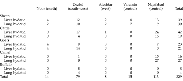
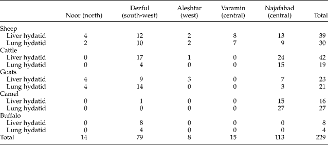
Liver and lung samples were obtained from 229 animals, including sheep, cattle, goats, camels and buffalo, in different geographical and ecological areas of Iran during June 2007 to April 2009 (table 1). Because of the large size of the country, in order to consider all the different areas of Iran and possible diversity of E. granulosus, five regions were selected: Mazandaran province (Noor County), Khozestan province (Dezful County), Lorestan province (Aleshtar County), Tehran province (Varamin County) and Isfahan province (Najafabad County).
The geographical origin and number of animals from which cysts were obtained.
Cysts were processed separately and in this study an isolate represents protoscoleces collected from an individual hydatid cyst. The aspirated contents were examined under light microscopy for the presence of protoscoleces. These were rinsed in physiological saline and then fixed and preserved in 70% ethanol until DNA isolation, and transported to the Foodborne and Diarrhoeal Diseases Laboratory for analysis. The protoscoleces were washed several times with sterile distilled water to remove the ethanol prior to DNA extraction and stored at − 20°C until further processing.
DNA extraction and molecular analysis
DNA was purified from protoscoleces by using a modified phenol chloroform method, as reported previously (Rostami Nejad et al., Reference Rostami Nejad, Nazemalhosseini Mojarad, Nochi, Fasihi Harandi, Cheraghipour, Mowlavi and Zali2008).
Amplification and identification of G1 and G6 genotypes of E. granulosus (G1 and G6 PCR)
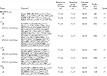
All samples were initially assigned to either the G1 or the G6 genotype as follows. Primers suitable for polymerase chain reaction (PCR) and sequencing were designed based on conserved regions of 12S rRNA sequence published for E. granulosus (AF297617 for G1, AB208063 for G6) (Rostami Nejad et al., Reference Rostami Nejad, Nazemalhosseini Mojarad, Nochi, Fasihi Harandi, Cheraghipour, Mowlavi and Zali2008). These primers amplify mitochondrial 12S ribosomal RNA sequence and are specific for the E. granulosus G1 and G6 strains, producing amplicons of very different lengths to facilitate identification in an agarose gel. Partial sequences of 12S rRNA for G1 (259 bp) and G6 (676 bp) were amplified and then sequenced by ABI 3130X Genetic analyser (Applied Biosystems, Foster City, California, USA). The PCR reaction mixture for amplification of DNA contained 10 × PCR buffer, 1.5 mm MgCl2, 0.2 mm of each deoxynucleoside triphosphate (dNTP), 0.04 U/μl Ampli-Taq DNA polymerase (Fermentas, Lithuania), 1.6 μ m of each of the forward and reverse primers, G1 FWD and G1 REV specific for G1strain and G6 FWD and G6 REV specific for G6 strain, in a 25 μl final volume, separately (table 2). Amplification was carried out for 35 cycles as follows: denaturation at 94°C for 30 s, annealing at 57°C (for G1) and 59°C (for G6) for 30 s and elongation at 72°C for 40 s.
Primers and profiles used for amplification of the G1 and G6 genotypes, cox1, nad1, atp6.

a Sequences are provided from 5′ to 3′ end.
PCR amplicons underwent electrophoresis in a 1.2% agarose gel with Tris-acetate electrophoresis buffer and were stained with ethidium and visualized under a UV transilluminator. Positive controls to monitor PCR success and negative controls to check for false-positive results that may have arisen from carryover contamination were also included in all experiments.
Amplification of mitochondrial nad1, cox1 and atp6
In these steps, targets were complete or near-complete sequences of the nad1, cox1 and atp6 genes. Published mtDNA sequences of cox1, nad1 and atp6 (AF297617 for G1, AB208063 for G6) genes were used for comparison.
For each sequenced isolate, three different genes were amplified by PCR, using primers specific for Echinococcus mtDNA. The sequences of primers, annealing temperatures, numbers of cycles, and sizes of products were individually determined for different primer pairs (table 2). The PCR reactions were prepared and carried out as described above. PCR products were purified and sequenced with an ABI 3130X Genetic analyser. Approximately 1 μg of test DNA was sequenced. Sequences were determined and analysed using the Chromas version 2.33 software (Technelysium Ltd, Australia). All sequences were subjected to a BLAST search to determine their identities and similarities to those in GenBank. Sequences were aligned with published 12S rRNA, cox1, nad1 and atp6 genes of genotypes G1 and G6 for E. granulosus by using multiple sequence alignment (http://multalin.toulouse.inra.fr/multalin/multalin.html).
Results
Two hundred and twenty-nine liver and lung hydatid isolates were acquired from sheep, cattle, camels, goats and buffalo in five different geographical and ecological areas of Iran (tables 1 and 3). For all species except the camel, the majority of samples were found in the liver. PCR amplifications for each of the desired four genes were performed on all 229 samples but, due to the large number of samples, only 108 isolates from each animal/area were selected and sequenced.
The number of isolates examined per animal species and strain/genetic variant.

Genotype 1 is most common in the studied population. 91.3% of sheep, 84% of cattle, 87% of goat and 88.3% of camel isolates but only 34% of buffalo samples belonged to the G1 strain. Out of 43 camel isolates only 5 were of the G6 genotype. G6 was not detected in the rest of the studied samples (table 3).
PCR was not successful for 29 samples and none of them belonged to G1 and G6 genotypes (10 cattle isolates, 8 buffalo, 6 sheep and 5 goats). Thus they might belong to other, different, genotypes or may be due to low DNA concentration, degeneration of DNA and inhibitors.
12S rRNA gene sequence data
The G1-PCR amplified the G1 genotype of E. granulosus with a specific band of 259 bp. Seven isolates showed 99% sequence identity with a 12S rRNA gene of E. granulosus G1 strain retrieved from GenBank (accession no. AF297617), while the rest of the isolates showed 100% sequence identity with the published 12S rRNA gene of E. granulosus G1 strain.
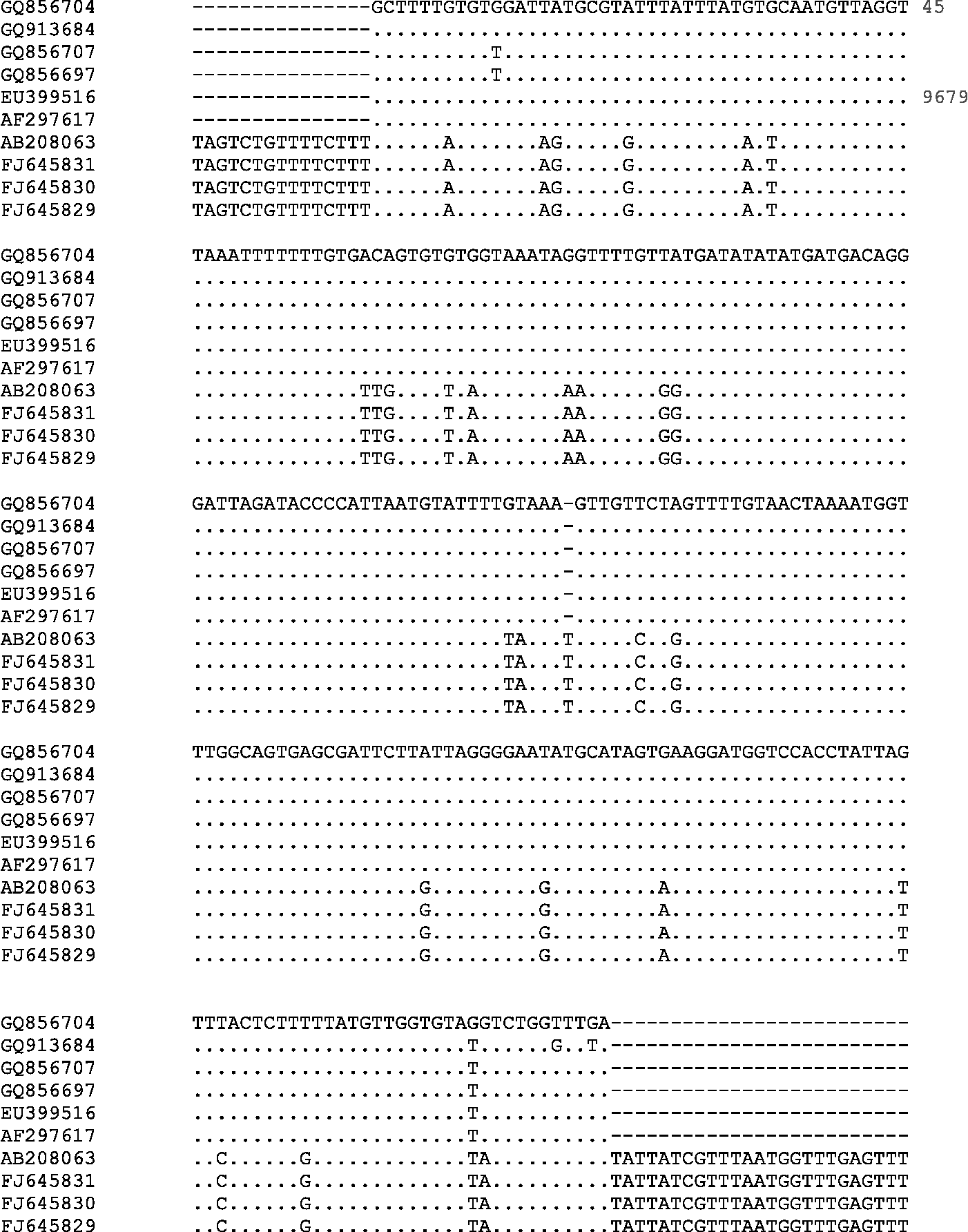
The G6-PCR amplified the G6 genotype of E. granulosus with a specific band of 676 bp. Five isolates (camel origin from Isfahan province) displayed 99% sequence identity with a 12S rRNA gene of G6 E. granulosus strain (accession no. AB208063). The alignments for the G6 genotype compared to the G1 genotype are given in fig. 1.
Comparison of 12S rRNA gene sequences of genotypes 1 and 6 in obtained sequences with those published in GenBank (accession numbers AF297617 (for G1)/AB208063 (for G6)) (conserved sequences are not mentioned).
Sequence data
The sequences obtained in this study have been deposited in the GenBank database under the accession numbers EU399513–EU399519, EU275231–EU275233, FJ010974–FJ010975, GQ913678–GQ913687 and GQ856694–GQ856717 for the G1 genotype and FJ645829–FJ645831 for the G6 genotype.
NADH dehydrogenase subunit I gene (nad1)
Complete nucleotide sequences (1157 bp for G1 genotype and 1054 bp for G6 genotype) were also obtained for the nad1 gene of all randomly selected E. granulosus isolates from different hosts. The sequences were aligned with published sequences for the G1 and G6 genotypes. Eight isolates of cattle, sheep and camel origin had a 98–99% identity with the common domestic sheep strain (G1 genotype, AF297617). A further 22 isolates had an absolute identity to the published nad1 sequences for the G1 strain.
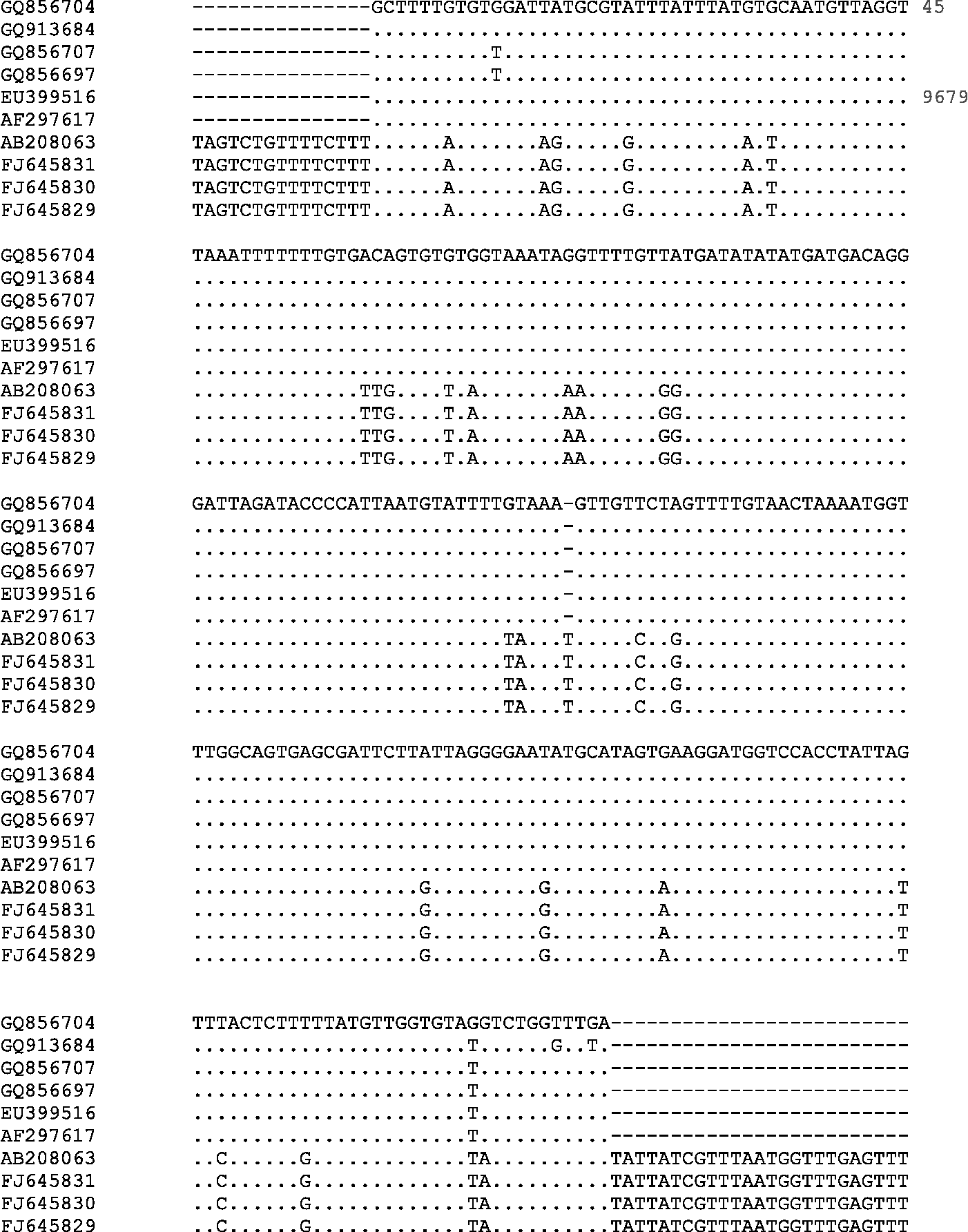
Two of four isolates (camel origin from Isfahan province) showed identical sequence to the well-characterized camel strain (AB208063) and other two isolates displayed 99% sequence identity with nad1 sequence for the G6 strain (fig. 2).
Comparison of nad1 gene sequences of genotypes 1 and 6 in obtained sequences with those published in GenBank (accession numbers AF297617/AB208063) (conserved sequences are not mentioned).
The nucleotide sequence data for this gene were submitted to GenBank under accession numbers FJ010971–FJ010973, GQ357999–GQ358013 and HM055617–HM055628 for G1 and accession numbers HM749615–HM749618 for G6.
ATPase subunit 6 (atp6)
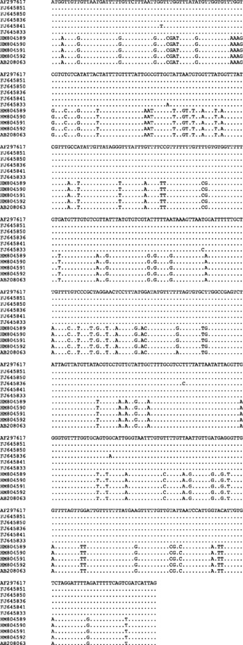
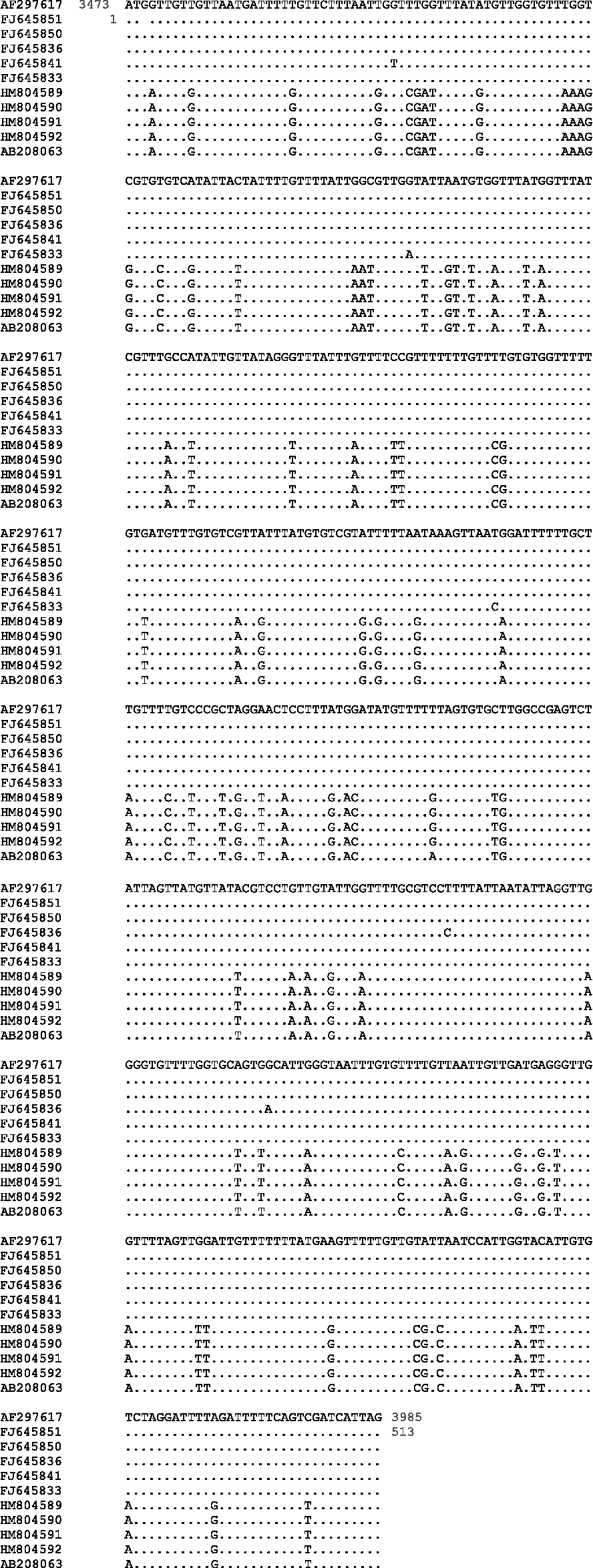
With the mt atp6 marker, we obtained a 708 bp complete sequence for G1 strain and 705 bp for G6 strain in all randomly selected samples. Eleven of 22 isolates had high levels of identity (98–99%) in the atp6 sequence compared with the reference G1 genotype. Nucleotide substitutions were seen only in one or two nucleotide positions. Sequence analyses of the atp6 gene in the other 11 specimens showed complete identity with the reference atp6 sequence for the G1 genotype. Four isolates displayed 99% sequence identity with the atp6 gene of G6 of E. granulosus. The alignments for atp6 sequences for the G6 compared to G1 are given in fig. 3. The same results have been obtained by the atp6 and nad1 gene sequence analysis for G1 and G6.
Comparison of atp6 gene sequences of genotypes 1 and 6 in obtained sequences with those published in GenBank (accession numbers AF297617/AB208063) (conserved sequences are not mentioned).

The G1 and G6 atp6 gene sequences were registered in GenBank under accession numbers FJ645832–FJ645851, EU667993–EU667994 and HM804589–HM804592.
Cytochrome c oxidase I gene (cox1)
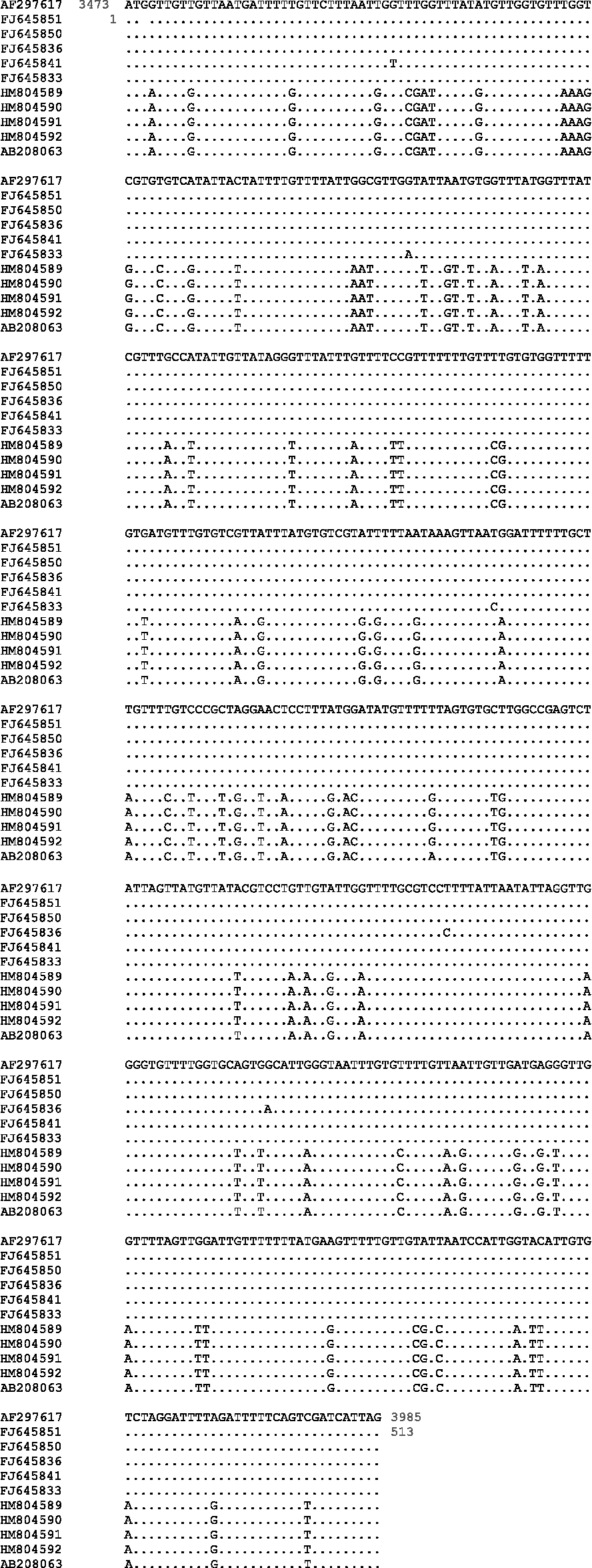
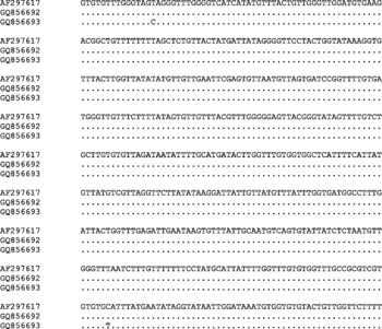
The sequencing of the mitochondrial cox1 gene of randomly selected isolates obtained from different hosts produced unambiguous sequences of 1830 bp and 1856 bp for G1 and G6 genotypes, respectively. These fragments were purified with a commercially available kit (Fermentas, Lithuania) according to the manufacturer's instructions, but only two isolates of the cox1 gene were successfully sequenced. A cattle isolate (GenBank GQ856693) was very similar to the above G1 genotypic sequence. The other isolate was completely identical to the G1 reference sequence. The alignments for cox1 are given in fig. 4. These two cox1 gene sequences were registered in GenBank as well (GQ856692 and GQ856693).
Comparison of cox1 gene sequences of genotype 1 in obtained sequences with those published in GenBank (accession no. AF297617) (conserved sequences are not mentioned).

Discussion
In the present study, 229 hydatid individuals of cattle, sheep, goat, camel and buffalo origin were collected in five different geographical and ecological areas of Iran. Extensive intra-specific variation in E. granulosus may influence life-cycle patterns, development rate, host specificity and transmission dynamics of the parasite (Utuka et al., Reference Utuka, Simsekb, Koroglub and McManus2008). Definitive identification of E. granulosus genotypes requires a combination of approaches for DNA characterization (Utuka et al., Reference Utuka, Simsekb, Koroglub and McManus2008). Many investigators have used either or both molecular and morphological studies in order to discriminate different Echinococcus strains/species (Kumaratilake et al., Reference Kumaratilake, Thompson and Eckert1986; Hope et al., Reference Hope, Bowles, Prociv and McManus1992; Wachira et al., Reference Wachira, Bowles, Zeyhle and McManus1993; Zhang et al., Reference Zhang, Eslami, Hosseini and McManus1998; Fasihi Harandi et al., Reference Fasihi Harandi, Hobbs, Adams, Mobedi, Morgan-Ryan and Thompson2002; Ahmadi & Dalimi, Reference Ahmadi and Dalimi2006; Badaraco et al., Reference Badaraco, Ayala, Bart, Gottstein and Haag2008; Sharbatkhori et al., Reference Sharbatkhori, Mirhendi, Jex, Pangasa, Campbell, Kia, Eshraghian, Fasihi Harandi and Gasser2009; Snábel et al., Reference Snábel, Altintas, D'Amelio, Nakao, Romig, Yolasigmaz, Gunes, Turk, Busi, Hüttner, Sevcová, Ito, Altintas and Dubinský2009; Nakao et al., Reference Nakao, Li, Han, Ma, Xiao, Qiu, Wang, Yanagida, Mamuti, Wen, Moro, Giraudoux, Craig and Ito2010).
A few mitochondrial and nuclear DNA analyses have been carried out successfully on Iranian isolates. The first study was based on regions of the mitochondrial cox1 and nad1 genes, on 16 isolates of E. granulosus and showed the presence of two genotypes, G1 and G6 (Zhang et al., Reference Zhang, Eslami, Hosseini and McManus1998). A second study using both morphological and molecular analysis (polymerase chain reaction–restriction fragment length polymorphism (PCR-RFLP) of the internal transcribed spacer (ITS1) region), showed that sheep and camel strains are cross-transmitted between camel, sheep, cattle and humans as well (Fasihi Harandi et al., Reference Fasihi Harandi, Hobbs, Adams, Mobedi, Morgan-Ryan and Thompson2002).
In a further study using PCR-RFLP of ITS1 of the rDNA, at least two distinct PCR-RFLP profiles of DNA of the human, common sheep and camel isolates were demonstrated (Ahmadi & Dalimi, Reference Ahmadi and Dalimi2006).
Shahnazi et al. (Reference Shahnazi, Hejazi, Salehi and Andalib2011) sequenced livestock and human isolates using the same method as in the previous study and showed that that the sheep strain was the most common genotype of E. granulosus, affecting humans, sheep, cattle, goats and, occasionally, camels; this result was confirmed by the study of Kia et al. (Reference Kia, Rahimi, Sharbatkhori, Talebi, Fasihi Harandi and Mirhendi2010).
In the study of 112 E. granulosus isolates from different livestock species in Iran, intra-genotype variation was investigated using random amplification of polymorphic DNA-polymerase chain reaction (RAPD-PCR). RAPD analysis based on 15 separate primers showed 7–14 bands of 200–3000 bp for strain G1. No differences were observed among isolates from different hosts and between livers and lungs (Sharbatkhori et al., Reference Sharbatkhori, Mirhendi, Fasihi Harandi, Rezaeian, Mohebali, Eshraghian, Rahimi and Kia2010).
The present study is the first genotypic analysis of E. granulosus infecting sheep, camel, buffalo, cattle and goat intermediate hosts using four mt markers – 12S rRNA and complete or near-complete sequences of nad1, cox1 and atp6. This study presents for the first time atp6 sequence data from Iranian hydatid isolates. The PCR system presented here was specifically designed as a screening tool for molecular epidemiological studies on echinococcosis in Iran.
Most of the sheep and cattle isolates had considerable similarity with the common domestic sheep strain (G1) referenced in GenBank (AF297617). Four isolates showed absolute identity with the published nad1 sequences for G6 strain. For atp6, the sequences of 11 isolates were near-identical to the G1 genotype and four camel isolates were closely similar to the G6 genotypic sequence. The PCR amplification of cox1 was successful just in two isolates. One of them was very similar to the G1 genotypic sequence and the other one was completely identical to the G1 strain.
The negative results of other samples may presumably be due to the low DNA concentration, degeneration of DNA, inhibitors or the presence of other non-target genotypes, etc. The DNA sequence variation within regions of the mitochondrial nad1, atp6 and cox1 genes of selected isolates of E. granulosus has demonstrated the presence of two E. granulosus genotypes in Iran: the common sheep/dog (G1) and camel/dog (G6) forms. Those isolates that were negative either for G1 or G6 genotypes could carry other genotypes, such as G3, especially in the camel strain, as Sharbatkhori et al. (Reference Sharbatkhori, Fasihi Harandi, Mirhendi, Hajialilo and Kia2011) recently reported. Therefore, exploring other genotypes by using universal primers is suggested for negative samples. On the other hand, the present study provides further evidence for the predominant presence of the G1 genotype of E. granulosus in all animal hosts in Iran. The results of this study show that, in addition to the 12S rRNA gene, atp6 and nad1 genes are good molecular markers for identifying phylogenetic analysis in E. granulosus isolates from a range of hosts in Iran.
Acknowledgements
This study has been financially supported by the Research Center of Gastroenterology and Liver disease (RCGLD), Shahid Beheshti University of Medical Science, Tehran, Iran. We acknowledge the very helpful assistance of Professor David Blair from the School of Marine and Tropical Biology, James Cook University, Townsville, Queensland, Australia. Also the authors would like to thank Mrs M. Azimi Rad, Mr M. Chiani, Mr E. Goldoust and Mr B. Damavand for their sincere cooperation in the molecular laboratory.