Cardiac rhabdomyomas are the most common cardiac tumours in foetal life, accounting for up to 60–86% of all primary cardiac tumours.Reference Isaacs 1 , Reference Holley, Martin and Brenner 2 Owing to improvements in foetal echocardiography, prenatal diagnosis has become more common. Association between cardiac rhabdomyomas and tuberous sclerosis has been reported.Reference Isaacs 1 The course in utero and the perinatal outcome depend upon growth, size, and location of the tumour; in fact, larger masses can cause haemodynamic and electrophysiological impairment and are associated with a poor prognosis.Reference Chao, Chao and Wang 3
It is important for clinicians to counsel on the basis of the natural course and outcome of affected pregnancies in order to reduce the cases of pregnancy termination after a prenatal diagnosis of cardiac tumour.
We present the case of a large rhabdomyoma of the interventricular septum diagnosed prenatally, presenting with supraventricular arrhythmias and right bundle branch block, which spontaneously regressed during childhood.
Case report
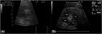
A 32-week primigravida was referred to our Prenatal Diagnosis Service for echocardiographic examination because of abnormal four-chamber view findings observed during obstetric ultrasound scan. The male foetus presented a round, homogeneous, hyperechogenic cardiac mass of 1.5 × 1.2 cm consistent with a single cardiac rhabdomyoma, located in the interventricular septum, involving the atrioventricular junction and resulting in a reduction of the right ventricular cavity, without signs of obstruction, arrhythmias, or haemodynamic impairment (Fig 1). Colour Doppler study showed minimal turbulence across the right ventricular outflow tract. The other cardiac structures, valves, and chambers were free from any other tumour mass or abnormality. There was no family history for tuberous sclerosis. The family received an adequate counselling and decided to continue the pregnancy.
Foetal echocardioghraphy findings at 32 weeks of gestation: ( a ) a single round echogenic mass consistent with a rhabdomyoma 12.71 × 15.78 mm in size; ( b ) a four-chamber view of the cardiac rhabdomyoma located in the interventricular septum, protruding in the right ventricle without signs of obstruction. LA = left atrium; LV = left ventricle; RA = right atrium; RV = right ventricle.

Follow-up ultrasound examinations were performed every 2 weeks until delivery. At 38 weeks of gestational age, the mass appeared to have grown to 2.5 × 1.6 cm without signs of outflow or inflow obstruction; however, short runs of supraventricular arrhythmias were recorded.
Delivery was planned at 40 weeks of gestational age: the weight of the newborn was 4085 g and Apgar score was 9–10. His heart rate was 180 beats/minute, blood pressure was 60/40 mmHg, and oxygen saturation was 90–95%. The ECG showed supraventricular arrhythmias with a complete right branch bundle block.
The transthoracic echocardiographic study confirmed the presence of a single large rhabdomyoma (2.6 × 1.6 cm) located in the interventricular septum with slight signs of right inflow obstruction. The baby was screened with physical examination, Wood's lamp skin examination, brain magnetic resonance and genetic test, all of which had negative results excluding the association with tuberous sclerosis.
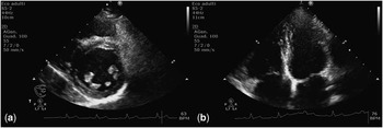
Owing to the extensive intramural myocardial involvement and the absence of clinical impairment, surgical resection of the mass was excluded and the patient was followed up by medical therapy. He was discharged from the hospital after several days of observation and was haemodynamically stable and asymptomatic. By 6 months of age, the patient continued to do well clinically. His cardiac rhabdomyoma remained large but decreased somewhat in size. At 1 year of age, the cardiac mass size was unchanged but supraventricular arrhythmias disappeared and no more evidence of right inflow obstruction was observed. Over the next 6 years, he underwent cardiac evaluation annually including echocardiography, 24-hour Holter monitoring, and cardiac stress test, and a progressive reduction in mass size was seen. At 7 years of age, ultrasound examinations documented the complete regression of the tumour. In the later follow-up, the child remained asymptomatic and the only echocardiographic finding was a hyperechogenic appearance of the septal wall in the area originally occupied by the tumour consistent with fibrosis (Fig 2). Left ventricular function and shortening fraction were normal. ECG showed a complete right bundle block.
Echocardiographic findings at 7 years of age: both short-axis ( a ) and apical four-chamber views ( b ) showing a complete regression of the mass with hyperechogenic aspect of the septal wall.

Discussion
We have described the case of a very large interventricular rhabdomyoma presenting with supraventricular arrhythmias and right bundle branch block, which regressed spontaneously during childhood. Primary cardiac tumours have been identified in 0.02–0.04% of the paediatric population.Reference Padalino, Vida and Boccuzzo 4 Most of them are benign rhabdomyomas closely associated with tuberous sclerosis complex. Other cardiac tumours include myxoma, fibroma, and teratoma. An important characteristic of cardiac rhabdomyomas is spontaneous regression, and this occurs in almost 50% of all cases.Reference Black, Kadletz, Smallhorn and Freedom 5 , Reference Günther, Schreiber, Noebauer, Eicken and Lange 6 Surgical resection is indicated in patients with inflow and outflow obstruction, which leads to haemodynamic instability, valvular dysfunction, and dysrhythmias, because of the risk of sudden death.Reference Stiller, Hetzer and Meyer 7 However, patients with giant tumour masses compressing or infiltrating the heart frequently cannot undergo complete resection. For these patients, preservation of sufficient heart function is the primary goal. Moreover, surgery is not usually recommended in patients with stable haemodynamics because of the possibility of spontaneous regression of rhabdomyomas.Reference Freedom, Lee, MacDonald and Taylor 8 Nevertheless, Cina et al.Reference Cina, Smialek, Burke, Virmani and Hutchins 9 reported that of 103 benign tumours that caused sudden death 9 (8.7%) were due to rhabdomyomas. The risk of sudden death with foetal arrhythmias and rhabdomyomas has been recently emphasised.Reference Nguyen, Smythe and Baranchuk 10
At birth, our patient presented with a very large rhabdomyoma with arrhythmias, which could represent an indication for surgical treatment. However, because of extensive intramural myocardial involvement and stable haemodynamics the patient was not considered candidate for surgical resection. The size and the location of the tumour played a major role in the prognosis of the patient. Chao et al.Reference Chao, Chao and Wang 3 presented a meta-analysis of 266 antenatally diagnosed cardiac rhabdomyoma. The smallest mass detectable was 4 mm and the biggest was 52 mm in diameter. The authors emphasised how these tumours might slowly increase their size in utero and identified tumour size ≥20 mm and foetal dysrhythmias as causes of neonatal morbidity. Although histologically benign, larger tumours carry a greater risk of causing haemodynamic derangement and dysrhythmias, which could result in poorer outcome at the foetal stage. This contribution shows how even larger masses, such as our case of 2.6 × 1.6 cm presenting with arrhythmias, can be associated with excellent long-term outcome, in the absence of haemodynamic impairment. Seriate echocardiography may represent the best conservative approach in the management of patients with cardiac masses who are haemodynamically stable. If the tumour does not regress and signs of haemodynamic instability persist, a new treatment with Everolimus has recently been proposed and successfully applied in critically ill patients who cannot be operated upon.Reference Demir, Ekici, Yazal Erdem, Emir and Tunç 11
We suggest that a conservative initial approach be justified even in large rhabdomyomas, particularly in cases without a family history of tuberous sclerosis complex.
Ethical Standards
An informed consent was obtained.