Introduction
Congenital choanal atresia is a rare malformation which causes airway obstruction in neonates and infants; it has an incidence of one in 7000 to 8000 births. It seems to occur more commonly in girls than boys, and to be more frequently unilateral and right-sided than bilateral. The nature of the obstructing atretic plate has been described as 90 per cent bony and 10 per cent membranous. Modern imaging techniques have revealed that 30 per cent of such malformations consist of a purely bony obstruction, and 70 per cent of a mixed bony-membranous obstruction.Reference Brown, Smith, Armstrong and Grundfast1
Most cases of congenital choanal atresia involve isolated malformations, but association with other congenital deformities is not exceptional (as in the ‘CHARGE’ anomaly syndrome, which includes coloboma, heart disease, congenital choanal atresia, retarded development, genital hypoplasia and ear anomalies).Reference Van Den Abbeele, Francois and Narcy2
There are currently four hypotheses regarding the embryological origin of congenital choanal atresia, but none has been proved. These include the persistence of the nasobuccal membrane or of the foregut buccopharyngeal membrane, abnormal mesodermal adhesions in the nasal choanae, and a misdirection of mesodermal flow due to local factors.Reference Hengerer and Marshall3
Congenital choanal atresia is characterised by hypertrophy of the posterior vomer, together with ossification of the posterior portion of the lateral nasal wall at the level of the sphenopalatine region adjacent to the pterygoid plates.Reference Wiatrak4
Many approaches have been used to repair congenital choanal atresia. Four classical surgical approaches have been described: transnasal, transpalatal, trans-septal and transantral.Reference Pardo Romero, Mogollón Cano-Cortés, Pando Pinto, Trinidad Ruiz, González Palomino and Pantoja Hernández5 The transpalatal technique is the method preferred by most surgeons, due to its excellent surgical visibility and success rates of approximately 80 to 90 per cent.Reference Van Den Abbeele, Francois and Narcy2 The transnasal approach has fallen from favour because of its high failure rate, requiring revision.Reference Richardson and Osguthorpe6
However, the development of rigid endoscopes and powered instrumentation has led to reconsideration of the transnasal approach.Reference Kamel7 This technique, performed endoscopically, is suitable for young children (even neonates) with bilateral congenital choanal atresia, and also for revision cases in which previous transnasal or transpalatal approaches have failed.Reference Van Den Abbeele, Francois and Narcy2 This endoscopic approach allows a shorter hospital stay and reduced blood loss, compared with other, more traditional techniques.Reference Pasquini, Sciarretta, Saggese, Cantaroni, Macrì and Farneti8
Patients and methods
This study included 10 neonates with bilateral congenital choanal atresia, aged between 3 and 27 days (mean, 11.2 days), seen at the otorhinolaryngology department of Mansoura University Hospital between March 2005 to February 2008. Prior to commencement, the relevant institutional review board approved the research, and appropriate consent was obtained from the parents of all patients.
Clinical evaluation included a complete physical examination, to assess for other congenital anomalies, and a complete nasal and nasopharyngeal examination, to assess the deformity. All patients underwent computed tomography (CT) scanning of the nose and paranasal sinuses to assess the location and type of atresia. The atresias were either bony (in six patients), or composed of mixed bony and membranous elements (four patients) (Figure 1).
Axial computed tomography scan showing bilateral congenital choanal atresia.

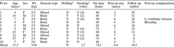
All patients were treated by the transnasal endoscopic surgical technique, using conventional ‘biting’ instruments. However, the transnasal endoscopic drill-out technique was added in four cases. Table I shows the patients' ages at the time of treatment, sex, weight, type of stenosis (bony, membranous or mixed), use of drilling and stenting, follow-up results, and post-operative complications.
Patient variables

Pt no = patient number; wt = weight; wks = weeks; op time = operation duration; post-op = post-operative; mths = months; F = female; M = male; Y = yes; N = no; L = left
Oxymetazoline hydrochloride spray was applied to the nasal cavity 15 minutes before transporting the patient to the operating theatre. On arrival, general anaesthesia was induced.
Using 4.0 mm 0° and 30° telescopes (Karl Storz, Tuttlingen, Germany), both sides of the nasal cavity were inspected, and the atretic plate was carefully identified. Otoscopic suction tubes were used to remove discharge and to palpate the atretic plate.
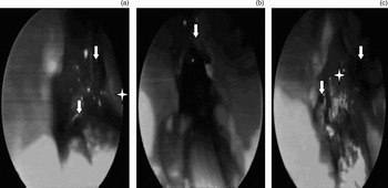
Under endoscopic visualisation, a vertical crossover incision was placed at the cartilaginous–bony junction, a few millimetres superior to the maxillary crest, over the posterior bony septum in the widest part of the nasal cavity. The vomer was then removed piecemeal using a dissector and paediatric endoscopic forceps with 2- and 4-mm blades (straight, 45° and 90°). The two nasal cavities could be reached via this septal window, and this enabled bilateral endoscopic visualisation, together with the facility to place the telescope on one side and the dissecting instrument on the other side in order to facilitate bone removal. The posterior vomer and the mucosa over the atretic plate were carefully removed. Using the suction tube, the atretic plate was punctured at its weakest point, to establish a reference point into the nasopharynx. Using conventional forceps, the atretic plates were then removed bilaterally, working in a medial to lateral direction, in order to open the choanae. Each choana was then enlarged circumferentially, avoiding superior manipulation, grasping, and blind, vigorous manoeuvres (Figure 2). The inferior aspect of the middle turbinate was used as a guide for the superior limit of dissection.
(a) Endoscopic view of the septal window, showing two posterior choanae (arrows) and septum (star). (b) Endoscopic view of right choana (arrow) after removal of atretic plate. (c) Endoscopic view of the septal window, showing two posterior choanae (arrows) and posterior vomer bone (star).

In four cases, drilling of the atretic plates was required in order to achieve sufficient choanal opening. Drilling was employed only when required by the thickness of the atretic plate, in order to achieve complete resection. Drilling was commenced inferiorly and medially and continued until the choana opened; then conventional instrumentation was used to widen the choana. Excess superior or blind drilling was avoided; drilling stayed posterior, inferior and medial in the nasal cavity, along the posterior maxillary crest.
The mean operating time was 74 minutes. When surgery was completed, seven patients were fitted with a custom-made, soft nasal stent created from a size 3.5 polyethylene tracheal tube, placed in the neochoanae and secured anteriorly with a trans-septal Vicryl 3-0 suture (Figure 3). The nasopharyngeal position of the stent was ensured by oropharyngeal examination, and stent patency was tested with a saline injection.
Customised soft nasal stent.

Antibiotic medication and paediatric isotonic sodium chloride nasal drops were administered post-operatively. Stents remained in situ for a period ranging from one to four weeks (mean, 2.7 weeks) (Table I).
Results
The success rate of the primary procedure was 70 per cent, as seven of the 10 patients had patent choanae after one transnasal endoscopic procedure. The endoscopic aspect of the choanae looked almost normal in five of these cases, and was slightly narrowed with a small amount of granulation or adhesions in two cases (Figure 4). All patients were clinically and endoscopically monitored for nasal obstruction and healing. The follow-up period ranged from five to 35 months (mean, 18 months) (Table I).
Endoscopic view taken during ‘second look’ endoscopy, showing nasal adhesions in the left posterior choana.

No major post-operative complications occurred; however, one case showed post-operative unilateral vestibular stenosis. In this case, drilling and stenting had been used. Excessive post-operative bleeding occurred in one case, which was managed by stent removal and packing for two days. No other post-operative complications (e.g. distress, infection, bleeding or nasal deformity) were encountered. Normal feeding was possible on the day after surgery. The mean post-operative hospital stay was 6.4 days (Table I).
Nasal stents (fashioned from size 3.5 endotracheal tubes) were used in seven patients, and were removed between one to four weeks after surgery. ‘Second look’ endoscopic evaluation was performed under general anaesthesia two to four weeks post-operatively (Table I). Of the seven patients with stenting, five remained patent, while two required minor debridement of granulation tissue, with a patent result after the second procedure. Of the three patients without stenting, two remained patent, while one required minor debridement of granulation tissue, with a patent result after the second procedure.
Discussion
Newborn children are obligate nasal breathers. Bilateral congenital choanal atresia is characterised by respiratory distress at birth, although Wiatrack observed that respiratory distress can also be present in cases of unilateral choanal atresia within the first week of life.Reference Wiatrak4 Pre-operative radiological assessment, by means of a CT scan, is very important for the success of surgical treatment; CT scans accurately characterise the nature and thickness of the atresia, the narrowing of the posterior nasal cavity, and the thickening of the vomer.Reference Brown, Smith, Armstrong and Grundfast1
Surgical management of choanal atresia is controversial. The evolution of optical telescopes and other instrumentation, and the development of powered microdebriders with drill attachments, have revolutionised the transnasal approach.Reference Wiatrak4 The transnasal endoscopic approach has been successfully used for the treatment of congenital choanal atresia.Reference Kamel7, Reference Anderhuber and Stammberger9, Reference Lazar and Younis10 This technique enables a direct approach to the atretic area, with the advantage of an angled vision, good illumination and magnification of the operative site. The current study modified this technique somewhat by using a transnasal approach under endoscopic guidance with a 120° Hopkins rod telescope, in order to visualise the posterior choanae from the nasopharynx.Reference Elloy, Cochrane and Albert11 In the current series, the operative technique was similar to that of Stankiewicz, who reported that resection of the vomer by means of backbiting forceps, together with drilling out of the lateral bone, helped to create a large opening, which was crucial to the success of the procedure.Reference Stankiewicz12 Kamel reported using a cruciate incision for exposure of the mucoperiosteum covering the atretic plate, then a diamond burr to create a hole at the level of the inferomedial portion of the plate (considered the thinnest and safest area); the neochoana was then widened laterally at the level of the pterygoid plate and palatine bone, medially at the septum, and superiorly at the sphenoid sinus.Reference Kamel7
The technique of mucosal preservation and neochoana resurfacing, used in the trans-septal approach to congenital choanal atresia repair, is useful in reducing the post-operative recurrence rate and also the period of post-operative stenting.Reference Andrieu, Guitrancourt and Stipon13 However, in this study flap preservation and elevation was technically inapplicable in this patients' age group. Furthermore, the shortened stenting period and the use of soft stents diminished the risk of granulation tissue formation and post-operative infection.Reference Pasquini, Sciarretta, Saggese, Cantaroni, Macrì and Farneti8
The benefits and risks of stenting after congenital choanal atresia repair have been hotly debated. Stents cause discomfort, localised infection and ulceration, circumferential scar tissue, and injury to surrounding tissue.Reference Friedman, Mitchell, Bailey, Albert and Leighton14 Once removed, circumferential restenosis is common. Indwelling nasal stents are difficult to manage, easily crust and clog (causing a functional stenosis or blockage during the period of stenting), and may migrate or break. However, regardless of the risks, some authors believe that stenting is necessary in all cases, while others believe that stenting is only needed in bilateral cases. One case series used long-term stenting for at least three to four weeks, while another used short-term stenting for less than one week.Reference Pasquini, Sciarretta, Saggese, Cantaroni, Macrì and Farneti8, Reference Samadi, Shah and Handler15 Another study used no stents at all.Reference Cedin, Peixoto Rocha, Deppermann, Moraes Manzano, Murao and Shimuta16 The use of stents is advocated by some and avoided by others.Reference Van Den Abbeele, Francois and Narcy2, Reference Holland and McGuirt17–Reference McLeod, Brooks and Mair19 In this study seven patients with stenting, five remained patent and two (28.5 per cent) required minor debridement of granulation tissue, with a patent result. Of three patients without stenting, two remained patent and one (33.3 per cent) required minor debridement of granulation tissue, with a patent result. However, one patient with transnasal endoscopic choanoplasty, who received stenting and drilling, developed vestibular stenosis and adhesions. In this series, stenting was preferred in hospitalised cases treated in humidicribs, and in cases in which extensive mucosal laceration and/or drilling had been required. The duration of stenting varied from one to four weeks (mean, 2.7 weeks).
In the new era of miniaturised telescopes, high-speed protected drill bits and microdebriders, lateral drilling is eliminated or minimised. Focusing on careful removal of the posterior septal–vomer segment reduces the need to protect denuded tissue in order to prevent the development of obstructive synechiae and exuberant granulation tissue. Schoem supports the notion that stenting may not be necessary in the transnasal endoscopic repair of both unilateral and bilateral choanal atresia.Reference Schoem20 All patients in this author's study were treated without stents, and patency results compared favourably with those of studies using stents.
Transnasal endoscopic repair of congenital choanal atresia is a safe and effective procedure. This technique is suitable for neonates with bilateral congenital choanal atresia.Reference Van Den Abbeele, Francois and Narcy2 The endoscopic approach allows a shorter hospital stay and fewer complications, compared with other, more traditional techniques.Reference Pasquini, Sciarretta, Saggese, Cantaroni, Macrì and Farneti8, Reference Mumbuç, Karataş, Durucu, Ozer and Kanlıkama21, Reference Yaniv, Hadar, Shvero, Stern and Raveh22 In the current series, early experience with transnasal endoscopic choanal atresia repair showed a primary success rate of 70 per cent; after second look endoscopic debridement, the success rate was 100 per cent, without any major complications. Post-operative failure of the endoscopic approach may be a result of prolonged mucosal trauma from stenting, rather than any deficiency inherent in the surgical technique.Reference Schoem20
Factors which appear to favourably affect the outcome of bilateral congenital choanal atresia surgery include the absence of associated facial anomalies, weight at the time of surgery (i.e. greater than 2.3 kg), stent size (i.e. greater than 3.5 mm) and duration of stenting.Reference Friedman, Mitchell, Bailey, Albert and Leighton14 In the current study, the most important operative and post-operative prognostic factors were: avoidance of excessive mucosal injury; early stent removal; avoidance of unnecessary drilling; second look endoscopic debridement; and close follow up.
• Transnasal endoscopic repair of bilateral congenital choanal atresia is a safe and successful technique in neonates
• The use of powered instrumentation in the neonatal age group requires appropriate experience and a sizable drill
• Stenting with a nasal tube for a suitable period is favourable, especially in hospitalised patients
New interventions are being introduced to improve the outcome of endoscopic repair of congenital choanal atresia, including the use of mitomycin C, potassium-titanyl-phosphate laser and diode laser (which also manages recurrences effectively).Reference Kubba, Bennett and Bailey23, Reference Saetti, Santoro and Silvestrini24 The use of an optical, CT data based navigation system has been described for surgical planning and intra-operative guidance, in order to improve treatment outcome.Reference Westendorff, Dammann, Reinert and Hoffmann25 However, in the current series the use of conventional endoscopic instruments, with a 4-mm endoscope, was effective in completing the procedure. When indicated, drilling was performed as precisely as possible using suitably sized burrs, which were covered completely except for the cutting head in order to avoid mechanical and thermal injury to the important surrounding nasal and cranial structures.
Conclusion
The endoscopic, transnasal, trans-septal approach for the treatment of bilateral congenital choanal atresia is a safe, effective, easily applicable procedure for which extensive prior experience is unnecessary.
In the current study, drilling was employed only in cases in which the thickness of the atretic plate required its use in order to achieve complete resection of the atretic plate and formation of adequate neochoanae bilaterally. Flap preservation and elevation were technically inapplicable in our patient age group.
Stenting is preferred in the neonatal age group, especially in cases with extensive mucosal lacerations and/or drilling, and in hospitalised patients, to avoid restenosis and reoperation. In this study of seven patients with stenting, five remained patent, while two required minor debridement of granulation tissue, with a patent result thereafter. Of three patients without stenting, two remained patent, while one required minor debridement of granulation tissue, with a patent result thereafter. However, prolonged stenting should be avoided, in order to prevent adhesions, vestibular stenosis and infection.
An adequate post-operative follow-up period and second look endoscopy are crucial to the success of this type of surgery. Despite the controversies and debates regarding transnasal endoscopic repair of bilateral congenital choanal atresia, this technique is still target-specific, easily applicable and widely accepted.