INTRODUCTION
A well-recognized and highly conserved aspect of the life cycles of digenetic trematodes is their use of molluscs, mainly snails, as first intermediate hosts, and within which all species undergo a complex process of asexual reproduction. Infections of the molluscan host are initiated by entry of the ciliated miracidial stage where, once inside the host, they typically shed their ciliated epidermal plates, transforming to the primary sporocyst. Germinal cells within sporocysts then begin to divide forming multicellular embryos, each of which are destined to become either secondary (daughter) sporocysts or rediae (depending on the trematode species), thus constituting the next larval generation. This asexual sporocystogenic or redial-generating reproductive cycle may repeat itself several times, but eventually gives rise to the cercarial stage, which represents the next free-living stage responsible for transmitting the infection to the next host, either a second intermediate or a definitive host. Clearly the molluscan developmental phase of trematode life cycles is critically important, serving to greatly amplify the infective cercarial population that, in turn, dramatically increases the probability of successful transmission to the next host, even if the prevalence of infection within a given snail population is low (Basch, Reference Basch1991).
As alluded to above, the intramolluscan phase of development is complex and, currently, little information is available regarding the underlying mechanisms regulating successful transition of the free-swimming miracidium to the parasitic primary sporocyst stage, or the biochemical and physiological requirements for establishing and maintaining an ongoing parasite infection. In general, digeneans exhibit very narrow ranges of snail host specificity, suggesting that the physiological parameters dictating larval survival may be quite different between parasite species or their hosts. In the present review we attempt to summarize a growing body of research that focuses on the in vitro manipulation of early intramolluscan trematode stages, emphasizing the blood flukes Schistosoma spp. Topics include the current status of in vitro culture as an investigative tool, gene discovery in model digeneans, gene and protein expression profiling during in vitro larval development, and the development and application of larval transgenesis and RNA interference (RNAi) as functional genomic complements to gene discovery efforts. The results of these studies not only give important insights into fundamental questions about how host specificity is regulated and/or what physiological factors dictate infection success, but also provide a basis for the identification and possible targeting of critical molecular or biochemical pathways for disruption by chemical or biological interventions leading to termination of infections within the molluscan host and thus, transmission to humans or domestic animals.
In vitro cultivation – essential tool for gene profiling and functional genomic investigations
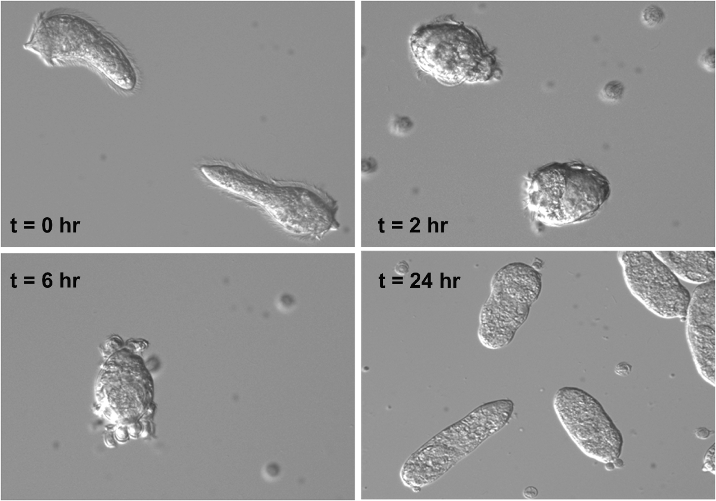
Earlier studies on the human blood flukes, Schistosoma mansoni (Yoshino and Laursen, Reference Yoshino and Laursen1995) and S. japonicum (Coustau et al. Reference Coustau, Ataev, Jourdane and Yoshino1997; Coustau and Yoshino, Reference Coustau and Yoshino2000), revealed that exposure of freshly-hatched miracidia to snail isotonic saline alone, e.g. Chernin's balanced salt solution (CBSS; Chernin, Reference Chernin1963) would trigger the shedding of ciliated miracidial epidermal plates, and the concomitant formation of the primary (=mother) sporocyst tegument under in vitro conditions (Fig. 1). Thus, it appeared that the change in environmental osmolarity (from ∼10 mOs/L in freshwater to ∼140 mOs/L in snail saline=osmolarity of snail hemolymph) was sufficient to mimic the initial penetration and early larval development in the snail intermediate host. Other trematode species including echinostomes (Echinostoma paraensei, E. caproni) (Loker et al. Reference Loker, Cimino and Hertel1992; Ataev et al. Reference Ataev, Fournaier and Coustau1998), and the common liver fluke Fasciola hepatica (Gourbal et al. Reference Gourbal, Guillou, Mitta, Sibille, Theron, Pointier and Coustau2008) also appear to share in part this osmotic ‘transformation signal’, although under in vitro conditions, transformation may not involve the release or ‘shedding’ of ciliated epidermal plates typical of S. mansoni and S. japonicum miracidia. In fact, E. caproni cultivated in vitro for 24 h or more in serum-free medium or snail cell- conditioned medium, although ceasing swimming activity and losing some cilia from epidermal plates (Ataev et al. Reference Ataev, Fournaier and Coustau1998), do not ‘shed’ their plates. Thus, it appears that the physico-chemical cues required by E. caproni, and possibly other Echinostoma spp. or fasciolids, for fully transforming to sporocysts in vitro differ from the ‘simple’ osmotic cues of schistosomes.
Schistosoma mansoni miracidia (t=0 h) as they undergo morphological transformation to the primary sporocyst stage (t=24 h). Miracidia, cultured in isotonic snail saline (Chernin's balanced salt solution, CBSS; Chernin, Reference Chernin1963), first cease swimming, assume a rounded shape (t=2 h) as expansion of intercellular ridges force ciliated epidermal plates to round up and detach from the larval surface (t=6 h). ‘t’ indicate the approximate time elapsed from initial introduction of miracidia into culture.

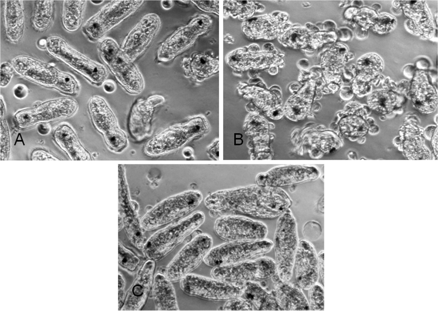
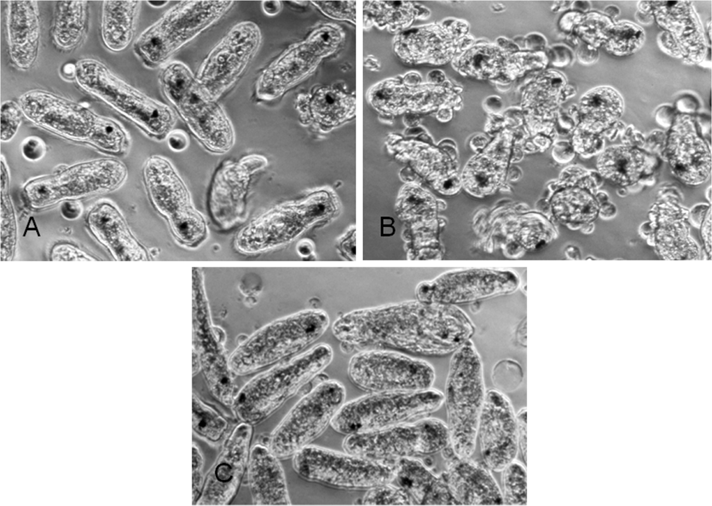
The deer liver fluke Fascioloides magna provides an informative case to illustrate the complexities of miracidial transformation signals. Miracidia of F. magna, like E. caproni, do not spontaneously transform to primary sporocysts when incubated in snail ringers (CBSS) or serum-containing medium (Laursen and Yoshino, Reference Laursen and Yoshino1999). Although miracidia stop swimming within a few hours in culture, ciliated plates remained firmly attached to the majority of larvae at 24 h post-cultivation (Fig. 2A). However, when F. magna miracidia were cultured in CBSS that had been preconditioned by incubation with the Biomphalaria glabrata embryonic (Bge) cell line, miracidium-to-sporocyst transformation was readily induced (Fig. 2B, C). Moreover, treatment of the Bge cell-conditioned medium with heat (56°C for 30 min; 100°C for 10 min) or proteinase K significantly reduced the transformation-stimulating activity of the conditioned medium (Laursen and Yoshino, Reference Laursen and Yoshino1999). Based on these findings it appears that F. magna miracidia require an additional signal(s), likely a protein or protein-associated factor, from the snail host in addition to or in place of an osmotic signal in order to trigger transformation. Supporting these findings, Campbell and Todd (Reference Campbell and Todd1955) reported that F. magna miracidial transformation in vitro (i.e. shedding of epidermal plates) only took place after miracidia had made physical contact with the snail, attempting to penetrate but failing, implying the requirement of a snail host molecular signal to initiate this process.
In vitro cultivation of Fascioloides magna (Fasiciolidae) miracidia in untreated CBSS (Chernin's balanced salt solution; Chernin, Reference Chernin1963). Within 24 h, most miracidia cease swimming activity but the majority will not fully transform (i.e. will retain their ciliated epidermal plates (A). However, miracidia cultured in CBSS, pre-conditioned by cultivation with snail Biomphalaria glabrata embryonic (Bge) cells, are induced to transform (rounding and shedding of ciliated epidermal plates (B) to become fully-transformed primary sporocysts (C).
Clearly, the signals involved in early miracidium-to-sporocyst development are complex and undoubtedly differ between trematode groups. However, in vitro culture systems, while not mimicking precisely the environment within the natural host, do provide a means of tracking changes in gene expression that accompany early larval development that may provide critical insights into the role of specific genes or gene families in the establishment and maintenance of infections within the snail host. This is particularly true for in vitro systems capable of supporting transitions to the first intramolluscan parasitic stage (primary or mother sporocyst) and, in the case of schistosome species and F. magna, advanced development to subsequent daughter sporocyst (Yoshino and Laursen, Reference Yoshino and Laursen1995; Coustau et al. Reference Coustau, Ataev, Jourdane and Yoshino1997; Ivanchenko et al. Reference Ivanchenko, Lerner, McCormick, Toumadje, Allen, Fischer, Hedstrom, Helmrich, Barnes and Bayne1999) or redial (Laursen and Yoshino, Reference Laursen and Yoshino1999) stages. With the rapid expansion of gene/protein sequence databases for schistosomes and other trematode species, the incorporation of in vitro culture systems into experimental designs represents an invaluable tool for evaluating molecular changes linked to parasite development or for exploring gene function through manipulating transcript expression.
GENOMICS, TRANSCRIPTOMICS, AND PROTEOMICS OF LARVAL TREMATODES – SO MUCH DATA, SO LITTLE INFORMATION
Because of the recognized global importance of schistosomiasis as a significant public health concern, it is not surprising that the majority of molecular research has focused on Schistosoma spp.. In part due to the limited success of traditional methods in controlling schistosomiasis, the scientific community joined efforts in 1993 to begin generating genomic sequence data from Schistosoma mansoni (Franco et al. Reference Franco, Adams, Soares, Simpson, Venter and Pena1995) with funding support from the WHO/TDR (UNICEF-UNDOWorld Bank-WHO Special Programme for Research and Training in Tropical Diseases) (Oliveira et al. Reference Oliveira, Franco and Verjovski-Almeida2008). It was envisioned at the time that the genomic data generated would promote the acceleration of gene identification strategies and data analysis, which in turn could then be translated into new tools for fighting infection and disease including discovery of new drug and vaccine targets, improved diagnostics and clinical management approaches, and, importantly, tools for dissecting the basic biological underpinning of the complex host-parasite interactions (Franco et al. Reference Franco, Valadão, Azevedo and Rabelo2000; El-Sayed et al. Reference El-Sayed, Bartholomeu, Ivens, Johnston and LoVerde2004; Wilson et al. Reference Wilson, Ashton, Braschi, Dillion, Berriman and Ivens2006; Brindley and Pearce, Reference Brindley and Pearce2007; Hokke et al. Reference Hokke, Fitzpatrick and Hoffmann2007).
By means of whole-genome shotgun sequencing approaches the entire assembled genomes for S. mansoni and S. japonicum recently have been completed, revealing estimates of genome sizes of approximately 363 MB for S. mansoni and 397 MB per haploid genome for S. japonicum, and for both species, ∼13 000 expressed transcripts, encoding ∼11 000 genes (Haas et al. Reference Haas, Berriman, Hirai, Cerqueira, LoVerde and El-Sayed2007; Berriman et al. Reference Berriman, Haas, Loverde, Wilson, Dillon, Cerqueira, Mashiyama, Al-Lazikani, Andrade, Ashton, Aslett, Bartholomeu, Blandin, Caffrey, Coghlan, Coulson, Day, Delcher, Demarco, Djikeng, Eyre, Gamble, Ghedin, Gu, Hertz-Fowler, Hirai, Hirai, Houston, Ivens, Johnston, Lacerda, Macedo, McVeigh, Ning, Oliveira, Overington, Parkhill, Pertea, Pierce, Protasio, Quail, Rajandream, Rogers, Sajid, Salzberg, Stanke, Tivey, White, Williams, Wortman, Wu, Zamanian, Zerlotini, Fraser-Liggett, Barrell and El-Sayed2009; The Schistosoma japonicumGenome Sequencing and Functional Analysis Consortium, Reference Zhou, Zheng, Chen, Zhang, Wang, Guo, Huang, Zhang, Huang, Jin, Dou, Hasegawa, Wang, Zhang, Zhou, Tao, Cao, Li, Vinar, Brejova, Brown, Li, Miller, Blair, Zhong, Chen, Liu, Hu, Wang, Zhang, Song, Chen, Xu, Xu, Ju, Huang, Brindley, McManus, Feng, Han, Lu, Ren, Wang, Gu, Kang, Chen, Chen, Chen, Wang, Yan, Wang, Li, Jin, Wang, Pu, Zhang, Zhang, Hu, Zhu, Wang, Yu, Wang, Yang, Ning, Berriman, Wei, Ruan, Zhao, Wang, Liu, Zhou, Wang, Lu, Zheng, Brindley, McManus, Blair, Zhang, Zhong, Wang, Han, Chen, Wang, Han and Chen2009). Genes are inserted into 7 pairs of autosomes and one pair of sex chromosomes (female=ZW, male=ZZ), each chromosome ranging in size from 18 to 73 MB (Simpson et al. Reference Simpson, Sher and McCutchan1982; LoVerde et al. Reference LoVerde, Hirai, Merrick, Lee and El-Sayed2004; http://lifecenter.sgst.cn/schistosoma/en/genomeProject.do; www.chgc.sh.cn/japonicum). Moreover, several transcriptomic analyses have been reported for S. mansoni (Franco et al. Reference Franco, Adams, Soares, Simpson, Venter and Pena1995, Reference Franco, Rabelo, Azevedo, Pena, Ortega, Santos, Meira, Rodrigues, Dias, Harrop, Wilson, Saber, Abdel-Hamid, Faria, Margutti, Parra and Pena1997; Santos et al. Reference Santos, Johnston, Azevedo, Ridgers, Martinez, Marotta, Santos, Fonseca, Ortega, Rabelo, Saber, Ahmed, Romeih, Franco, Rollinson and Pena1999; Verjovski-Almeida et al. Reference Verjovski-Almeida, DeMarco, Martins, Guimarães, Ojopi, Paquola, Piazza, Nishiyama, Kitajima, Adamson, Ashton, Bonaldo, Coulson, Dillon, Farias, Gregorio, Ho, Leite, Malaquias, Marques, Miyasato, Nascimento, Ohlweiler, Reis, Ribeiro, Sá, Stukart, Soares, Gargioni, Kawano, Rodrigues, Madeira, Wilson, Menck, Setubal, Leite and Dias-Neto2003; Oliveira et al. Reference Oliveira, Rodrigues, Romanha and Bahia2004), and S. japonicum (Hu et al. Reference Hu, Yan, Shen, Liu, Zhu, Song, Xu, Wang, Rong, Zeng, Wu, Zhang, Wang, Xu, Wang, Fu, Zhang, Wang, Brindley, McManus, Zue, Feng, Chen and Han2003; Peng et al. Reference Peng, Chen, Wang and Lun2003; McManus et al. Reference McManus, Hu, Brindley, Feng and Han2004), contributing to a total of 205 892 expressed sequence tags (ESTs) from S. mansoni and 103 725 for S. japonicum in the dbEST database (dbEST; release 071009 – July 10, 2009), the vast majority of which are still unannotated. Given that schistosomes, like all other digenean species, exhibit complex life cycles involving multiple developmental stages, it is expected that at least 1000 genes would be uniquely expressed in a stage-specific manner (Wilson et al. Reference Wilson, Ashton, Braschi, Dillion, Berriman and Ivens2006), although this estimate has yet to be experimentally verified. In terms of identifying genes expressed in the intramolluscan larval stages, transcriptomic analysis of S. mansoni has produced the most comprehensive datasets on gene expression in which 45 367 of 163 000 open-reading frame ESTs (ORESTs) were derived from miracidia, sporocysts (germballs) and cercariae (Verjovski-Almeida et al. Reference Verjovski-Almeida, DeMarco, Martins, Guimarães, Ojopi, Paquola, Piazza, Nishiyama, Kitajima, Adamson, Ashton, Bonaldo, Coulson, Dillon, Farias, Gregorio, Ho, Leite, Malaquias, Marques, Miyasato, Nascimento, Ohlweiler, Reis, Ribeiro, Sá, Stukart, Soares, Gargioni, Kawano, Rodrigues, Madeira, Wilson, Menck, Setubal, Leite and Dias-Neto2003). For S. japonicum, 84 499 total ESTs were generated, but unfortunately only 569 ESTs were derived from miracidia (433) and cercariae (136), with the rest originating from non-molluscan parasite stages including adult worms and eggs (Peng et al. Reference Peng, Chen, Wang and Lun2003; Liu et al. Reference Liu, Lu, Hu, Wang, Cui, Chi, Yan, Wang, Song, Xu, Wang, Zhang, Zhang, Wang, Xue, Brindley, McManus, Yang, Feng, Chen and Han2006).
With the exception of S. mansoni and S. japonicum, the accumulated genomic and transcript sequence data on other schistosome species or non-schistosome flukes are sparce indeed. For the third major human-infecting species of blood fluke, S. haematobium, dbEST lists only 6 ESTs, while GenBank entries (NCBI) total 127, with 21 sequences from cercariae and the rest from adult worms or unknown sources. Fasciola hepatica (common liver fluke) and F. gigantica (intestinal fluke), causative agents of fascioliasis in sheep and cattle (occasionally humans), are distributed worldwide and represent significant sources of economic losses in the livestock industry (McManus and Dalton, Reference McManus and Dalton2006). To date, there are only 324 and 149 nucleotide sequences in the NCBI gene database and 242 and 91 protein sequence entries in the NCBI protein database for F. hepatica and F. gigantica, respectively, and most of these originated from adult worm sources. Another model trematode that has received considerable attention are the echinostomes, which currently lists ∼400 ESTs for Echinostoma spp. in the NCBI dbEST. Most of these are from the miracidium and primary sporocyst, but very few have been annotated.
Although there is an impressive amount of gene sequence data for schistosomes and databases for other non-schistosome species continue to grow, those genes specifically expressed during larval stage development, with the exception of cercariae, have been largely under-represented or unidentified in large genome or transcriptome investigations compared to adult worm-, schistosomula- and egg-derived sequences (Verjovski-Almeida et al. Reference Verjovski-Almeida, DeMarco, Martins, Guimarães, Ojopi, Paquola, Piazza, Nishiyama, Kitajima, Adamson, Ashton, Bonaldo, Coulson, Dillon, Farias, Gregorio, Ho, Leite, Malaquias, Marques, Miyasato, Nascimento, Ohlweiler, Reis, Ribeiro, Sá, Stukart, Soares, Gargioni, Kawano, Rodrigues, Madeira, Wilson, Menck, Setubal, Leite and Dias-Neto2003; Peng et al. Reference Peng, Chen, Wang and Lun2003; Liu et al. Reference Liu, Lu, Hu, Wang, Cui, Chi, Yan, Wang, Song, Xu, Wang, Zhang, Zhang, Wang, Xue, Brindley, McManus, Yang, Feng, Chen and Han2006). Bioinformatic searches of the NCBI's UNIGENE database revealed 10 219 and 9395 sequence clusters for S. mansoni and S. japonicum, respectively. Of total S. mansoni clusters, 3392 contain identified miracidial sequences, 3128 sporocyst cDNA sequences, and 2206 cercarial sequences, although many of the raw larval sequences are unknown/unannotated. For S. japonicum, only 352 and 137 sequence clusters contain cDNA sequences (transcripts) for miracidia and cercariae, respectively, and none for sporocysts (last update in the Unigene database 09/04/2008). Similarly few intramolluscan larval sequences are specifically identified from other non-schistosome trematode species.
Despite the considerable progress made in completing the sequencing and assembly of the S. mansoni and S. japonicum genomes, gene identification and annotation are far from complete. During annotation of the S. mansoni and S. japonicum transcriptomes approximately 55% and 35%, respectively (Verjovski-Almeida et al. Reference Verjovski-Almeida, DeMarco, Martins, Guimarães, Ojopi, Paquola, Piazza, Nishiyama, Kitajima, Adamson, Ashton, Bonaldo, Coulson, Dillon, Farias, Gregorio, Ho, Leite, Malaquias, Marques, Miyasato, Nascimento, Ohlweiler, Reis, Ribeiro, Sá, Stukart, Soares, Gargioni, Kawano, Rodrigues, Madeira, Wilson, Menck, Setubal, Leite and Dias-Neto2003; Hu et al. Reference Hu, Yan, Shen, Liu, Zhu, Song, Xu, Wang, Rong, Zeng, Wu, Zhang, Wang, Xu, Wang, Fu, Zhang, Wang, Brindley, McManus, Zue, Feng, Chen and Han2003) of sequences shared no similarity with known genes. Given that approximately half of putative genes are classified as unknown or predict hypothetical proteins based on possession of apparent ORFs and/or conserved domains, there is a critical need for continued efforts in identifying and annotating these ‘unknown’ genes with the future goal of assessing their expression and functional roles throughout parasite development. Because the intramolluscan larval stages, with the exception of cercariae, have generally been neglected as specific subjects of detailed gene analyses, an updating of gene sequence identification of, and expression within, the miracidium, primary sporocysts and secondary sporocysts are needed. Another limitation of the current schistosome transcriptomes is that expression profiles represent a composite of the whole organism and not genes expressed in specific tissues/organs within these complex multi-cellular animals. Consequently, although assumed functions of certain transcripts using computational analyses may provide hints as to possible cellular localization, the actual tissue in which specific genes are expressed cannot be predicted with accuracy, rendering the data much less useful in terms of aggregated information (Dillon et al. Reference Dillon, Illes, Isaacs and Wilson2007).
Finally, protein analyses (proteomics) of early developing larvae are just beginning and amino acid sequence data are slowly accumulating in the NCBI database. Currently there are only ∼46 entries for S. mansoni sporocysts in the schistosome protein db, and of those, 36 are mucins (∼83% of the total) reported by Roger et al. (Reference Roger, Mitta, Mone, Bouchut, Rognon, Grunau, Boissier, Theron and Gourbal2008a). The same pattern is observed for other larval stages like miracidia and cercariae representing all schistosome species, in which only 13 miracidial and 62 cercarial sequences have been deposited to date. In S. japonicum, approximately 900 protein sequences were reported in miracidia by Liu et al. (Reference Liu, Lu, Hu, Wang, Cui, Chi, Yan, Wang, Song, Xu, Wang, Zhang, Zhang, Wang, Xue, Brindley, McManus, Yang, Feng, Chen and Han2006). Overall, however, protein sequence data for molluscan schistosome larval forms is highly under-represented, further emphasizing the need for continued efforts to explore the larval proteome if we are to achieve a better understanding of the biology of these and other trematode groups. As detailed below, recent proteomic analyses of in vitro-cultured S. mansoni primary sporocysts and proteins released during miracidial transformation in culture represent significant additions to the larval protein database.
Regardless of the difficulties of manipulating and analysing the vast amounts of genomic and transcriptomic data in schistosomes, the mining of these data has stimulated the conduct of many spin-off investigations centering on intramolluscan larval stages including the study of SNPs and microsatellites (Valentim et al. Reference Valentim, LoVerde, Anderson and Criscione2009), large-scale profiling of larval gene expression by microarray analysis (Vermeire et al. Reference Vermeire, Taft, Hoffmann, Fitzpatrick and Yoshino2006) and SAGE (Williams et al. Reference Williams, Sayed, Bernier, Birkeland, Cipriano, Papa, McArthur, Taft, Vermeire and Yoshino2007; Taft et al. Reference Taft, Vermeire, Bernier, Birkeland, Cipriano, Papa, McArthur and Yoshino2009), stage-associated proteome analyses (Curwen et al. Reference Curwen, Ashton, Johnston and Wilson2004, Reference Curwen, Ashton, Sundaralingam and Wilson2006; Knudsen et al. Reference Knudsen, Medzihradszky, Lim, Hansell and McKerrow2005; Roger et al. Reference Roger, Gourbal, Grunau, Pierce, Galinier and Mitta2008b; Wu et al. Reference Wu, Sabat, Brown, Zhang, Taft, Peterson, Harms and Yoshino2009) and assessments of the larval glycome (Robijn et al. Reference Robijn, Wujrer, Kornelis, Deelder, Geyer and Hokke2005; Lehr et al. Reference Lehr, Beuerlein, Doenhoff, Grevelding and Geyer2008; Peterson et al. Reference Peterson, Hokke, Deelder and Yoshino2009). An update on the status of gene and protein expression during larval trematode development using stage-specific profiling approaches is discussed below.
GENE AND PROTEIN PROFILING DURING INTRAMOLLUSCAN LARVAL DEVELOPMENT
An important first step in evaluating the functional significance of gene expression is to identify genes whose expression is associated with specific developmental stages and localize the encoded protein products within parasite cells/tissues to help infer potential biological activities or roles. For early intramolluscan developmental stages (miracidia, sporocysts, germballs, cercariae) such gene expression profiling has been accomplished using several approaches. In each of these cases, in vitro cultivation systems have provided a critical means of identifying stage-associated expression of various molecules, from genes to carbohydrates, which may have functional significance for that particular stage of development.
In recent years, investigations of gene and protein expression in different schistosome intramolluscan larval stages have become increasingly common as more sophisticated questions are being asked regarding the regulation of larval development, growth and asexual reproduction. A number of molecular approaches, such as cDNA subtraction ( Sargent and Dawid, Reference Sargent and Dawid1983), suppression subtractive hybridization (SSH, Diatchenko et al. Reference Diatchenko, Lau, Campbell, Chenchik, Moqadam, Huang, Lukyanov, Lukyanov, Gurskaya, Sverdlov and Siebert1996) and mRNA differential display (Liang and Pardee, Reference Liang and Pardee1992), have been used in other systems to compare gene expression repertoires between different populations, and have now been applied to larval trematodes (e.g. Adema et al. Reference Adema, Léonard, DeJong, Day, Edwards, Burgett, Hertel and Loker2000; Coppin et al. Reference Coppin, Lefebvre, Caby, Cocquerelle, Vicogne, Coustau and Dissous2002; Nowak and Loker, Reference Nowak and Loker2005). These techniques principally evaluate transcript presence vs. absence and as such, generally reveal little in the way of quantification of gene expression between populations under study. Now with the accumulation of robust databases of gene sequences, especially for schistosome species, high-throughput methods for both qualitative and quantitative profiling of multiple transcript expression have made it possible to evaluate ‘global’ gene expression in stage-associated comparative analyses. Two such approaches, microarray analysis and serial analysis of gene expression (SAGE), illustrate how quantitative, multi-gene expression profiling can provide valuable insights into the regulation of early developing intramolluscan larval stages.
Microarray analyses
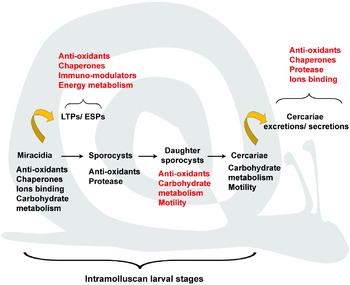
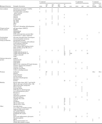
The cDNA/oligonucleotide microarray has recently served as an important tool for the rapid (high-throughput) quantitative analyses of gene expression patterns in the intramolluscan stages of schistosome development including miracidia, sporocysts and cercariae. Based on mRNA hybridization to its complementary cDNA template, two different cDNA populations (targets) are synthesized, usually with Cy-dye fluorescent-labeled nucleotides (cDNA populations labeled with different dyes), from RNA samples of interest. Recently, dye-labeled target cDNAs can be created from very small cell/tissue samples by incorporating high-fidelity amplified RNA (aRNA) (Petalidis et al. Reference Petalidis, Bhattacharyya, Morris, Collins, Freeman and Lyon2003; Tang et al. Reference Tang, Barbacioru, Wang, Nordman, Lee, Xu, Wang, Bodeau, Tuch, Siddiqui, Lao and Surani2009), which has greatly facilitated gene expression comparisons between scarce or low-abundance tissues or between parasite stages. Typically, pair-wise comparisons of relative transcript abundances are made by simultaneous hybridization of labeled cDNA populations to an array of pre-selected known genes affixed or spotted to a glass slide or silicon chip, allowing for the quantification of relative gene expression patterns by detection and measurement of the intensity of the fluorescent labels hybridizing to each spotted gene. In the last several years, microarray techniques have been used to identify stage-specific expression patterns of transcripts in the different S. mansoni and S. japonicum intramolluscan larval stages including miracidia, primary sporocysts (Vermeire et al. Reference Vermeire, Taft, Hoffmann, Fitzpatrick and Yoshino2006; Gobert et al. Reference Gobert, Moertel, Brindley and McManus2009), secondary sporocysts (germballs) (Jolly et al. Reference Jolly, Chin, Miller, Baghat, Lim, DeRisi and McKerrow2007), cercariae (Fitzpatrick et al. Reference Fitzpatrick, Johnston, Williams, Williams, Freeman, Dunne and Hoffmann2005; Jolly et al. Reference Jolly, Chin, Miller, Baghat, Lim, DeRisi and McKerrow2007; Gobert et al. Reference Gobert, Moertel, Brindley and McManus2009) and even gender-associated transcript expression pattern in S. mansoni cercariae (Fitzpatrick et al. Reference Fitzpatrick, Protasio, McArdle, Williams, Johnston and Hoffmann2008). A subset of the reported up-regulated transcripts is summarized in Supplemental Table 1, organized by sequence-derived biological functions based on gene ontology (GO) annotation. Although this table is only a qualitative compilation of gene undergoing expression changes, quantitative comparisons are presented in the referenced citations. Taken together, the changes in relative transcript levels associated with specific larval stages are interpreted to represent the parasite's response to the snail host environment, as well as its own changing needs for further growth and development. Indeed, there is a predominance of up-regulated transcripts related to specific biological functions for each of the intramolluscan larval stages (Fig. 3). For example S. mansoni miracidia show enrichment in transcripts encoding proteins related to functions like refolding/chaperoning (p40, HSP40, HSP70, HSP90), energy production (triosephosphate isomerase, phosphoenolpyruvate carboxykinase PEPCK), motility and calcium binding (SME16, calcineurin and Ca binding dynein-like protein), primary sporocysts have higher gene expression levels for anti-oxidants (e.g. peroxiredoxins, GST26/28 and Cu/Zn SOD), proteases (cathepsin, elastase, preprocathepsin and haemoglobinase) and protein synthesis/degradation (40 S and L37a ribosomal proteins, ubiquitins), while cercariae are enriched in proteins associated with energy production (ATP-synthase, lipid-binding protein, NADH dehydrogenase 4/5/7, ATPase subunit 6) and motility transcripts (tubulin, actin, myosin light and heavy chain).
Schematic representation of Schistosoma intramolluscan larval stages; miracidia, primary (mother) sporocysts, secondary (daughter) sporocysts and cercariae. For each larval stage, specific biological functions corresponding to predominant up-regulated transcripts (black lettering) or proteins (red lettering) are described. Functional categories of differentially-expressed excretory-secretory proteins/products (ESPs) and larval transformation proteins (LTPs) released inside the molluscan intermediate host during miracidium-to-sporocysts transformation, and cercarial ESPs release during and following emergence from the snail host are also listed (red lettering).

These latter findings of higher transcript levels involving motility and energy production transcripts in both schistosome miracidia and cercariae were not unexpected considering that both are highly motile with accompanying high energy requirements. Similarly, although anti-oxidant transcripts are observed throughout the different intramolluscan larval stages, their expression is higher in miracidia and both primary and secondary sporocysts, as these are the first larval stages to confront a potentially hostile oxidative haemolymph environment of the snail host (Bender et al. Reference Bender, Bixler, Lerner and Bayne2002), as well as possible encounter with the snail's immune defence system. In response to host reactive oxygen species (ROS), elevated anti-oxidant levels seen in early larvae suggest a potential role in protection against oxidative stress generated by the snail host, in addition to the need for larvae to maintain their own internal redox balance (Zelck and Von Janowsky, Reference Zelck and Von Janowsky2004; Vermeire and Yoshino, Reference Vermeire and Yoshino2007). A similar transcript pattern is observed for Sm16, an anti-inflammatory glycoprotein (Ramaswamy et al. Reference Ramaswamy, Salafsky, Potluri, He, Li and Shibuya1995), previously found in the cercarial secretory glands and believed to be involved in penetration into the mammalian host, as well as induction of apoptosis in responding immune cells (Curwen et al. Reference Curwen, Ashton, Sundaralingam and Wilson2006). Elevated steady-state Sj16 transcript levels in S. japonicum miracidia and sporocysts might suggest a similar role for this protein in host entry and/or defence against snail immune cells (Gobert et al. Reference Gobert, Moertel, Brindley and McManus2009). Sporocyst stages also appear to be significantly up-regulated in protease transcripts (cathepsin, elastase) and ribosomal genes. Proteases are believed to play an important role in the degrading of host tissue proteins, while elevated ribosomal proteins are indicative of high protein synthetic activity expected in these rapidly growing larval stages. These first multigene transcriptome analyses of different S. mansoni and S. japonicum intramolluscan stages provide valuable information supporting numerous hypotheses of the involvement of selected genes or gene families in parasite development, growth and survival within the snail host.
Gene expression in schistosome cercariae, although representing the ‘end-product’ of intramolluscan development, is of particular interest because sex-differentiation is presumed to occur during cercarial development within daughter sporocysts. Fitzpatrick et al. (Reference Fitzpatrick, Protasio, McArdle, Williams, Johnston and Hoffmann2008) used a microarray referenced with genomic DNA to attempt a gender characterization of morphologically identical S. mansoni cercariae. By analyzing specific transcripts known to be gender-related, these authors successfully established specific transcriptional patterns distinguishing ‘male’ and ‘female’ cercariae. As would be expected, transcripts related to egg biology or to male germ cell development and reproduction were highly expressed in female and male cercariae, respectively. Moreover, genes encoding proteins involved in glucose transport, protein degradation (proteases) and immune response modulation dominate expression in ‘male’ cercariae, supporting the existing hypothesis that males are more capable when it comes to infection and survival within the mammalian host. The microarrays performed in S. mansoni and S. japonicum allowed the discovery of distinct patterns of gene expression that are assumed to reflect major biological functions for each of the specific intramolluscan larval stages. This suggests that regulation of transcriptional activity is highly dependent, not only on preset genetic programmes (e.g. those associated with sex-determination and activity), but certainly on the environment presented by the host. Microarray analyses allow a rapid, quantitative and simultaneous assessment of multiple gene expression in different populations at a specific developmental time points, or parasites maintained under different experimental conditions. However, one of the drawbacks of this technique is that construction of microarrays or the gene ‘chips’ themselves require prior knowledge of and sequence information for the genes of interest, thus limiting the study to the spectrum of genes for which sequence information is known.
Serial analysis of gene expression (SAGE)
An alternative to microarray analysis as an expression profiling tool is SAGE or serial analysis of gene expression (Velculescu et al. Reference Velculescu, Zhang, Vogelstein and Kinzler1995). Unlike microarray, SAGE can be performed without prior pre-selection of specific transcripts, and allows for greater flexibility and potential transcripts coverage in its quantitative analysis of expressed transcripts. SAGE is based on similar principles as EST sequencing, with the additional notion that short tags (10–14 mers) of specific locations are sufficient to identify a specific transcript. In LongSAGE methods, tags are usually ∼21 nt in length, a sequence length that is predicted through theoretical modelling to have a >99·8% chance of matching only once in a genome the size of humans (Saha et al. Reference Saha, Sparks, Rago, Akmaev, Wang, Vogelstein, Kinzler and Velculescu2002). To create SAGE libraries, copies of cDNA are synthesized from target mRNA previously trapped on polyT-coated beads. Once synthesized, bead-immobilized cDNAs undergo a series of specific digestions, resulting in short nucleotide sequences (tags) that, in turn, are bound end-to-end, generating concatemers of multiple tags that are finally cloned and sequenced. Given the availability of the complete transcriptome of the organism of interest, this analysis permits the identification of expressed transcripts (genes), and by enumerating the SAGE tags for a given transcript, a quantitative comparison of specific gene expression between multiple sample libraries also can be ascertained (Williams et al. Reference Williams, Sayed, Bernier, Birkeland, Cipriano, Papa, McArthur, Taft, Vermeire and Yoshino2007). Verification that differentially expressed tags in fact represent differential expression of the genes of interest can be determined by real-time q-PCR or semi-quantitative reverse transcriptase PCR. In addition, SAGE allows for the identification of anti-sense transcripts, which have been hypothesized to function in post-transcriptional gene regulation in vertebrate cells (Luther et al. Reference Luther, Haase, Hohaus, Beckmann, Reich and Morano1998; Hastings et al. Reference Hastings, Ingle, Lazar and Munroe2000). The inverse-proportionality of sense-to-antisense tag counts in larval S. mansoni, and overall high representation of antisense tags (35%) (Taft et al. Reference Taft, Vermeire, Bernier, Birkeland, Cipriano, Papa, McArthur and Yoshino2009) is consistent with high anti-sense transcript content reported in other parasites (Patankar et al. Reference Patankar, Munasinghe, Shoaibi, Cummings and Wirth2001; Radke et al. Reference Radke, Behnke, Mackey, Radke, Roos and White2005), and suggests a potentially novel mechanism for post-translational gene regulation in larval schistosomes (Gunasekera et al. Reference Gunasekera, Patankar, Schug, Eisen, Kissinger, Roos and Wirth2004).
To date, only two LongSAGE analyses in S. mansoni have included intramolluscan larval stages; the first, by Williams et al. (Reference Williams, Sayed, Bernier, Birkeland, Cipriano, Papa, McArthur, Taft, Vermeire and Yoshino2007), produced an average of 60 000 tags from each of the 10 stage-specific SAGE libraries, and more recently, a detailed follow-up analysis of 21 440 unique SAGE tags from 5 intramolluscan larval libraries reported by Taft et al. (Reference Taft, Vermeire, Bernier, Birkeland, Cipriano, Papa, McArthur and Yoshino2009). In both studies, 5 groups of S. mansoni larvae were analyzed; 1 miracidial library, and 4 different primary sporocyst libraries from larvae maintained in in vitro culture for 6 or 20 days (representing early and late developing primary sporocysts, respectively) in 2 separate media supplemented with or without Biomphalaria glabrata embryonic (Bge) cell pre-conditioned medium. Larval cultivation in the presence/absence of conditioned Bge cell medium was an attempt to mimic the snail's internal environment, and any resulting alterations in the parasite transcriptomic response. Although both SAGE studies identified numerous relevant transcripts specifically-associated with miracidial and sporocyst stages, the resulting transcript repertoire identified proteins that were highly consistent with previous microarray findings: namely a predominance of proteins/enzymes related to anti-oxidants, chaperones, egg antigens, carbohydrate metabolism and calcium-binding functions in miracidia, and anti-oxidant-, protease-, and ribosomal protein (protein synthesis)-related transcripts highly expressed in sporocysts. In general, this set of transcripts exhibiting enhanced expression appears to reflect parasite stages that are adjusting to abrupt physiological changes associated with the transition from a free-living to parasitic existence. By the same token, elevated steady-state transcript levels seen in sporocysts at 6 and 20 days of culture also were rich in growth and development-related genes, especially those involved in protein synthesis/metabolism (60 S acidic ribosomal protein P, 40 S ribosomal protein S19, elongation factor 1α, Golgi membrane protein, ubiquitin, HSP90 chaparonin) and energy production (ATP synthase B, ATPase) (Taft et al. Reference Taft, Vermeire, Bernier, Birkeland, Cipriano, Papa, McArthur and Yoshino2009). Although not providing information as to the precise roles being played by the specific genes identified in these profiles, these studies can give an excellent impression of the major types of activities that are presumed to be important to larval stages at a particular point in their development.
Proteomic analyses
More information regarding the identity, functionality and origins of intramolluscan larval proteins would permit a better understanding of important physiological processes, in particular those associated with parasite defence and potential immune evasion mechanisms. A series of proteomic studies was recently performed focusing on: (1) miracidium-to-sporocyst transformation excretory-secretory (ES) proteins (Guillou et al. Reference Guillou, Roger, Mone, Rognon, Grunau, Theron, Mitta, Coustau and Gourbal2007; Gourbal et al. Reference Gourbal, Guillou, Mitta, Sibille, Theron, Pointier and Coustau2008), also termed larval transformation proteins (Wu et al. Reference Wu, Sabat, Brown, Zhang, Taft, Peterson, Harms and Yoshino2009); (2) protein differential expression between compatible and incompatible S. mansoni sporocysts (Roger et al. Reference Roger, Mitta, Mone, Bouchut, Rognon, Grunau, Boissier, Theron and Gourbal2008a, b) and (3) proteins contained within or released by schistosome cercariae (Knudsen et al. Reference Knudsen, Medzihradszky, Lim, Hansell and McKerrow2005; Liu et al. Reference Liu, Lu, Hu, Wang, Cui, Chi, Yan, Wang, Song, Xu, Wang, Zhang, Zhang, Wang, Xue, Brindley, McManus, Yang, Feng, Chen and Han2006; Yang et al. Reference Yang, Lu, Hu, He, Li, Zhang, Zheng, Li, Yu, Fung and Wu2009).
Using mass spectrometry (MS) techniques, recent studies have focused on identifying proteins released/secreted during in vitro miracidium-to-sporocyst transformation in several trematode species: S. mansoni (Guillou et al. Reference Guillou, Roger, Mone, Rognon, Grunau, Theron, Mitta, Coustau and Gourbal2007; Wu et al. Reference Wu, Sabat, Brown, Zhang, Taft, Peterson, Harms and Yoshino2009), E. caproni (Guillou et al. Reference Guillou, Roger, Mone, Rognon, Grunau, Theron, Mitta, Coustau and Gourbal2007) and F. hepatica (Gourbal et al. Reference Gourbal, Guillou, Mitta, Sibille, Theron, Pointier and Coustau2008). In their proteomic study, Wu et al. (Reference Wu, Sabat, Brown, Zhang, Taft, Peterson, Harms and Yoshino2009) suggested a change in terminology from ‘excretory-secretory proteins/products’ (ESPs), commonly used to characterize proteins released during larval transformation in culture medium, to ‘larval transformation proteins’ (LTPs) in characterizing this group of molecules. The reason for the suggested change was based on their finding that approximatively 60% of the proteins identified as ‘ES’ transformation proteins lacked identifiable signal peptides or characteristics of non-classically-secreted proteins (Nickel, Reference Nickel2005), strongly suggesting that many proteins are being released by leakage or breakdown of ciliary epidermal plates during in vitro miracidia-to-sporocyst transformation. Interestingly, however, despite the fact that these studies focused on miracidia-to-sporocyst transformation in three different species and used different methods for preparing samples prior to and during MS analyses, they all identified proteins with similar functions. For example, as in the previously described transcriptomic studies, chaperones/stress proteins, anti-oxidants, and calcium-binding proteins were well represented in the proteome of these digenean species. In addition, the presence of various proteases and protease inhibitors are indicative of active digestive/nutritional processes or, combined with the presence of anti-oxidants and immune modulators, suggest LTP involvement in the protection of the parasite against ROS generated from plasma or circulating immune cells (haemocytes). The presence of functional antioxidant enzymes and their activity (Vermeire and Yoshino, Reference Vermeire and Yoshino2007; Wu et al. Reference Wu, Sabat, Brown, Zhang, Taft, Peterson, Harms and Yoshino2009) and proteases/protease inhibitors that are shared between invading miracidia and the snail host suggest at least two types of LTP-mediated functions; (1) aggressive counter-measures to the host's hostile environment or haemocyte responses or (2) more passive mechanisms whereby the parasite avoids host recognition and immune elimination by disrupting immune recognition or mimicking host molecular structures (Guillou et al. Reference Guillou, Roger, Mone, Rognon, Grunau, Theron, Mitta, Coustau and Gourbal2007; Wu et al. Reference Wu, Sabat, Brown, Zhang, Taft, Peterson, Harms and Yoshino2009).
Recent studies by Roger and colleagues (Reference Roger, Mitta, Mone, Bouchut, Rognon, Grunau, Boissier, Theron and Gourbal2008a, Reference Roger, Gourbal, Grunau, Pierce, Galinier and Mittab, Reference Roger, Grunau, Pierce, Hirai, Gourbal, Galinier, Emans, Cesari, Cosseau and Mittac), further support a passive interference hypothesis of host-parasite compatibility, as they identified differentially expressed sporocyst proteins between incompatible (IC, Guadeloupian) and compatible (C, Brazilian) S. mansoni strains. After clearly showing an antigenic variation between C and IC larvae, their differential proteomic study revealed an increase of a few proteins in C larval strain and the presence of 9 structurally-related, but highly polymorphic mucin proteins (Sm poMuc), differentially expressed between the 2 strains. Mucin-like proteins are believed to be involved in host-parasite interaction in various helminth species (Theodoropoulos et al. Reference Theodoropoulos, Hicks, Corfield, Miller and Carrington2001), and are speculated to function as a potential smoke-screen or decoy against host defence (Roger et al. Reference Roger, Grunau, Pierce, Hirai, Gourbal, Galinier, Emans, Cesari, Cosseau and Mitta2008c). In addition to differential expression poMuc in C and IC larvae, their presence in apical glands and in LTPs further support a potential protective role of these proteins in the intramolluscan stages of S. mansoni. As suggested earlier by Yoshino and Boswell (Reference Yoshino, Boswell and Lackie1986), the sharing of schistosome-specific glycotopes (Lehr et al. Reference Lehr, Beuerlein, Doenhoff, Grevelding and Geyer2008; Peterson et al. Reference Peterson, Hokke, Deelder and Yoshino2009) also represents another type of mimetic mechanism in which larval stages express host-like molecules, thus avoiding recognition.
Acetabular gland contents and ESPs from S. mansoni cercariae and LTPs from transforming miracidia also exhibit similar expression patterns, with proteins belonging to the same predicted functional groups described previously (Appendix A; Supplementary Table 1). However, a few proteins appeared specific to cercarial ES, such as elastase, the 20·8/21·7 kDa Ca-binding proteins and HSP60, whereas S. mansoni LTPs were found strongly enriched in a variety of venom allergen-like proteins, suspected of being potentially involved in immuno-modulation and larval infection. Both miracidial LTPs and cercarial ES proteins (released upon simulated invasion) clearly show a similar pattern in functionality, which can be explained by the fact that these two larval stages are responsible for active penetration and host entry, migration/movement within host tissues, maintaining energy production, and aggressive counter-measures against oxidative stress and immune reactivity. A similar case can be made for S. japonicum carcariae, in which UV treatment-induced abrogation of proteins involved in motility, energy metabolism and protein transport (chaparones) were shown to be important for parasite infection and survival (Yang et al. Reference Yang, Lu, Hu, He, Li, Zhang, Zheng, Li, Yu, Fung and Wu2009). Indeed as shown by Liu et al. (Reference Liu, Lu, Hu, Wang, Cui, Chi, Yan, Wang, Song, Xu, Wang, Zhang, Zhang, Wang, Xue, Brindley, McManus, Yang, Feng, Chen and Han2006), like S. mansoni, cercariae and miracidia of S. japonicum share a common proteome when it comes to expression of proteins involved in host entry (calpain, calreticulin), motility (α- and β-tubulins, dynein light chain, myosin heavy chain, actin), energy metabolism (ATP synthase, ADP/ATP carrier protein, MDH, GAPDH, PepCK, fructose bisphosphate aldolase) and stress responses (several HSP homologues, thioredoxin peroxidase).
Although cercarial ES proteins are believed to potentially originate from the secretory glands and cytosolic component of the tegument (Knudsen et al. Reference Knudsen, Medzihradszky, Lim, Hansell and McKerrow2005), the origins of miracidial LTPs have only recently been explored. Polyclonal antibodies generated against S. mansoni LTPs and poMuc in two different studies (Wu et al. Reference Wu, Sabat, Brown, Zhang, Taft, Peterson, Harms and Yoshino2009; Roger et al. 2008 b, respectively), revealed differential immunolocalization patterns. Anti-LTPs antibodies showed no specific vesicle or gland cross-reactivity, but strong reactivity to ciliated epidermal plates of miracida and the sporocyst tegument. This supports the hypothesis that, similar to cercarial ES proteins, a subset of miracidial LTPs probably originate from the tegument. Anti-epidermal plate reactivity likely is a result of proteins released into LTP from plates undergoing lysis following larval detachment. Antibodies against poMuc, however, immunolocalized to miracidial and sporocyst apical glands, which are believed to be involved in the production of secretions essential to snail host invasion. Regardless of their specific function or localization, both the miracidium and the cercaria release an impressive variety of proteins whose main functions are likely targeted larval survival, defence, and/or development.
Over the past decade, there has been an impressive collection and archiving of gene and protein sequence data focused mainly on two schistosome species, S. mansoni and S. japonicum. Due to our current ability to culture the early intramolluscan stages of these digeneans we can now qualitatively and/or quantitatively assess protein and transcript patterns across the intramolluscan larval stages, and as a result, are beginning to have a glimpse of the complex mechanisms used by trematode larvae during invasion, survival, and development within its intermediate host. With the next generation 454 pyrosequencing technology (Nordstrom et al. Reference Nordstrom, Gharizadeh, Pourmand, Nyren and Ronaghi2001; Vera et al. Reference Vera, Wheat, Fescemyer, Frilander, Crawford, Hanski and Marden2008) it is now possible to simultaneously evaluate multi-transcriptomic samples both qualitatively (gene discovery) and quantitatively (gene expression), and with the completion of genome sequencing, assembly and annotation of both S. mansoni and its snail intermediate host, Biomphalaria, ultra-sequencing using pyrosequencing methodologies will provide valuable insights into snail host-larval gene interactions (‘interactome’) (Barakat et al. Reference Barakat, DiLoreto, Zhang, Smith, Baier, Powell, Wheeler, Sederoff and Carlson2009). Despite the creation of these ever-growing gene/protein databases, a full understanding of the functional role played by identified genes and gene products ultimately will depend on our ability to manipulate their expression within the living, intact parasite, and this represents the next critical challenge facing molecular helminthology investigators.
IN VITRO MANIPULATION OF GENE EXPRESSION AND FUNCTION IN INTRAMOLLUSCAN LARVAL STAGES
As amply demonstrated above, gene and protein discovery efforts have resulted in an ever-growing accumulation of molecular and biochemical data cataloguing numerous genes/gene products and their expression in various digenetic trematodes species, most notably the schistosomes, fasciolids and echinostomes. Moreover, for two of the major human fluke pathogens, S. mansoni and S. japonicum, life cycle stage-associated gene and protein expression profiling provides important clues as to the potential involvement of identified proteins or putative protein homologues in the myriad of molecular activities driving the development and/or maintenance of a given developmental stage. Unfortunately, this kind of information provides only limited understanding or insight into the actual functional role(s) of specifically-expressed genes or their protein products within the intact organism. Thus, effective approaches for predictably manipulating endogenous expression of specific genes within trematodes still represent a formidable barrier to exploring gene functions as they relate to the biology of these complex, multi-stage parasites and their interactions with their intermediate and definitive hosts.
Gene manipulation in Trematoda: development of methodologies
Methodologies for manipulating genes and their expression in flukes are still in its infancy, although significant progress is being made in the areas of transgenesis (i.e. introduction and expression of heterologous or homologous gene constructs using DNA plasmid vectors, DNA transposons, retrotransposons or replication-defective retroviruses) and RNA interference (RNAi). The work involving trematode transgenesis has focused almost exclusively on the schistosome blood flukes and has emphasized both snail and mammalian host stages of development as experimental models. Likewise, functional gene studies using RNAi also have emphasized the schistosomes, with a focus mainly on the mammalian stages of infection. Recent excellent reviews have summarized in detail the technologies underlying the construction, delivery and expression analyses of the variety of gene constructs used in demonstrating transient transfection and viral transformation within the Schistosoma spp. (Grevelding, Reference Grevelding, Maule and Marks2006; Beckmann et al. Reference Beckmann, Wippersteg, El-Bahay, Hirzmann, Oliveria and Grevelding2007; Brindley and Pearce, Reference Brindley and Pearce2007; Kalinna and Brindley, Reference Kalinna and Brindley2007; Mann et al. Reference Mann, Morales, Kines and Brindley2008; Han et al. Reference Han, Brindley, Wang and Chen2009), and gene-specific knockdown in flukes by RNAi (Skelly, Reference Skelly, Maule and Marks2006; Geldhof et al. Reference Geldhof, Visser, Clark, Saunders, Britton, Gilleard, Berriman and Knox2007; Kalinna and Brindley, Reference Kalinna and Brindley2007; Pearce and Freitas, Reference Pearce and Freitas2008). In keeping with the theme of this review, the following discussion will focus on how these technologies have been applied specifically to evaluating gene function in the intramolluscan developmental stages of trematodes and the critical role being played by in vitro culture systems in the manipulation of specific larval genes.
Manipulation of gene expression in larval trematodes
Methods for the in vitro cultivation of intramolluscan larval stages, from the free-living miracidium to multi-generation sporocysts or rediae, have now evolved to the point where specific larval stages can be produced in vitro for use as potential targets of genetic manipulation, making it possible to begin addressing the role of specific genes during larval development. As pointed out previously, an added feature of transgenic approaches (silencing or over-expressing genes of interest) in the miracidium or sporocyst stages is the likelihood of effecting changes within embryonic germ cells, which then have the potential of being passed on to progeny (assuming expression is not deleterious) through the normal asexual reproductive process. Live, genetically-transformed miracidia or sporocysts carrying altered genes can then be transferred to compatible uninfected snails through natural infection or surgical transplantation, respectively. Although RNAi provides only transient knockdown effects, this method currently represents the only approach in which specific endogenous genes can be manipulated and larvae assessed for potential functional defects. Advances in double-stranded RNA delivery by transgenic approaches, as well as precautions in the set-up, execution and interpretation of findings in RNAi-type experiments also will be discussed.
Transgenesis in larval trematodes
Transgenesis, or the introduction and induction of expression of exogenous or foreign genes, has enjoyed considerable success in its application to trematode larval stages, particularly in the schistosomes. Although all stages of schistosomes to date have been subjects of attempted transfection, we will mainly focus this review on approaches involving the intramolluscan larval stages. Arguably, these stages represent the most desirable targets for transfection due to their ease of in vitro manipulation, the abundance of germinal cells readily accessible to transgene targeting while carrying the possibility for germline introduction and genomic integration, and finally the ability to propagate transgene-carrying progeny by natural infection or surgical transfer of transfected asexually-reproducing stages (miracidia or sporocysts) into suitable snail hosts (Kapp et al. Reference Kapp, Coustau, Wippersteg, Jourdane, Kunz and Grevelding2003; Grevelding, Reference Grevelding, Maule and Marks2006; Kalinna and Brindley, Reference Kalinna and Brindley2007).
The miracidium and in vitro-cultured primary sporocyst have attracted considerable attention as targets of transgenesis for reasons mentioned above. The earliest attempts to successfully deliver and express exogenous transcripts or transgenes utilized particle bombardment (biolistics) in which the gold particles coated with DNA encoding the green fluorescent protein (GFP) ORF flanked by S. mansoni HSP70 promoter and terminator sequences, were introduced into live S. mansoni sporocysts with a particle accelerator or ‘gene gun’ (Wippersteg et al. Reference Wippersteg, Kapp, Kunz, Jackstadt, Zahner and Grevelding2002a). The presence of GFP transcripts (by RT-PCR) in sporocyst extracts and GFP protein expression (by confocal fluorescent microscopy) provided evidence for transient larval transfection. Follow-up experiments showed that biolistic introduction of GFP flanked with promoter/termination sequences of the cysteine proteinase ER60, localized and expressed in lateral glands and cytons of the protonephridia, tissues with putative proteinase activity and known excretory-secretory function (Wippersteg et al. Reference Wippersteg, Kapp, Kunz and Grevelding2002b, Reference Wippersteg, Ribeiro, Liedtke, Kunz and Grevelding2003). These seminal studies showed the tractability of introducing and expressing foreign DNA in living sporocysts and opened up the possibility of tissue-specific targeting of transgene expression. The next important breakthrough in larval transgenesis was the demonstration that transgenes introduced into miracidia were expressed in developing sporocysts following natural infection of snails (Heyers et al. Reference Heyers, Walduck, Brindley, Bleizß, Lucius, Dorbic, Wittig and Kalinna2003). Biolistics was used to deliver an enhanced GFP (eGFP) construct flanked by an HSP70 promoter/terminator into S. mansoni miracidia, followed by infection of B. glabrata snails. Histological localization of gold particles near germinal cells in primary sporocysts of snails with 14-day-old infections and the finding of eGFP mRNA expression by nested PCR in infected, but not uninfected, snail tissues demonstrated the feasibility of transgene delivery and expression in miracidia, and importantly, the possibility of continuing propagation of germline-altered progeny (sporocysts, cercariae) and establishment of transgenic schistosome lines. This latter senario recently was eloquently demonstrated by Beckmann et al. (Reference Beckmann, Wippersteg, El-Bahay, Hirzmann, Oliveria and Grevelding2007), who introduced plasmid GFP constructs driven by 1·5 kb promoter fragments of the S. mansoni actin gene (Sm Act1) into miracidia (F0 generation) using biolistics, and detected GFP transcript expression through the F0 and F1 generations of the entire life cycle; F0 miracidia→F0 cercariae→F0 adult worms generated in hamsters→F1 miracidia from F0 adults→F1 cercariae→F1 adults. Loss of transcript detection in F2 stages indicated that, although clearly being carried in germline cells, the GFP transgene was not stably integrated, if at all, in the germinal cell genome (Beckmann et al. Reference Beckmann, Wippersteg, El-Bahay, Hirzmann, Oliveria and Grevelding2007).
A successful alternative approach for delivery and expression of foreign or altered genes in the various schistosome stages explores the use of transposons, retrotransposons and pseudo-typed retroviruses as schistosome-transducing agents (Mann et al. Reference Mann, Morales, Kines and Brindley2008). Applications to asexually developing larval stages have been limited, although work reported by Kines et al. (Reference Kines, Mann, Morales, Shelby, Kalinna, Gobert, Chirgwin and Brindley2006) appears to show promise. In vitro-cultured S. mansoni sporocysts were infected with S. mansoni promoter-flanked eGFP sequence incorporated into a Moloney murine leukaemia retroviral vector packaged into a non-replicative VSV-pseudotyped retrovirus. Larval exposure of the retroviral construct in polybrene resulted in infection and viral transduction of sporocysts as evidenced by localization of virions at the larval surface, integration of proviral sequences into sporocyst genomic DNA, and detection of reporter gene expression in virally-infected, cultured larvae. Whether or not retroviral transformation will prove to be a practical method for producing genetically-modified schistosome lines has yet to be determined. However, early results show considerable promise.
RNA interference as a functional genomics tool in trematodes
The post-transcriptional silencing of specific messenger RNA expression by RNA interference (RNAi) in parasitic helminths, including trematodes, is a significant advance in parasite postgenomics, as it represents the only approach to date for experimentally manipulating expression of specifically-targeted endogenous genes, thereby providing insight into putative gene function. Although there is some controversy as to whether helminth parasites, in particular parasitic nematodes, possess a fully-functional RNAi mechanism (Geldhof et al. Reference Geldhof, Visser, Clark, Saunders, Britton, Gilleard, Berriman and Knox2007; Knox et al. Reference Knox, Geldhof, Visser and Britton2007; Viney and Thompson, Reference Viney and Thompson2008), accumulating evidence for trematodes strongly supports a functional system for the regulation of mRNA expression by small interfering (si) RNA and/or microRNA (Verjovski-Almeida et al. Reference Verjovski-Almeida, DeMarco, Martins, Guimarães, Ojopi, Paquola, Piazza, Nishiyama, Kitajima, Adamson, Ashton, Bonaldo, Coulson, Dillon, Farias, Gregorio, Ho, Leite, Malaquias, Marques, Miyasato, Nascimento, Ohlweiler, Reis, Ribeiro, Sá, Stukart, Soares, Gargioni, Kawano, Rodrigues, Madeira, Wilson, Menck, Setubal, Leite and Dias-Neto2003; Xue et al. Reference Xue, Sun, Zhang, Wang, Huang and Pan2008; Krautz-Peterson and Skelly, Reference Krautz-Peterson and Skelly2008; Gomes et al. Reference Gomes, Cabral, Jannotti-Passos, Carvalho, Rodriques, Baba and Sa2009). Because several recent reviews and primary reports cover the topic of RNAi applications in trematodes, mainly emphasizing the mammalian stages of schistosomes (Skelly, Reference Skelly, Maule and Marks2006; Geldhof et al. Reference Geldhof, Visser, Clark, Saunders, Britton, Gilleard, Berriman and Knox2007; Kalinna and Brindley, Reference Kalinna and Brindley2007; Brindley and Pearce, Reference Brindley and Pearce2007; Ndegwa et al. Reference Ndegwa, Krautz-Peterson and Skelly2007; Morales et al. Reference Morales, Rinaldi, Gobert, Kines, Tort and Brindley2008; Pereira et al. Reference Pereira, Pascoal, Marchesini, Maia, Magalhaes, Zanotti-Magalhaes and Lopes-Cendes2008; Rinaldi et al. Reference Rinaldi, Morales, Alrefaei, Cancela, Castillo, Dalton, Tort and Brindley2009; Krautz-Peterson et al. Reference Krautz-Peterson, Bhardwaj, Faghiri, Tararam and Skelly2009 – in current special issue) and the liver fluke Fasciola hepatica (Rinaldi et al. Reference Rinaldi, Morales, Cancela, Castillo, Brindley, Jose and Tort2008), we will focus our attention on its application to stages within the snail intermediate host; namely miracidia and primary sporocysts.
The possible existence of an RNAi-like mechanism in trematodes was first demonstrated in S. mansoni, initially in adult worms (Skelly et al. Reference Skelly, Da'dara and Harn2003) and then in the primary sporocyst (Boyle et al. Reference Boyle, Wu, Shoemaker and Yoshino2003). In these studies, isolated cercariae and miracidia were treated in vitro with dsRNAs synthesized from transcripts of target genes by incubating (soaking) parasites for 6 days in dsRNA-containing media, during which time juvenile or larval stages transformed to their successive stages of development: cercariae→schistosomula and miracidia→sporocysts. These first attempts at silencing the cathepsin B gene in schistosomula (Skelly et al. Reference Skelly, Da'dara and Harn2003) and a glucose transporter (SGTP1) gene in primary sporocysts (Boyle et al. Reference Boyle, Wu, Shoemaker and Yoshino2003) have now led to a number of follow-up studies investigating a variety of target transcripts, dsRNA/siRNA delivery systems and other experimental parameters aimed at optimizing the specificity and efficacy of gene knockdown (Skelly, Reference Skelly, Maule and Marks2006; Dinguirard and Yoshino, Reference Dinguirard and Yoshino2006; Krautz-Peterson et al. Reference Krautz-Peterson, Radwanska, Ndegwa, Shoemaker and Skelly2007; Ndegwa et al. Reference Ndegwa, Krautz-Peterson and Skelly2007; Mourão et al. Reference Mourão, Dinguirard, Franco and Yoshino2009). As a result of the convergence of technologies involving in vitro cultivation, stage-associated gene discovery/expression and gene manipulation through reverse-genetics approaches, we anticipate that functional genomic studies will not only continue in the schistosomes at an accelerating pace, but incorporate a diversity of other trematode species as well.
Assessing gene function in the development or activities of the miracidium of S. mansoni has been accomplished by introduction of gene-specific double-stranded (ds) RNA into eggs recovered from adult female worms or isolated from infected livers. Freitas et al. (Reference Freitas, Jung and Pearce2007) demonstrated that a S. mansoni Inhibin/Activin gene (SmInAct; member of the TGF-β signaling family) was a key regulator of miracidial embryogenesis by soaking eggs, newly deposited by cultured female worms, in SmInAct dsRNA and quantifying inhibition of larval development in ovo. A similar methodological approach was used to confirm previous pharmacological findings (Xu and Dresden, Reference Xu and Dresden1986) that S. mansoni leucine aminopeptidases (SmLAP) produced by miracidia in ovo were required for egg hatching. Rinaldi et al. (Reference Rinaldi, Morales, Alrefaei, Cancela, Castillo, Dalton, Tort and Brindley2009) exposed liver-isolated eggs to dsRNA of the two SmLAP isoforms (1 and 2) for 7 days at 37 C, after which time they were induced to hatch. Results demonstrated that egg hatching in the presence of SmLAP1 dsRNA, SmLAP2 dsRNA or both, was reduced by ∼80% compared to both irrelevant dsRNA-treated and untreated control groups, and was comparable to groups treated with the LAP inhibitor bestatin. Concomitant with this hatching phenotype were significant reductions in specific LAP transcripts and enzymatic activity in dsRNA-treated eggs, indicating a clear relationship between LAP gene expression and hatching (Rinaldi et al. Reference Rinaldi, Morales, Alrefaei, Cancela, Castillo, Dalton, Tort and Brindley2009). These studies not only provide a valuable method for screening potential drug targets for chemotherapeutic intervention, but also can be used for investigating the potential functions of genes involved in miracidial development, behaviour (e.g. ciliary activity, phototaxis), and larval transformation.
Application of RNAi methods to primary (mother) sporocyst stages of Schistosoma spp. also has taken advantage of in vitro culture methods that have permitted development of the miracidial to cercarial stages (Ivanchenko et al. Reference Ivanchenko, Lerner, McCormick, Toumadje, Allen, Fischer, Hedstrom, Helmrich, Barnes and Bayne1999). As noted above, the first application of RNAi to sporocysts involved incubation (soaking) of freshly-isolated miracidia in snail ringers (CBSS) containing dsRNA and allowing them to transform to sporocysts followed by cultivation for 6 days in the presence of dsRNA (Boyle et al. Reference Boyle, Wu, Shoemaker and Yoshino2003). Using dsRNA for the S. mansoni glucose transporter (SGTP1) and glyceraldehyde-3- phosphate dehydrogenase (GAPDH), they demonstrated specific knockdown of SGTP1 transcripts upon exposure to SGTP1 dsRNA, using GAPDH dsRNA exposure as a specificity control. The gene knockdown effect was dependent on the presence of dsRNA during miracidial transformation and persisted in larvae maintained for up to 28-days in culture. Importantly, tritiated-glucose uptake by sporocysts treated with SGTP1 dsRNA, but not those exposed to GAPDH dsRNA, was significantly reduced, indicating a functional role of SGTP1, or a structurally related homologue (Skelly et al., Reference Skelly, Kim, Cunningham and Shoemaker1994), in sporocyst glucose acquisition. A second study, using essentially the same protocols as Boyle et al. (Reference Boyle, Wu, Shoemaker and Yoshino2003), examined the role of a sporocyst-expressed CD36-like scavenger receptor (SR) gene in modified (acetylated) low-density lipoprotein (LDL) binding to the sporocyst tegument and larval growth (Dinguirard and Yoshino, Reference Dinguirard and Yoshino2006). Consistent with previous findings, exposure of S. mansoni miracidia to SR dsRNA during in vitro transformation to sporocysts and incubation for 6-days, resulted in a significant decrease in SR transcripts, decreases in the prevalence and intensity of acetylated-LDL binding to sporocysts, and reduction in larval length. These examples illustrate the types of functional information that may be gained by applying RNAi-type approaches to in vitro developing schistosome larvae.
Universality of gene-specific RNAi-like effects in cultured larval schistosomes
In our experience investigating dsRNA-mediated gene silencing we noted that not all genes employed in RNAi experiments yielded consistent and/or specific knockdowns. This prompted us to examine a wider range of different genes for their abilities to produce RNAi-like effects both at the transcriptional and phenotypic levels (Mourão et al. Reference Mourão, Dinguirard, Franco and Yoshino2009). Briefly, in vitro transforming miracidia of S. mansoni were treated with dsRNAs (Boyle et al. Reference Boyle, Wu, Shoemaker and Yoshino2003) generated from 32 genes expressed in primary sporocysts, including those encoding anti-oxidants, signaling proteins, transcription factors, metabolic enzymes and the like, and screened for morphological changes in larval phenotype. After 7 days in culture, a reduction in sporocyst size (length), similar to that reported by Dinguirard and Yoshino (Reference Dinguirard and Yoshino2006), was the only morphological phenotype noted, and this was observed for 11 of the 32 genes examined. Of these 11 genes associated with the ‘size’ phenotype, 6 exhibited consistent knockdown in their steady-state transcript levels by real-time quantitative PCR, one (SOD) was highly elevated compared to the irrelevant GFP dsRNA-treatment control, while the remaining 4 phenotype-presenting genes were unchanged in transcript levels. The remaining 21 genes tested did not produce a consistent ‘size’ phenotype, although significant transcript knockdown was demonstrated for half of these genes (Mourão et al. Reference Mourão, Dinguirard, Franco and Yoshino2009).
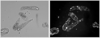
Results of this profiling study were enlightening as it pointed out several aspects of RNAi-type experiments incorporating larval schistosomes that require careful consideration: (1) The choice of gene to be examined – For reasons yet unknown transcript knockdown appears to be depend on the target gene or the specific gene segment being used to template dsRNA synthesis. Genes of interest should be pre-tested to ensure consistent, significant knockdown effects (Mourão et al. Reference Mourão, Dinguirard, Franco and Yoshino2009). (2) Off-target effects – Some genes or gene segments may produce non-specific or off-target effects that are not consistent with the predicted interaction with and/or degradation of specific target transcripts, but instead appear to affect phenotype-associated non-target genes. Off-target effects are one of the common unwanted side-effects of RNAi manipulation (Kulkarni et al. Reference Kulkarni, Booker, Silver, Friedman, Hong, Perrimon and Mathey-Prevot2006), although steps can be taken to minimize these effects (Reynolds et al. Reference Reynolds, Leake, Boese, Scaringe, Marshall and Khvorova2004). (3) Optimizing dsRNA delivery and timing – Although we use miracidial incubation (soaking) in dsRNA for 6 days as part of our standard delivery protocol for gene knockdown in sporocysts, others have incorporated square-wave electroporation as effective alternative dsRNA delivery methods for mammalian stages of schistosomes (Correnti et al. Reference Correnti, Brindley and Pearce2005; Osman et al. Reference Osman, Niles, Verjovski-Almeida and LoVerde2006; Sayed et al. Reference Sayed, Cook and Williams2006; Krautz-Peterson et al. Reference Krautz-Peterson, Radwanska, Ndegwa, Shoemaker and Skelly2007; Morales et al. Reference Morales, Rinaldi, Gobert, Kines, Tort and Brindley2008; Zhao et al. Reference Zhao, Lei, Liu, Zhu, Ren, Wang and Shen2008) and fasciolids (Rinaldi et al. Reference Rinaldi, Morales, Cancela, Castillo, Brindley, Jose and Tort2008). In preliminary electroporation experiments we have found that over a range of delivered voltages (65–230 V) and capacitances (25–100 mF), 65–70% maximum sporocysts survival rates were attained at 72 h post-treatment resulting in an apparent consistency of dsRNA delivery (Fig. 4). Follow-up studies are now needed to determine if, in fact, specific gene silencing in surviving larvae is achieved by this approach and how knockdown rates and long-term larval viability compare to soaking or other delivery methods. Consistent with previous finding in schistosomula (Skelly et al. Reference Skelly, Da'dara and Harn2003), in general, sporocysts did not tolerate lipofection-type reagents used within recommended concentration ranges.
Example of Schistosoma mansoni primary sporocysts subjected to square-wave electroporation in the presence of rhodamine-labeled double-stranded (ds)RNA. Localization of electroporated dsRNA is mainly tegumental in discrete ‘patches’ (right panel; epifluorescence). Intact sporocysts are shown in the left panel.

SUMMARY AND CONCLUDING REMARKS
With the dramatic advancements in DNA and amino acid sequencing technologies made over the last decade, combined with the powerful analytical tools of bioinformatics, identification of genes and their products in diverse organisms, including model parasitic helminths, has become almost routine. However, it should come as no surprise that despite the rapid accumulation of gene/protein databases to date, our knowledge, and therefore understanding, of the role or function of identified genes/proteins is lagging far behind. This is especially evident in parasitic metazoans such as the digenetic trematodes, in which the entire genomes of selected species are only now completed. Although parasite expression profiling leading to the identification and activities of specific genes or gene networks can have immediate practical applications involving drug targeting or protective immune interventions (Brindley and Pearce, Reference Brindley and Pearce2007; Pearce and Freitas, Reference Pearce and Freitas2008; Han et al. Reference Han, Brindley, Wang and Chen2009), experimental approaches for addressing fundamental questions of how genes may be functioning in the biology of parasites undergoing complex life cycle changes are urgently needed. As summarized in this review, part of this need is being met by the establishment of larval isolation and in vitro cultivation methods capable of generating and maintaining the major intramolluscan stages for trematodes of several prominent families (Schistosomatidae, Fasciolidae, Echinostomatidae), in combination with gene discovery efforts that have already yielded completed draft genomes of S. mansoni and S. japonicum, and ever-growing stage-associated transcriptomic and proteomic databases for these and other digenean species. The next major challenge for trematode biologists is to effectively combine these different approaches to investigating the functional role of expressed genes and their products in the maintenance, growth and development these parasites as they undergo morphological and physiological changes from one stage to the next. Recent exciting and promising research into gene transfer techniques (in vitro transgenesis and viral transduction) with the prospect of germline transfection, and reverse-genetic approaches using RNAi to manipulate endogenous gene function offer novel approaches for meeting this challenge as we truly advance into the post-genomics era.
ACKNOWLEDGEMENTS
Selected published and unpublished data cited in this review were supported by NIH grants AI061436 and AI015503 (TPY). MMM was supported by a CAPES (Process no. 4753/06-2) and MCT/CNPq 02/2006 Universal (Process no. 475151). We also thank Maria Castillo, Andrew Taft and 2 anonymous reviewers for their valuable input.
APPENDIX A:
Qualitative representation of predominant ‘up-regulated’ transcripts (vertical arrows) organized by specific biological functions in intramolluscan larval stages of S. mansoni miracidia, 4 day-sporocysts (mira and spo-4d, respectively; Vermeire et al. Reference Vermeire, Taft, Hoffmann, Fitzpatrick and Yoshino2006), 6- and 20-day old sporocysts (spo-6d and spo-20d, respectively; Williams et al. Reference Sayed, Cook and Williams2006; Taft et al. Reference Taft, Vermeire, Bernier, Birkeland, Cipriano, Papa, McArthur and Yoshino2009), secondary sporocysts and cercariae (2nd spo and cerc, respectively; Jolly et al. Reference Jolly, Chin, Miller, Baghat, Lim, DeRisi and McKerrow2007), S. japonicum miracidia, sporocysts and cercariae (Gobert et al. Reference Gobert, Moertel, Brindley and McManus2009), and female (cerc-fem) and male (cerc-male) cercariae of S. mansoni (Fitzpatrick et al. Reference Fitzpatrick, Protasio, McArdle, Williams, Johnston and Hoffmann2008).