Implications
Geese grow slowly and inefficiently after 8 weeks of age, but are not typically marketed until 12 to 14 weeks of age. Consequently, reducing the cost of feed during fattening period is a critical topic for the goose farming industry. Time-restricted feeding has been proven to increase feed efficiency (FE) in certain animals. In this study, we demonstrated that time-restricted feeding in the morning is superior to feeding in the afternoon regarding FE. We also found that time-restricted feeding in the morning greatly reduces feed intake and weight gain, but substantially improves FE compared with ad libitum feeding.
Introduction
Geese grow rapidly and demonstrate a high level of feed efficiency (FE) during their growth period (from 0 to 8 weeks of age). In contrast, geese grow slowly and exhibit a low level of FE during the fattening period (Chen et al., Reference Chen, Hsu, Shih, Lio and Chen2003). The marginal economic profit during the fattening period is typically negative. However, geese are not typically sent to market at 12 to 14 weeks of age. On the basis of daily gain (DG), carcass weight, pin feathers on the carcass, muscle panel scores and rheological properties, Chen et al. (Reference Chen, Hsu, Shih, Lio and Chen2003) suggested that the optimal marketing age for White Roman geese and Chinese geese was 13 and 14 weeks, respectively. Considering the characteristics of growth curve, increasing DG and FE during the fattening period is difficult; thus, attempting to decrease feed costs by reducing feed consumption during this period is worthwhile.
Quantity-restricted feeding is commonly practiced during the fattening period for pigs, and the rearing and laying periods of broiler breeders. However, in large-group feeding situations, quantity-restricted feeding may lead to underfeeding in certain inferior animals and overfeeding in certain superior animals. Feeding ad libitum for an adequate but limited duration offers an alternative strategy, where all animals are allowed to ingest as much feed as desired at meal times, although their daily feed intake (DFI) is reduced. Time-restricted feeding has been found to improve FE in broilers (Su et al., Reference Su, Sorensen and Kestin1999; Svihus et al., Reference Svihus, Sacranie, Denstadli and Choct2010). Furthermore, broilers that were fed ad libitum but denied feed on the 3rd and 5th days of the week showed a 30% decrease in feed intake. Their DG and FE substantially exceeded that of broilers subjected to a 30% quantitative feed reduction, and were similar to that of broilers subjected to a 15% quantitative feed reduction (Benyi and Habi, Reference Benyi and Habi1998). Feeding on alternate days also improved FE (Boa-Amponsem et al., Reference Boa-Amponsem, Dunnington and Siegel1991). Therefore, time-restricted feeding can potentially reduce feed costs.
The nutrient metabolism, body weight and FE of certain species have been found to be affected by meal time. In humans, glucose tolerance decreased in the evening and night (Morgan et al., Reference Morgan, Ross, Mercer and Barrett2003). Pigs that were fed identical meals at 12-h intervals followed a clear diurnal biorhythm in protein anabolism, exhibiting a greater amino-acid utilization and lower plasma urea response in the morning compared with the evening (Koopmans et al., Reference Koopmans, van der Meulen, Dekker, Corbijn and Mroz2006). Feeding experiments have shown that meal time significantly affects the weight gain of black-headed buntings (Emberiza melanocephala; Kumar et al., Reference Kumar, Singh, Misra and Malik2001), mice (Nelson et al., Reference Nelson, Scheving and Halberg1975), rats (Philippens et al., Reference Philippens, Von Mayersbach and Scheving1977) and catfish (Sundararaj et al., Reference Sundararaj, Nath and Halberg1982; Noeske-Hallin et al., Reference Noeske-Hallin, Spieler, Parker and Suttle1985), although the optimal time varies among species. In this study, we compared the fattening traits of White Roman geese fed in the morning with those fed in the afternoon in Trial I, and compared the parameters between ad libitum and time-restricted feeding regimens in Trial II. Because plasma glucose and triacylglycerol levels reflect the absorption, de novo synthesis and catabolism balance for carbohydrates and lipids, and the plasma uric acid level is an index of amino-acid oxidation, plasma levels of the metabolites were also determined in both trials of this study.
Material and methods
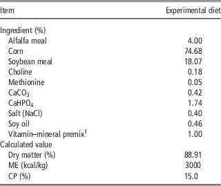
The experimental protocols used in this study were approved by the Experimental Animal Care and Use Committee of Tunghai University. This study comprises two trials. During the experimental periods, geese were housed in a windowed barn enclosed with netting and raised on an elevated plastic mesh floor without litter. In addition, the geese were fed a fattening diet (Table 1), formulated to meet the nutrient requirements according to the National Research Council (NRC, 1994) and water was provided ad libitum.
The composition of the experimental fattening diet for both trials

1 Vitamin–mineral premix provided per kilogram of diet: vitamin A, 18 000 IU; vitamin D3, 2250 IU; vitamin E, 22.5 IU; vitamin K, 4.5 mg; vitamin B1, 1.5 mg; vitamin B2, 9 mg; vitamin B6, 1.5 mg; vitamin B12, 15 μg; pantothenic acid 22.5 mg; niacin 105 mg; folic acid, 1.2 mg; biotin, 0.15 mg; Mn, 120 mg; Cu, 45 mg; Zn, 120 mg; Fe, 12.5 mg; I, 0.3 mg; Mo, 0.3 mg; Co, 0.15 mg; Se, 0.15 mg.
Trial I
In this trial, 24 White Roman geese aged 8 weeks of each sex were randomly assigned to 12 pens (1.8×1.2 m). Each pen housed two male and two female geese (six replicates per treatment). Before initiating the trial, geese were fed a grower diet twice per day during the growing period. During the experimental period, the geese experienced a natural photoperiod and day length of ∼14.5 h, and the daily mean ambient temperatures ranged between 23.7°C and 33.5°C. From 8 to 12 weeks of age, the geese were allowed free access to the fattening diet for 1 h either in the morning (morning-feeding group; from 0700 to 0800 h) or in the afternoon (afternoon-feeding group; from 1700 to 1800 h). At 12 weeks of age, hourly blood samples were collected from three male and three female geese per group for 4 h, beginning 1 h after feeding to determine the plasma levels of glucose, triacylglycerols, uric acid and urea by using an automatic blood biochemistry analyzer (VITROS® DT60 II Chemistry System, Johnson & Johnson Ortho Clinical Diagnostics, Raritan, New Jersey, USA). Feed intake was recorded daily, and body weight immediately before feeding was recorded weekly. The area under the plasma concentration v. time curve (AUC), which is commonly used to indicate the persistence of a metabolite in plasma after a meal, was calculated by using the trapezoidal rule to assess the postprandial response of each analyzed plasma metabolite.
Trial II
In this trial, 24 White Roman geese aged 8 weeks, which were fed a grower diet ad libitum during the growing period, were randomly assigned to 12 pens (1.8×1.2 m), with equal numbers of male and female geese in each pen. Each treatment comprised six pens. During the experimental period, the geese experienced a natural photoperiod and day length of 11.6 to12.1 h, and the daily mean temperature ranged between 15.6°C and 29.5°C. The geese were allowed access to a fattening diet either ad libitum (control group) or for 2 h in the morning (restricted group; from 0600 to 0800 h) from 8 to 13 weeks of age. Their feed intake was recorded daily, and their body weight was recorded weekly. At 13 weeks of age, all geese were fasted for 24 h and blood samples were collected to determine the fasting plasma levels of glucose, triacylglycerols and uric acid by using an automatic blood biochemistry analyzer (VITROS® DT60 II Chemistry System, Johnson & Johnson Ortho Clinical Diagnostics, Raritan, New Jersey, USA). Subsequently, the geese were killed to evaluate their carcass traits. Their live body weights were recorded immediately before bleeding, and carcass weight was defined as the weight after exsanguination, defeathering and viscera removal. The abdominal and visceral fat, liver and empty digestive tract weights were expressed as % body weight.
Statistics
Data were analyzed using the general linear model of SAS statistical software (SAS Institute Inc., Cary, NC, USA). In Trial I, all data were analyzed using one-way analysis of variance (ANOVA). In Trial II, the carcass traits were analyzed using two-way ANOVA (treatment×sex); other traits were analyzed using one-way ANOVA.
Results
Trial I
The DFI during the last week before the start of the trial was 257 g/day per bird. At the start of the trial, DFI was low but then gradually increased (over ∼10 days) before reaching a plateau in both groups (data not shown). The DFI of the morning-feeding group was typically lower than that of the afternoon-feeding group. The average DFI throughout the 4-week fattening period for the morning-feeding group was 8.3% lower (P<0.05) than that for the afternoon-feeding group (Table 2). However, the average DG throughout the experimental period in the morning-feeding group was 30.1% higher (P<0.05) than that in the afternoon-feeding group. Consequently, the FE for the morning-feeding group was 41.7% higher than that for the afternoon-feeding group (Table 2). Postprandial plasma levels of urea were insufficient for detection (<0.1 mg/dl) in all blood samples. The AUC (arbitrary unit) of postprandial plasma glucose (584 and 603 for the morning-feeding and afternoon-feeding groups, respectively), triacylglycerols (280 and 334 for the morning-feeding and afternoon-feeding groups, respectively) and uric acid (12.9 and 14.3 for the morning-feeding and afternoon-feeding groups, respectively) exhibited by each group were similar (P>0.05).
The effect of feeding time on the fattening performances in White Roman geese (Trial I)

RMSE=root mean square error.
1 Geese were allowed access to fattening diet for 1 h either in the morning (morning-feeding group; from 0700 to 0800 h) or afternoon (afternoon-feeding group; from 1700 to 1800 h) during the experimental period (from 8 to 12 weeks of age).
2 n=6 for each mean.
Trial II
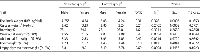
The DFI during the last week before the start of Trial II was 280 g/day per bird. At the start of Trial II, the DFI for the restricted group was extremely low, but then gradually increased in the first 2 weeks before reaching a plateau of approximately two-thirds the DFI for the control group (data not shown). The average DFI throughout the 5-week fattening period for the restricted group was only 63% for the control group (Table 3). The DG for the restricted group was significantly lower (P<0.05) than that for the control group throughout the experimental period (Table 3). The low level of weight gain for the restricted group was primarily caused by weight loss during the 1st week of the experiment (data not shown). The weight gain during the remaining experimental period for the restricted group (1.08 kg/bird; from 3.91 kg/bird at 9 weeks of age to 4.99 kg/bird at 13 weeks of age) did not differ from that for the control group (0.96 kg/bird; from 4.20 kg/bird to 5.16 kg/bird). Because the extent of feed intake reduction was greater than the DG, the FE for the restricted group was significantly higher (P<0.05) than that for the control group (Table 3). In addition, the levels of fasting plasma glucose (185 and 174 mg/dl for the restricted and control groups, respectively) and uric acid (1.98 and 2.44 mg/dl for the restricted and control groups, respectively) did not differ (P>0.05) between groups, although the fasting plasma triacylglycerol level of the restricted group was significantly lower (P<0.05) than that of the control group (61.2 v. 78.8 mg/dl). The live and carcass weights did not differ (P>0.05) between groups; however, the males of both groups were significantly heavier (P<0.05) than the females (Table 4). The dressing percentage did not differ (P>0.05) between groups or sexes (Table 4). The restricted group exhibited lower relative abdominal and visceral fat weights (% live body weight) and higher relative liver and empty digestive tract weights than those of the control group (P<0.05; Table 4). The relative fat, liver and empty digestive weights did not differ (P>0.05) between sexes (Table 4).
The effect of time-restricted feeding on the fattening performances in White Roman geese (Trial II)

RMSE=root mean square error.
1 Geese were fed either ad libitum (control group) or in the morning for 2 h only (restricted group; from 0600 to 0800 h) during the experimental period (from 8 to 13 weeks of age).
2 n=6 for each mean.
Effect of time-restricted feeding on carcass traits in White Roman geese (Trial II)

RMSE=root mean square error.
1 Geese were fed either ad libitum (control group) or in the morning for 2 h (restricted group; from 0600 to 0800 h) during the experimental period (from 8 to 13 weeks of age).
2 Trt=treatment.
3 n=6 for each mean.
4 Carcass weight was defined as the weight after exsanguination, defeathering and viscera removal.
Discussion
In Trial I, although the DFI in the morning-feeding group was significantly less than that of the afternoon-feeding group, the level of reduction was only 8.3% (Table 2). A previous study showed that when implementing a twice-a-day feeding regimen, feed intake in the afternoon accounted for 56% of the DFI, and that in the morning accounted for only 44% (Ho, Reference Ho2010). In other words, feed intake in the morning equaled only 78.6% of that in the afternoon. This discrepancy in the level of feed intake reduction observed in the morning implies that circadian rhythm in the threshold of satiation can be modified with the feeding regimen.
A low DFI and high DG resulted in comparatively higher FE for the morning-feeding group. The higher FE caused by feeding in the morning may be attributed to low-maintenance requirements, efficient nutrient accumulation or an alteration in body composition. Circadian rhythms of cell proliferation, intestinal enzymes, monosaccharide transport, and the height and width of villi in the intestinal tract are entrained (or synchronized) to the feeding schedule; however, numerous enzyme rhythms persist in fasting animals (Scheving et al., Reference Scheving, Tsai and Scheving1983). Pigs that were fed identical meals at 12-h intervals showed higher amino-acid utilization and lower plasma urea response in the morning than in the evening (Koopmans et al., Reference Koopmans, van der Meulen, Dekker, Corbijn and Mroz2006). In humans, the glucose tolerance typically decreases in the evening and at night (Morgan et al., Reference Morgan, Ross, Mercer and Barrett2003). These time-related processes suggest that meal times can influence the metabolism of nutrients. However, the AUC of glucose, triacylglycerols and uric acid did not differ between the morning-feeding and afternoon-feeding groups in this study. This result indicates that the postprandial metabolic pathways for carbohydrates, lipids and proteins did not differ between feeding times. Therefore, the superior DG and FE in the morning-feeding group may be attributable to lower energy expenditure after absorption compared with that of the afternoon-feeding group. Meal time affects weight gain in various animals. Among black-headed buntings subjected to a time-restricted feeding regimen, only those fed during the first 5 h of the day gained body weight, whereas those fed during the middle or last 5 h of the day lost body weight (Kumar et al., Reference Kumar, Singh, Misra and Malik2001). In addition, mice fed early in the day weighed more compared with mice fed early in the night (Nelson et al., Reference Nelson, Scheving and Halberg1975). Rats fed during the light phase gained less weight than those fed during the dark phase (Philippens et al., Reference Philippens, Von Mayersbach and Scheving1977). Body weight gain in presumably dark-active catfish appeared maximal when food was available in the middle or later part of the dark phase (Sundararaj et al., Reference Sundararaj, Nath and Halberg1982). Catfish that were fed in the morning exhibited substantially higher body weight and less fat compared with catfish fed in the afternoon (Noeske-Hallin et al., Reference Noeske-Hallin, Spieler, Parker and Suttle1985). These results indicate that gains in body weight are affected by feeding time, although the optimum time varies among species. In Trial I, feeding in the morning was superior to feeding in the afternoon regarding weight gain and FE in geese. Therefore, in Trial II, we evaluated whether time-restricted feeding in the morning improved FE compared with that of ad libitum feeding.
In Trial II, DFI in the 1st week for the restricted group failed to satisfy maintenance requirements because the geese decreased in body weight. The low feed intake of the restricted group may be attributable to decreased energy requirements for maintenance, although the physical limitation of digestive tract capacity cannot be excluded. The low DFI of the restricted group during the remaining experimental period was clearly sufficient to support a DG similar to that of the control group. Overall, the experiment results showed that time-restricted feeding not only improved FE but also greatly reduced feed intake. Thus, time-restricted feeding can greatly reduce the feed costs of fattening geese. The initial weights in Trial II were higher than those in Trial I. This difference primarily resulted from the feeding systems implemented during the growing period. Specifically, geese were fed twice per day in Trial I, whereas they were given ad libitum access to feed in Trial II.
A low weight gain and high FE caused by restricted feeding during the fattening period has also been observed in broilers (Benyi and Habi, Reference Benyi and Habi1998; Svihus et al., Reference Svihus, Sacranie, Denstadli and Choct2010) and pigs (Mullan et al., Reference Mullan, Trezona, D'Souza and Kim2009). The superior FE caused by time-restricted feeding may be attributable to low-maintenance requirements or efficient nutrient utilization. In cockerels and hens, restricted feeding decreased fasting and fed heat productions and maintenance energy requirement (MacLeod et al., Reference MacLeod, Tullett and Jewitt1979). In pigs, feed restriction decreased the total and resting heat production, as well as the short-term thermic effect of feeding compared with ad libitum feeding (Lovatto et al., Reference Lovatto, Sauvant, Noblet, Dubois and van Milgen2006). In pigeons, when food was consumed ad libitum, low-amplitude daily cycles in metabolic rates and body temperatures occurred. However, when food supply was restricted or the birds were deprived, high-amplitude cycles occurred, primarily resulting from nocturnal hypometabolism and hypothermia (Rashotte et al., Reference Rashotte, Basco and Henderson1995). In mice, the amplitudes of circadian variations in rectal temperatures, serum corticosterone and liver glycogen were higher when meal feeding was applied compared with ad libitum feeding (Nelson et al., Reference Nelson, Scheving and Halberg1975). These data imply that the energy requirements can be modified according to feed intake. An improvement in feed digestibility may also be implicated. Numerous studies have reported an improvement in feed digestibility, either during the restriction or the re-feeding period following restriction (Gidenne et al., Reference Gidenne, Combes and Fortun-Lamothe2012).
The sexual dimorphism in body size found in this study was also reported in a previous study (Lin et al., Reference Lin, Chang, Wu, Chen, Jea, Lee and Fan2007). This study found that the dressing percentage did not differ between sexes, as reported by Lin et al. (Reference Lin, Chang, Wu, Chen, Jea, Lee and Fan2007). This study and a previous study (Chen et al., Reference Chen, Hsu, Shih, Lio and Chen2003) found that adiposity did not differ between sexes in fattening geese. This is most likely the result of the low sex hormone levels in immature geese. The low relative abdominal and visceral fat weights observed for the restricted group in this study have been reported for broilers (Benyi and Habi, Reference Benyi and Habi1998; Ocak and Sivri, Reference Ocak and Sivri2008) and pigs (Mullan et al., Reference Mullan, Trezona, D'Souza and Kim2009) that underwent restricted feeding during the fattening period. However, time-restricted feeding resulted in comparatively higher liver and empty digestive tract weights in this study. Similar results were observed for both quantity- and time-restricted feeding in the late growing period of broilers (Ocak and Sivri, Reference Ocak and Sivri2008). The high percentage of viscera and low fat accumulation may also account for the superior FE in animals that underwent restricted feeding. The high-fasting plasma triacylglycerol levels observed for the control group imply comparatively higher hepatic lipogenesis compared with that of the restricted group. For obese women who enrolled in a weight loss program based on calorie restriction, their fasting plasma triacylglycerol levels and VLDL-triacylglycerol (the major plasma triacylglycerol during the postabsorptive condition) secretion rates were lower than those before weight loss (Mittendorfer et al., Reference Mittendorfer, Patterson and Klein2003). Therefore, high feed intake may result in high-fasting plasma triacylglycerol levels. In addition, the low glucose and high uric acid levels observed for the control group in this study suggest that a greater amount of glucose and amino acids were oxidized to meet the maintenance energy requirements.
In summation, time-restricted feeding in the morning leads to a comparatively lower feed intake but faster weight gain and higher FE compared with feeding in the afternoon. Time-restricted feeding in the morning reduces feed intake and improves FE compared with ad libitum feeding. Moreover, time-restricted feeding results in less fat and a heavier empty digestive tract and liver compared with ad libitum feeding. The improvement in FE and substantial reduction in feed intake indicate that time-restricted feeding in the morning can considerably reduce feed costs during the fattening period for geese.
Acknowledgments
This work was supported with a grant provided by the Council of Agriculture, Executive Yuan, Taiwan (97AS-2.1.3-AD-U1). The authors thank the graduates at the Meat Processing Laboratory of the Department of Animal Science and Biotechnology at Tunghai University for their assistance with carcass analysis.