INTRODUCTION
Facial and vocal expressions communicate rich information about others’ emotional states. Being able to process this information impacts on our behavior in everyday interactions and relates to personal and social adjustment. Higher emotion recognition abilities are associated with increased interpersonal well-being and lower depressive symptoms (Carton, Kessler, & Pape, Reference Carton, Kessler and Pape1999), as well as with traits such as empathy and affiliation, and better work-related skills (Hall, Andrzejewski, & Yopchick, Reference Hall, Andrzejewski and Yopchick2009). Emotion recognition abilities also mediate the capacity to inhibit verbosity in communication (Ruffman, Murray, Halberstadt, & Taumoepeau, Reference Ruffman, Murray, Halberstadt and Taumoepeau2010) and to judge the appropriateness of social behaviors (Halberstadt, Ruffman, Murray, Taumeopeau, & Ryan, Reference Halberstadt, Ruffman, Murray, Taumoepeau and Ryan2011). Difficulties in recognizing facial and vocal emotions are seen in several neurodevelopmental (e.g., Filipe, Frota, Villagomez, & Vicente, Reference Filipe, Frota, Villagomez, Vicente, Armstrong, Henriksen and Vanrell2016; Stewart, McAdam, Ota, Peppé, & Cleland, Reference Stewart, McAdam, Ota, Peppé and Cleland2013; Uljarevic & Hamilton, Reference Uljarevic and Hamilton2013), psychiatric (e.g., Dalili, Penton-Voak, Harmer, & Munafò, Reference Dalili, Penton-Voak, Harmer and Munafò2015; Hoekert, Kahn, Pijnenborg, & Aleman, Reference Hoekert, Kahn, Pijnenborg and Aleman2007), and neurological disorders (e.g., Gray & Tickle-Degnen, Reference Gray and Tickle-Degnen2010). Declining performance in these abilities is also a feature of healthy aging (e.g., Lima, Alves, Scott, & Castro, Reference Lima, Alves, Scott and Castro2014; Ruffman, Henry, Livingstone, & Phillips, Reference Ruffman, Henry, Livingstone and Phillips2008). A widespread neural network supports emotion recognition, including the amygdala, fusiform gyrus, superior temporal sulcus and gyrus, and medial and lateral prefrontal cortices (e.g., Frühholz, Trost, & Kotz, Reference Frühholz, Trost and Kotz2016; Schirmer & Adolphs, Reference Schirmer and Adolphs2017). Some of these regions represent zones of convergence across visual and auditory modalities, namely the medial prefrontal cortex and the posterior superior temporal sulcus (e.g., Peelen, Atkinson, & Vuilleumier, Reference Peelen, Atkinson and Vuilleumier2010). Other regions are modality-specific, however, with voices more readily engaging the superior temporal gyrus, and faces the medial temporal cortex (Schirmer, Reference Schirmer2018; Young, Frühholz, & Schweinberger, Reference Young, Frühholz and Schweinberger2020). This underlines the need to consider faces and voices as unique, and to study them in isolation as well as in combination.
In dementia, the emphasis has been on the study of cognitive impairments, but it is now established that socioemotional abilities can also be affected. In Alzheimer’s disease, a meta-analysis by Klein-Koerkamp, Beaudoin, Baciu, and Hot (Reference Klein-Koerkamp, Beaudoin, Baciu and Hot2012) indicated large deficits in emotion recognition abilities, observed across task types, modalities (visual and auditory), and emotions (see also McLellan, Johnston, Dalrymple-Alford, & Porter, Reference McLellan, Johnston, Dalrymple-Alford and Porter2008). These impairments become more severe with disease progression (Spoletini et al., Reference Spoletini, Marra, DiIulio, Gianni, Sancesario, Giubilei and Spalletta2008; Weiss et al., Reference Weiss, Kohler, Vonbank, Stadelmann, Kemmler, Hinterhuber and Marksteiner2008) and are accompanied by abnormal electrophysiological responses to the emotional stimuli (Fide et al., Reference Fide, Emek-Savaş, Aktürk, Güntekin, Hanoğlu and Yener2019). Facial emotion recognition is moreover predictive of quality of life and relationships among these patients (Phillips, Scott, Henry, Mowat, & Bell, Reference Phillips, Scott, Henry, Mowat and Bell2010; Shimokawa et al., Reference Shimokawa, Yatomi, Anamizu, Torii, Isono, Sugai and Kohno2001). Difficulties with emotion recognition might be a feature of a number of other neurodegenerative diseases too, such as behavioral variant frontotemporal dementia (Bora, Velakoulis, & Walterfang, Reference Bora, Velakoulis and Walterfang2016; Goodkind et al., Reference Goodkind, Sturm, Ascher, Shdo, Miller, Rankin and Levenson2015), primary progressive aphasia (Fittipaldi et al., Reference Fittipaldi, Ibanez, Baez, Manes, Sedeno and García2019), and progressive supranuclear palsy (Ghosh, Rowe, Calder, Hodges, & Bak, Reference Ghosh, Rowe, Calder, Hodges and Bak2009).
In the present study, we examined emotion recognition abilities in mild cognitive impairment (MCI). Individuals with MCI show greater cognitive impairment than expected for their age and education, but such decline does not significantly interfere with activities of daily life (Petersen et al., Reference Petersen, Smith, Waring, Ivnik, Tangalos and Kokmen1999, Reference Petersen, Roberts, Knopman, Boeve, Geda, Ivnik, Smith and Jack2009). This condition often represents a prodromal stage of dementia, but the impairment might similarly remain stable or improve (Gauthier et al., Reference Gauthier, Reisberg, Zaudig, Petersen, Ritchie, Broich and Cummings2006; Ward, Tardiff, Dye, & Arrighi, Reference Ward, Tardiff, Dye and Arrighi2013; Roberts et al., Reference Roberts, Knopman, Mielke, Cha, Pankratz, Christianson, Geda, Boeve, Ivnik, Tangalos, Rocca and Petersen2014). MCI subtypes include amnestic single domain, amnestic multidomain, nonamnestic single domain, and nonamnestic multidomain (Petersen et al., Reference Petersen, Roberts, Knopman, Boeve, Geda, Ivnik, Smith and Jack2009; Winblad et al., Reference Winblad, Palmer, Kivipelto, Jelic, Fratiglioni, Wahlund and Petersen2004). Those with amnestic multidomain MCI are at a greater risk of progressing to dementia (Hessen et al., Reference Hessen, Reinvang, Eliassen, Nordlund, Gjerstad, Fladby and Wallin2014; Michaud, Su, Siahpush, & Murman, Reference Michaud, Su, Siahpush and Murman2017; Petersen et al., Reference Petersen, Roberts, Knopman, Boeve, Geda, Ivnik, Smith and Jack2009; Roberts et al., Reference Roberts, Knopman, Mielke, Cha, Pankratz, Christianson, Geda, Boeve, Ivnik, Tangalos, Rocca and Petersen2014). MCI patients often develop Alzheimer’s dementia (Lee, Nho, Kang, Sohn, & Kim, Reference Lee, Nho, Kang, Sohn and Kim2019; Nordlund et al., Reference Nordlund, Rolstad, Klang, Edman, Hansen and Wallin2009; Palmer, Bäckman, Winblad, & Fratiglioni, Reference Palmer, Bäckman, Winblad and Fratiglioni2008), but the syndrome can result from Parkinson’s disease (Monastero et al., Reference Monastero, Cicero, Baschi, Davì, Luca, Restivo and Nicoletti2018), frontotemporal lobar degeneration (Hallam et al., Reference Hallam, Silverberg, LaMarre, Mackenzie and Feldman2008), or vascular pathology as well (Nordlund et al., Reference Nordlund, Rolstad, Klang, Edman, Hansen and Wallin2009). MCI neuropathology is complex and diverse: patients can present histopathological hallmarks of Alzheimer’s disease, including neuritic plaques in neocortical regions and neurofibrillary tangles in the hippocampus, entorhinal cortex, and amygdala; and other reported changes include vascular pathologies, synaptic loss, disturbed protein metabolism, neurochemical deficits, cerebral amyloid angiopathy, Lewy body pathology, and abnormalities in TAR DNA-binding protein of 43kDa (e.g., Markesbery, Reference Markesbery2010; Nag et al., Reference Nag, Yu, Capuano, Wilson, Leurgans, Bennett and Schneider2015; Stephan et al., Reference Stephan, Hunter, Harris, Llewellyn, Siervo, Matthews and Brayne2012). MRI studies show structural and functional impairment in medial temporal and posterior cingulate cortices (Markesbery, Reference Markesbery2010; Petersen et al., Reference Petersen, Caracciolo, Brayne, Gauthier, Jelic and Fratiglioni2014; Tabatabaei-Jafar, Shaw, & Cherbuin, Reference Tabatabaei-Jafari, Shaw and Cherbuin2015), with some studies reporting more widespread atrophy in temporal and frontal regions (Edmonds et al., Reference Edmonds, Eppig, Bondi, Leyden, Goodwin and Delano-Wood2016; Hamalainen et al., Reference Hämäläinen, Tervo, Grau-Olivares, Niskanen, Pennanen, Huuskonen and Soininen2007). This might explain why, in addition to the cognitive difficulties, affective symptoms are prevalent in MCI, including anxiety and depression (Gallagher, Fischer, & Iaboni, Reference Gallagher, Fischer and Iaboni2017; Geda et al., Reference Geda, Roberts, Knopman, Petersen, Christianson, Pankratz and Rocca2008; Rozzini et al., Reference Rozzini, Chilovi, Peli, Conti, Rozzini, Trabucchi and Padovani2009).
Studies on how MCI affects emotion recognition have focused primarily on facial expressions. Spoletini and colleagues (Reference Spoletini, Marra, DiIulio, Gianni, Sancesario, Giubilei and Spalletta2008) found deficits in the recognition of low-intensity fearful faces, but not for other emotions or high-intensity expressions. Henry and colleagues (Reference Henry, Thompson, Rendell, Phillips, Carbert, Sachdev and Brodaty2012) uncovered emotion recognition difficulties in facial expressions, but not in point-light animations of body motion. Other studies reported null effects, however. Bediou and colleagues (Reference Bediou, Ryff, Mercier, Milliery, Hénaff, D’Amato and Krolak-Salmon2009), for example, found no differences in facial expression recognition between MCI patients and controls or between MCI patients and those with mild dementia. Along the same lines, Dodich and colleagues (Reference Dodich, Cerami, Crespi, Canessa, Lettieri, Iannaccone and Cacioppo2016) found that MCI patients had intact performance in an emotion attribution task. In a recent attempt to summarize this literature, Bora and Yener (Reference Bora and Yener2017) reviewed 17 studies of facial emotion recognition in MCI and concluded that there is a medium-size impairment for the recognition of fear, sadness, and anger, but intact performance for disgust, happiness, and surprise. The impairments were moreover larger for multidomain versus single-domain MCI. Considered altogether, despite the inconsistency across individual studies, the available evidence thus suggests impaired facial emotion recognition in MCI, at least for some emotions. However, central questions remain unanswered: it is unclear how these impairments relate to neuropsychiatric symptoms and to cognitive and functional variables. Crucially, because current evidence is limited to faces, it remains unknown whether the impairments reflect specific difficulties with faces or general difficulties with emotion recognition across modalities. Given that the neuropathological profile of MCI can include widespread atrophy in temporal and frontal regions important for facial and vocal emotions (e.g., Edmonds et al., Reference Edmonds, Eppig, Bondi, Leyden, Goodwin and Delano-Wood2016; Hamalainen et al., Reference Hämäläinen, Tervo, Grau-Olivares, Niskanen, Pennanen, Huuskonen and Soininen2007), we could expect the impairment to extend across visual and auditory modalities. However, because medial temporal regions are particularly vulnerable in this condition (e.g., Duara et al., Reference Duara, Loewenstein, Potter, Appel, Greig, Urs and Potter2008; Sturm et al., Reference Sturm, Yokoyama, Seeley, Kramer, Miller and Rankin2013; Tabatabaei-Jafari et al., Reference Tabatabaei-Jafari, Shaw and Cherbuin2015), and these seem more important for facial emotions (Schirmer, Reference Schirmer2018), it could alternatively be that the difficulties are more salient for faces.
We compared MCI patients with healthy controls in two forced-choice emotion recognition tasks. One was focused on the ability to recognize emotions in facial expressions, and the other on the ability to recognize emotions in nonverbal vocalizations, such as laughter. This allowed us to address our primary question of interest: whether the emotion impairment in MCI is specific to faces or extends across sensory modalities. Nonverbal vocalizations represent a universal and efficient communicative channel (Lima, Anikin, Monteiro, Scott, & Castro, Reference Lima, Anikin, Monteiro, Scott and Castro2019; Sauter, Eisner, Ekman, & Scott, Reference Sauter, Eisner, Ekman and Scott2010; Scherer, Reference Scherer1995). Unlike speech, they lack linguistic content, thus closely mirroring the kind of information conveyed by faces (Scott, Sauter, & McGettigan, Reference Scott, Sauter and McGettigan2010). Participants also completed a neuropsychological assessment covering hearing ability, functional status, anxiety and depression symptoms, screening of global cognitive impairment, memory, executive functions, language, and visuospatial abilities. This allowed us to examine associations between these variables and emotion recognition. Given previous evidence that the severity of cognitive impairment relates to more pronounced emotion impairments in faces in MCI and Alzheimer’s disease (Pietschnig et al., Reference Pietschnig, Aigner-Wöber, Reischenböck, Kryspin-Exner, Moser, Klug and Lehrner2015; Spoletini et al., Reference Spoletini, Marra, DiIulio, Gianni, Sancesario, Giubilei and Spalletta2008; Weiss et al., Reference Weiss, Kohler, Vonbank, Stadelmann, Kemmler, Hinterhuber and Marksteiner2008), we predicted a correlation between cognitive and emotion recognition performance. As for functional status and neuropsychiatric symptoms, our analyses were exploratory.
METHOD
Participants
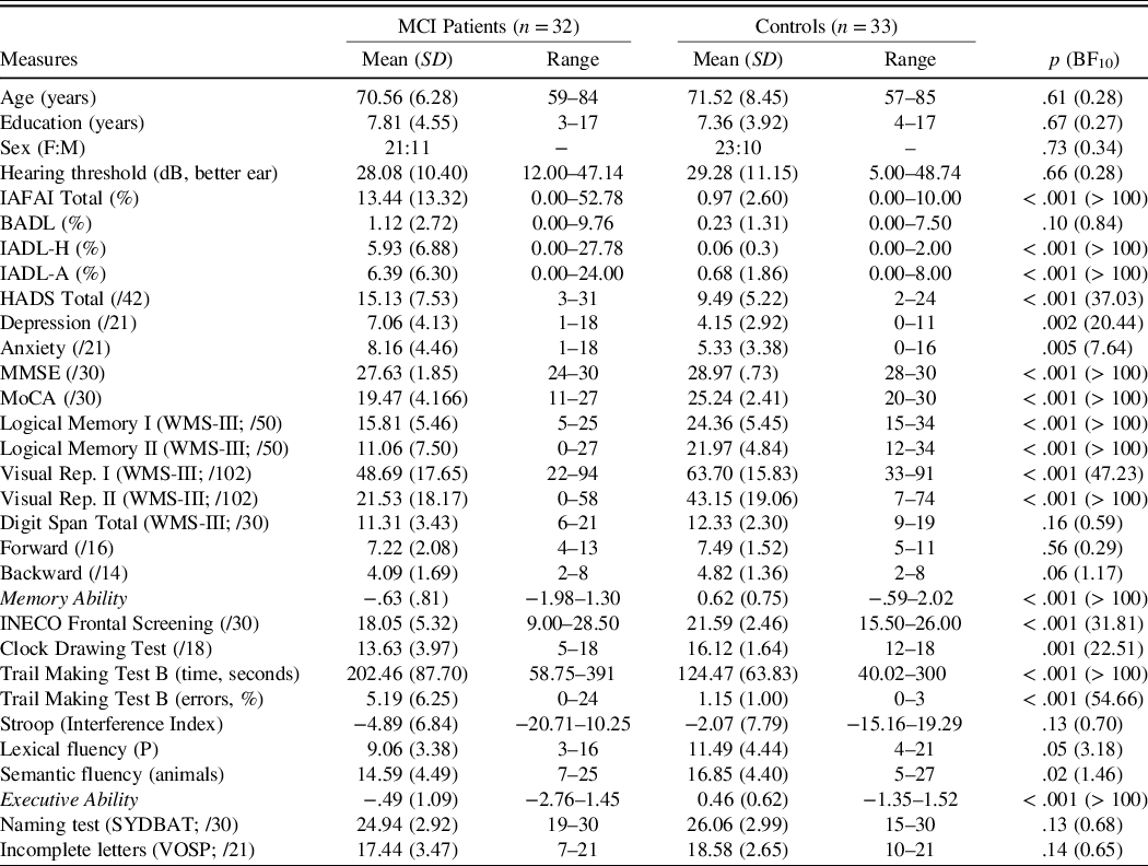
Thirty-two patients meeting the criteria for MCI (Petersen et al., Reference Petersen, Caracciolo, Brayne, Gauthier, Jelic and Fratiglioni2014) and 33 healthy controls were tested. As indicated in Table 1, the groups were matched for age, sex, and education. MCI patients were recruited from the Department of Neurology at Hospital de Braga and from the Neuropsychology Counselling Service at Universidade do Porto. Their diagnosis was confirmed by a neurologist and a neuropsychologist. All completed both MMSE and MoCA and had total scores at least 1.5 SD below the demographically corrected mean in at least one of these tests (Portuguese norms, MMSE, Freitas, Simões, Alves, & Santana, Reference Freitas, Simões, Alves and Santana2015; MoCA, Freitas, Simões, Alves, & Santana, Reference Freitas, Simões, Alves and Santana2013). All the patients were functionally independent and were diagnosed with multidomain amnestic MCI on the basis of standard criteria (Petersen, Reference Petersen2004; Petersen et al., Reference Petersen, Caracciolo, Brayne, Gauthier, Jelic and Fratiglioni2014; Winblad et al., Reference Winblad, Palmer, Kivipelto, Jelic, Fratiglioni, Wahlund and Petersen2004): all of them had objective memory impairment (scores at least 1.5 SD below the demographically corrected norms in one or more subtests of the Wechsler Memory Scale III; Wechsler, Reference Wechsler1997; Portuguese version, Wechsler, Reference Wechsler2008) and impairment in at least one more cognitive domain, namely executive functions, language, or visuospatial abilities. At the time of testing, their medication included: antidepressants (n = 15), anxiolytics-benzodiazepines (n = 12), antidyslipidemics (n = 10), acetylcholinesterase inhibitors (n = 7), antihypertensives (n = 7), and nutritional supplements (n = 5).
Background and neuropsychological characteristics of participants with mild cognitive impairment (MCI) and healthy controls

Note. p values correspond to the statistic of independent-samples t-tests (two-tailed); for sex, groups were compared using a Chi-squared test; BF= Bayes Factor; F = Female; M = Male; IAFAI = Adults and Older Adults Functional Assessment Inventory; BADL = Basic Activities of Daily Living; IADL-H = Instrumental Activities of Daily Living – Household; IADL-A = Instrumental Activities of Daily Living – Advanced; HADS = Hospital Anxiety and Depression Scale; MMSE = Mini-Mental State Examination; MoCA = Montreal Cognitive Assessment; Visual Rep. = Visual Reproduction; SYDBAT = Sydney Language Battery; VOSP = Visual Object and Space Perception Battery.
Healthy control participants were recruited from the community, and they all had intact cognitive functioning, as indicated by their MMSE and MoCA scores.
Exclusion criteria for both groups were history of alcoholism/substance abuse and diagnosis of dementia or other psychiatric (e.g., schizophrenia) or nonneurodegenerative neurological disorders (as well as neurodegenerative disorders in the case of controls). Participants with history of depression and anxiety disorders were not excluded, but we assessed depression and anxiety symptoms and considered them in the analyses that follow. Participants were European Portuguese native speakers and had normal or corrected-to-normal vision.
An a priori power analysis with G*Power 3.1 (Faul, Erdfelder, Buchner, & Lang, Reference Faul, Erdfelder, Buchner and Lang2009) indicated that for our main analysis, a sample size of at least 31 participants per group was required to detect a medium-to-large group effect (Bora and Yener, Reference Bora and Yener2017; McCade, Savage, & Naismith, Reference McCade, Savage and Naismith2011). This was calculated for a repeated-measures ANOVA with two groups and seven measurements (seven emotions), considering
 $\eta _p^2$
= .12, alpha level = .05, and power = .80.
Ethical approval was obtained from the local Ethics Committee, Hospital de Braga (CESHB 061/2016), and participants provided written informed consent according to the Declaration of Helsinki. They received no financial compensation for their participation.
Materials
Neuropsychological Measures
Participants completed a hearing test based on pure-tone audiometry, covering the frequencies 500 Hz, 1000 Hz, 2000 Hz, and 4000 Hz. Those with slight-to-moderate hearing loss were not excluded, given that this is a common condition among the elderly population (e.g., Lin, Niparko, & Ferrucci, Reference Lin, Niparko and Ferrucci2011) and that the volume of the emotional stimuli was individually adjusted to a comfortable level.
The ability to perform basic and instrumental activities of daily living (BADL and IADL, respectively) was evaluated using the Adults and Older Adults Functional Assessment Inventory (Inventário de Avaliação Funcional de Adultos e Idosos, IAFAI; Sousa, Simões, Pires, Vilar, & Freitas, Reference Sousa, Simões, Pires, Vilar and Freitas2008). The IAFAI assesses BADL such as feeding and dressing and two types of IADL: household IADL (IADL-H) such as preparation of meals and housekeeping tasks, and advanced IADL (IADL-A) such as comprehension and communication skills and health-related decision making.
Affective symptoms were assessed with the Hospital Anxiety and Depression Scale (HADS; Snaith & Zigmond, Reference Snaith and Zigmond1994; Portuguese version, Pais-Ribeiro et al., Reference Pais-Ribeiro, Silva, Ferreira, Martins, Meneses and Baltar2007).
Concerning the cognitive assessment, participants completed the MMSE (Folstein, Folstein, & McHugh, Reference Folstein, Folstein and McHugh1975; Portuguese version, Freitas et al., Reference Freitas, Simões, Alves and Santana2015) and MoCA tests (Nasreddine et al., Reference Nasreddine, Phillips, Bédirian, Charbonneau, Whitehead, Collin and Chertkow2005; Portuguese version, Freitas et al., Reference Freitas, Simões, Alves and Santana2013), as well as tests covering memory, executive functions, language, and visuospatial abilities. Verbal memory and visual memory were assessed with the Wechsler Memory Scale III (Wechsler,Reference Wechsler1997; Portuguese version, Wechsler, Reference Wechsler2008), subtests Logical Memory (I and II), Visual Reproduction (I and II), and Digit Span. Executive functions were assessed with the INECO Frontal Screening (Torralva et al., Reference Torralva, Roca, Gleichgerrcht, López and Manes2009; Portuguese version, Moreira, Lima, & Vicente, Reference Moreira, Lima and Vicente2014), Clock Drawing Test (Babins, Slater, Whitehead, & Chertkow, Reference Babins, Slater, Whitehead and Chertkow2008; Portuguese norms, Santana, Duro, Freitas, Alves, & Simões, Reference Santana, Duro, Freitas, Alves and Simões2013), Trail Making Test Part B (Army Individual Test Battery, 1994; Portuguese norms, Cavaco et al., Reference Cavaco, Goncalves, Pinto, Almeida, Gomes, Moreira and Teixeira-Pinto2013a), Stroop test (Golden & Freshwater, Reference Golden and Freshwater1978; Portuguese version, Fernandes, Reference Fernandes2013), and one-minute phonemic (letter P) and semantic (animals) verbal fluency tasks (Portuguese norms, Cavaco et al., Reference Cavaco, Gonçalves, Pinto, Almeida, Gomes, Moreira and Teixeira-Pinto2013b). Language was assessed with a Portuguese version of the naming test of the Sydney Language Battery (Savage et al., Reference Savage, Hsieh, Leslie, Foxe, Piguet and Hodges2013). Visuospatial abilities were assessed with the incomplete letters test of the Visual Object and Space Perception Battery (Warrington & James, Reference Warrington and James1991).
Emotion Recognition Tasks
Participants completed two emotion recognition tasks, one focusing on facial expressions and the other on vocalizations. Each task included 70 trials, with 10 different stimuli representing each of seven emotions: anger, disgust, fear, happiness, sadness, neutral, and pleasure in the case of vocalizations or surprise in the case of facial expressions. The stimuli were taken from validated corpora (nonverbal vocalizations: Lima, Castro, & Scott, Reference Lima, Castro and Scott2013; Sauter et al., Reference Sauter, Eisner, Ekman and Scott2010; facial expressions: Karolinska Directed Emotional Faces database, Goeleven, De Raedt, Leyman, & Verschuere, Reference Goeleven, De Raedt, Leyman and Verschuere2008) and have been used previously (e.g., Eisenbarth & Alpers, Reference Eisenbarth and Alpers2011; Lima et al., Reference Lima, Alves, Scott and Castro2014, Reference Lima, Brancatisano, Fancourt, Müllensiefen, Scott, Warren and Stewart2016, Reference Lima, Anikin, Monteiro, Scott and Castro2019; Strachan, Sebanz, & Knoblich, Reference Strachan, Sebanz and Knoblich2019). Nonverbal vocalizations were vocal sounds such as laughs or screams, as produced by male and female speakers. Their duration was approximately 1 s. Facial expressions were color photographs of male and female actors with no beards, moustaches, earrings, eyeglasses, or visible makeup. Each photograph was presented for 3 s.
Participants made an eight-alternative forced-choice judgment for each stimulus, selecting the emotion that was being expressed from a list including neutrality, anger, disgust, fear, happiness, pleasure/surprise, sadness, and other. They were instructed to select other whenever none of the remaining options reflected the emotional meaning of the stimuli. We ensured that the emotion labels were correctly understood by providing examples of everyday life situations in which the corresponding states are experienced. Participants also had the opportunity to familiarize themselves with the stimuli and task format in four practice trials. The 70 experimental trials that followed were randomized for each participant and divided into two blocks of 35 trials each. Each stimulus was presented once, and no feedback was given. Participants had no time limit to respond, but they were encouraged to provide fast and intuitive responses. The tasks had a total duration of about 45 min and were implemented in E-Prime 2.0 Professional.
Accuracy rates were calculated for each emotion and task, and they were corrected for response biases using unbiased hit rates or Hu (Wagner, Reference Wagner1993; see also, Isaacowitz et al., Reference Isaacowitz, Löckenhoff, Lane, Wright, Sechrest, Riedel and Costa2007). Hu values account for hits (number of times a given response category is correctly used) and false alarms (number of times a given response category is incorrectly used), and they vary between 0 and 1. Hu = 0 when no stimulus from a given emotion is correctly recognized, and Hu = 1 only when all the stimuli from a given emotion (e.g., happy prosody) are correctly recognized, and the corresponding response category (e.g., happiness) is always correctly used (i.e., there are no false alarms). These data were arcsine square-root transformed for inferential analyses. The response category other was selected in a small percentage of cases (facial expressions: 3.66% and 2.77% for MCI and controls, respectively; nonverbal vocalizations: 11.52% and 7.35% for MCI and controls).
Procedure
Participants were tested individually in a quiet room at Hospital de Braga or at the Neuropsychology Counselling Service at Universidade do Porto. They completed the demographic questionnaires, the background measures, and the emotion recognition tasks, in this order. Assessments lasted about 1.5/2 hours, and short breaks were allowed between tasks. The auditory stimuli were presented via high-quality headphones (Sennheiser HD 202), with the volume adjusted to a comfortable level.
Statistical Analysis
The data were evaluated based on standard frequentist and Bayesian approaches. In each analysis, a Bayes Factor (BF10) statistic was estimated, which considers the likelihood of the observed data given the alternative and null hypotheses. These analyses were conducted in JASP Version 0.10.2 (JASP Team, 2019), using the default priors. BFs10 values were interpreted following Jeffreys’ guidelines (Jarosz & Wiley, Reference Jarosz and Wiley2014), such that values between 1 and 3 correspond to anecdotal evidence for the alternative hypothesis, between 3 and 10 to substantial evidence, between 10 and 30 to strong evidence, between 30 and 100 to very strong evidence, and >100 to decisive evidence. A BF10 < 1 corresponds to evidence in favor of the null hypothesis: values between 0.33 and 1 correspond to anecdotal evidence, between 0.10 and 0.33 to substantial evidence, between 0.03 and 0.10 to strong evidence, between 0.01 and 0.03 to very strong evidence, and <0.01 to decisive evidence. Thus, one important advantage of Bayesian statistics over the frequentist approach is that they allow us to interpret null results and to formally draw inferences based on them.
RESULTS
Neuropsychological Data
The results of the background neuropsychological measures are summarized in Table 1. As for hearing ability, an independent-samples t test indicated that thresholds in the better hearing ear did not differ between patients and controls. According to the World Health Organization grades of hearing impairment, 11 patients and 12 controls had slight hearing loss (thresholds 26–40 dB), and 6 patients and 7 controls had moderate hearing loss (thresholds 41–60 dB).
As for the ability to perform activities of daily living, there were no group differences for BADL, but controls scored better than patients for both types of IADL.
Patients reported more depression and anxiety symptoms than controls. Considering a cutoff of ≥11 (Snaith, Reference Snaith2003), 7 patients and 2 controls had significant depression symptoms, and 10 patients and 1 control had significant anxiety symptoms.
Concerning cognitive performance, as expected, patients scored lower than controls in both MMSE and MoCA. Patients also scored lower across all memory measures, except for the forward Digit Span. For further analyses regarding memory abilities, we extracted a single latent variable (hereafter memory ability) based on a principal component analysis (varimax rotation), to reduce collinearity and the contribution of measure-specific error variance. The latent variable accounted for 62.33% of the variance in the original memory subtests, each of which loaded highly on the variable, rs > .59. The forward Digit Span was not included in this analysis because it was similar across groups and had a low loading on the latent variable, r = −.05.
Patients scored lower than controls across all executive measures, except for the Stroop test. We extracted a principal component (executive ability), which accounted for 59.37% of the variance in the original measures (loading on the variable, rs > .62). The Stroop test was not included because it was similar across groups and had a low loading on the latent variable, r = .07.
The two groups did not differ in language and visuospatial abilities.
Emotion Recognition Accuracy
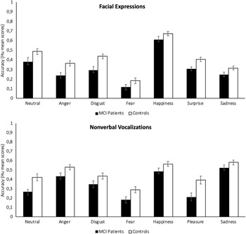
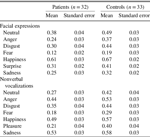
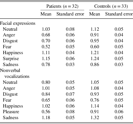
The average scores across all participants were .36 for faces (SD = .12; range = .06–.60) and .41 for vocalizations (SD = .14; range = .16–.72). One-sample t tests, conducted separately for MCI and control participants, confirmed that both groups performed above chance, for faces and for vocalizations (.13, i.e., 1/8 responses correct; patients: ps < .001, BF10 > 100; controls: ps < .001, BF10 > 100). Figure 1 shows accuracy rates for each emotion as a function of task and group (see also Appendices A and B). To assess group differences, we conducted mixed-design ANOVAs for each task, with the emotions as repeated-measures factor, and group as between-subjects factor (MCI vs. control participants). Greenhouse–Geisser corrections were applied when necessary (Mauchly’s sphericity test).
Accuracy rates for each emotion as a function of task and group. Error bars show standard errors.

For facial expressions, MCI patients (M = .32; SD = .13; range = .06–.60) performed worse that controls (M = .41; SD = .09; range = .21–.60), as indicated by a large main effect of group, F(1, 63) = 12.91, p < .001, η
2
= .17. Bayesian statistics indicated that the level of evidence for this effect was strong, BF10 = 10.30. Some emotions were more difficult to recognize than others, F(4.90, 308.88) = 53.30, p < .001,
$\eta _p^2$
= .46, BF10 > 100: post hoc comparisons (Bonferroni-corrected) indicated that performance was highest for happiness, neutrality, and surprise (with similarly high scores, ps > .2); significantly lower (ps < .001) for disgust, sadness, and anger (with similarly intermediate scores, ps = .1); and lowest for fear (ps < .001 in comparison to all other emotions). There was no interaction between group and emotion; however, p = .23, BF10 = 0.01.
For nonverbal vocalizations, again MCI patients (M = .34; SD = .13; range = .16–.66) performed worse that controls (M = .46; SD = .12; range = .19–.72). The effect was large, F(1, 63) = 17.52, p < .001, η
2 = .22, and the level of evidence was very strong, BF10 = 58.29. The main effect of emotion was significant, F(5.12, 322.80) = 28.35, p < .001,
$\eta _p^2$
= .31, BF10 > 100: performance was highest for sadness (ps < .02 in comparison to all other emotions), followed by happiness, anger, and neutrality (with similarly intermediate scores, ps = .07), and then by disgust and pleasure (p = .08), and finally fear (ps < .02 in comparison to all other emotions apart from pleasure, p = .1). There was no interaction between group and emotion, p = .07, BF10 = 0.46.
We thus found strong evidence that MCI is associated with impaired emotion recognition in visual and auditory expressions.Footnote 1 To compare the magnitude of the impairment across tasks, we conducted a follow-up ANOVA, with average performance on faces and vocalizations as repeated-measures factor, and group as between-subjects factor. This analysis confirmed the effect of group, F(1, 63) = 24.20, p < .001, η 2 = .28, BF10 > 100, but there was no effect of task, p = .19, BF10 = 0.41, or interaction between task and group, p = .73, BF10 = 0.28. The magnitude of the impairment was thus similar across visual and auditory expressions.
Effects of Background and Neuropsychological Variables
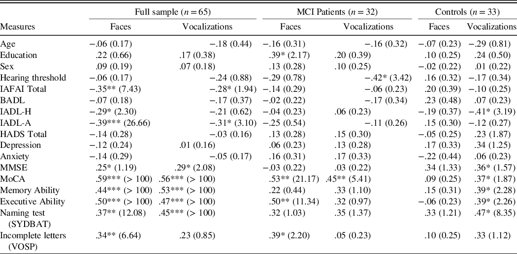
Table 2 presents zero-order correlations (Pearson’s r) between emotion recognition and background and neuropsychological variables for the full sample and separately for MCI and control participants. Emotion recognition was not associated with age, education, or sex. There was a positive association between education and facial emotion recognition in MCI patients, but Bayesian statistics indicated that the evidence was weak. We also found no association between emotion recognition and anxiety and depression symptoms.
Correlations between emotion recognition and background and neuropsychological measures. BF10 are indicated in brackets.

Note.*p < .05; **p < .01; ***p < .001;IAFAI = Adults and Older Adults Functional Assessment Inventory; BADL = Basic Activities of Daily Living; IADL-H = Instrumental Activities of Daily Living – Household; IADL-A = Instrumental Activities of Daily Living – Advanced; HADS = Hospital Anxiety and Depression Scale; MMSE = Mini-Mental State Examination; MoCA = Montreal Cognitive Assessment; SYDBAT = Sydney Language Battery; VOSP = Visual Object and Space Perception Battery.
As for hearing ability, there was a moderate correlation between higher hearing thresholds and worse vocal emotion recognition in MCI patients. Such correlation does not account for the effect of MCI in emotion recognition, though. This was confirmed in a multiple regression including the full sample and modeling vocal emotion recognition accuracy as a function of hearing thresholds and group (dummy coded). The model explained 26.01% of the variance, R = .51, F(2,64) = 10.90, p < .001, BF10 > 100. Hearing thresholds independently contributed to the model, partial r = −.29, p = .02, BF10 = 2.95, but there was also decisive evidence for an independent contribution of group, partial r = .45, p < .001, BF10 > 100.
Emotion recognition was not associated with the ability to perform activities of daily living in patients. There were associations with IADL in the full sample, but multiple regressions showed that they disappear when group effects are accounted for. For facial expressions, a model with three predictors (IADL-H, IADL-A, and group) accounted for 27.88% of the variance, R = .53, F(3,64) = 7.88, p < .001, BF10 > 100. An independent contribution was made by group, partial r = .38, p = .002, BF10 = 22.18, but not by IADL-H, partial r = .11, p = .38, BF10 = 0.41, or IADL-A, partial r = −.21, p = .11, BF10 = 0.94. A similar model for vocalizations accounted for 22.25% of the variance, R = .46, F(3,64) = 5.48, p = .002, BF10 = 16.21. An independent contribution was again evident for group, partial r = .36, p = .004, BF10 = 13.25, but not for IADL-H, partial r = .13, p = .33, BF10 = 0.49, or IADL-A, partial r = −.16, p = .13, BF10 = 0.62. Even when power is increased by considering the full sample, performance of IADL is not uniquely associated with emotion recognition.
Concerning cognitive variables, we found moderate-to-large correlations between MoCA scores and facial and vocal emotion recognition in patients, indicating that the cognitive impairment played a role in emotion recognition difficulties. As for specific cognitive domains, more severe impairments in executive and visuospatial abilities correlated with lower facial emotion recognition in patients. Such associations did not reach significance for vocalizations, however, and no associations were found between memory and language abilities and any of the emotion recognition tasks. Memory and language abilities correlated with emotion recognition in the full sample, but in these analyses cognitive and group effects are hardly separable, because the groups were formed based on cognitive measures.
DISCUSSION
MCI was associated with impaired recognition of facial and vocal expressions. The impairment was statistically large and similar across individual emotions and modalities. Furthermore, it could not be accounted for by impairments in hearing ability nor by affective symptoms. Emotion recognition difficulties did not correlate with patients’ ability to perform activities of daily living, but they did correlate with their global cognitive status. Patients with more severe cognitive decline had more trouble categorizing emotions.
Previous studies indicated that MCI can be associated with facial emotion recognition difficulties (Henry et al., Reference Henry, Thompson, Rendell, Phillips, Carbert, Sachdev and Brodaty2012; McCade et al., Reference McCade, Savage, Guastella, Lewis and Naismith2013; Spoletini et al., Reference Spoletini, Marra, DiIulio, Gianni, Sancesario, Giubilei and Spalletta2008; Teng, Lu, & Cummings, Reference Teng, Lu and Cummings2007). Results were mixed (e.g., Bediou et al., Reference Bediou, Ryff, Mercier, Milliery, Hénaff, D’Amato and Krolak-Salmon2009; Dodich et al., Reference Dodich, Cerami, Crespi, Canessa, Lettieri, Iannaccone and Cacioppo2016), however, and it remained unclear whether the impairments generalize across emotions. Our results corroborate the notion that MCI affects facial emotion recognition and suggest that these difficulties are not circumscribed to specific emotions. The discrepancy in relation to previous findings might stem from differences in sample characteristics or from differences in methods. Expressions of happiness and disgust are often easier to recognize than those of fear and sadness and sometimes even associated with ceiling effects (e.g., McCade et al., Reference McCade, Savage and Naismith2011; Moradi, Najlerahim, & Humphreys; Reference Moradi, Najlerahim and Humphreys2018; Richard-Mornas et al., Reference Richard-Mornas, Borg, Klein-Koerkamp, Paignon, Hot and Thomas-Antérion2012; Spoletini et al., Reference Spoletini, Marra, DiIulio, Gianni, Sancesario, Giubilei and Spalletta2008; Weiss et al., Reference Weiss, Kohler, Vonbank, Stadelmann, Kemmler, Hinterhuber and Marksteiner2008), and this could have masked group differences in previous work. Our stimuli were selected so that they would elicit intermediate levels of performance, and this might have made our tasks more sensitive to a wider range of impairments. Concerning sample characteristics, ours included only multidomain amnestic MCI patients. It remains unclear if emotion recognition differs across MCI subtypes, which would be relevant knowledge for clinical practice, but there is evidence that the magnitude of the impairment might be larger when cognitive deficits are widespread than when they are limited to memory (Bora & Yener, Reference Bora and Yener2017). Impairments might also differ across amnestic and nonamnestic MCI subtypes (McCade et al., Reference McCade, Savage, Guastella, Lewis and Naismith2013). Studying amnestic multidomain MCI is critical for its value in predicting conversion to dementia (Hessen et al., Reference Hessen, Reinvang, Eliassen, Nordlund, Gjerstad, Fladby and Wallin2014; Michaud et al., Reference Michaud, Su, Siahpush and Murman2017; Petersen et al., Reference Petersen, Caracciolo, Brayne, Gauthier, Jelic and Fratiglioni2014), but studies directly comparing MCI subtypes in emotion recognition are warranted. They will play a critical role in determining whether the impairment uncovered here is a general feature of MCI or whether impairments might be smaller when cognitive impairments are limited to one domain.
The results of the auditory emotion recognition task indicate that the emotion impairment in MCI extends beyond faces. The ability of patients to recognize vocal emotions was as impaired as their ability to recognize faces. This impairment could not be attributed to hearing difficulties, because hearing thresholds were similar across groups, and a multiple regression showed a significant group effect, after hearing threshold was held constant. Only a few studies examined auditory emotion recognition in neurodegenerative conditions such as Alzheimer’s disease (e.g., Bucks & Radford, Reference Bucks and Radford2004; Drapeau et al., Reference Drapeau, Gosselin, Gagnon, Peretz and Lorrain2009; Klein-Koerkampa et al., Reference Klein-Koerkamp, Beaudoin, Baciu and Hot2012; Park et al., Reference Park, Kim, Shin, Kim, Sohn, Park, Youn and Lee2017), and to our knowledge, no previous work has addressed this issue in MCI. MCI patients might have difficulties decoding affect from body postures and hand gestures in addition to faces (McCade et al., Reference McCade, Savage, Guastella, Lewis and Naismith2013), but all these cues pertain to the visual domain. Expanding current ideas on how MCI relates to socioemotional impairments, our findings suggest that difficulties in emotion recognition reflect a failure at supramodal levels of processing. Like faces, vocal cues are a major source of emotional information in social interactions (Brück, Kreifelts, & Wildgruber, Reference Brück, Kreifelts and Wildgruber2011; Schirmer & Kotz, Reference Schirmer and Kotz2006). Facial and vocal expressions engage distinct neural networks, namely for sensory/perceptual processes, but there are regions that respond to emotional stimuli regardless of modality, such as prefrontal systems and the posterior superior temporal sulcus (Peelen et al., Reference Peelen, Atkinson and Vuilleumier2010; Schirmer, Reference Schirmer2018; Schirmer & Adolphs, Reference Schirmer and Adolphs2017). Given that the profile and magnitude of impairments were similar across modalities, they plausibly result from a dysfunction in systems that support domain-general emotional processing, rather than in those that are modality-specific. This is consistent with the evidence of atrophy in temporal and prefrontal systems in MCI (Edmonds et al., Reference Edmonds, Eppig, Bondi, Leyden, Goodwin and Delano-Wood2016; Hämäläinen et al., Reference Hämäläinen, Tervo, Grau-Olivares, Niskanen, Pennanen, Huuskonen and Soininen2007), which might be particularly evident in patients with impairments in multiple cognitive domains (e.g., Whitwell et al., Reference Whitwell, Petersen, Negash, Weigand, Kantarci, Ivnik and Jack2007; Zhang et al., Reference Zhang, Sachdev, Wen, Kochan, Crawford, Brodaty and Trollor2012). Future MRI studies, including visual and auditory emotion processing tasks, will be necessary to directly address these questions.
Consistent with previous evidence, depression and anxiety symptoms were higher in MCI compared with control participants (e.g., Ismail et al., Reference Ismail, Elbayoumi, Fischer, Hogan, Millikin, Schweizer and Fiest2017; Steffens, Reference Steffens2012; Monastero, Mangialasche, Camarda, Ercolani, & Camarda, Reference Monastero, Mangialasche, Camarda, Ercolani and Camarda2009). While these neuropsychiatric symptoms can also be prevalent among healthy older individuals (e.g., Wang et al., Reference Wang, Wu, Lai, Long, Zhang, Li and Lin2017; Valiengo, Stella, & Forlenza, Reference Valiengo, Stella and Forlenza2016), they occur in approximately 40%–50% of MCI patients, and they might be a first manifestation of a neurocognitive disorder and predict more accelerated cognitive decline (e.g., Gallagher et al., Reference Gallagher, Fischer and Iaboni2017; Geda et al., Reference Geda, Roberts, Knopman, Petersen, Christianson, Pankratz and Rocca2008). Our findings suggest that anxiety and depression symptoms are unrelated to the emotion recognition impairment, though. In fact, Bayesian analyses provided substantial evidence for the null hypothesis. A similar lack of association between affective variables and emotion recognition has been obtained in the context of healthy aging (e.g., Lima et al., Reference Lima, Alves, Scott and Castro2014) and Parkinson’s disease (Lima, Garrett, & Castro, Reference Lima, Garret and Castro2013). The mechanisms underlying neuropsychiatric and socioemotional difficulties in MCI might be partly distinct. Several mechanisms underlie depression symptoms, such as hypercortisolaemia, reduced hippocampal volume, cerebrovascular disease, and increased deposition of beta-amyloid protein (e.g., Gallagher et al., Reference Gallagher, Fischer and Iaboni2017), and these could possibly have a less prominent role for emotion recognition, an idea that warrants direct examination.
Although emotion recognition difficulties were unrelated to affective symptoms, they were linked to global cognitive impairment in MCI participants. Patients with lower MoCA scores had lower performance in the facial and vocal emotion recognition tasks. This is consistent with results linking cognitive performance with facial emotion recognition in MCI (e.g., Henry et al., Reference Henry, Thompson, Rendell, Phillips, Carbert, Sachdev and Brodaty2012; Pietschnig et al., Reference Pietschnig, Aigner-Wöber, Reischenböck, Kryspin-Exner, Moser, Klug and Lehrner2015; Sarabia-Cobo, García-Rodríguez, Navas, & Ellgring, Reference Sarabia-Cobo, García-Rodríguez, Navas and Ellgring2015; Spoletini et al., Reference Spoletini, Marra, DiIulio, Gianni, Sancesario, Giubilei and Spalletta2008; Teng et al, Reference Teng, Lu and Cummings2007; Weiss et al., Reference Weiss, Kohler, Vonbank, Stadelmann, Kemmler, Hinterhuber and Marksteiner2008) and with associations uncovered in samples covering the full continuum of cognitive capacity (Virtanen et al., Reference Virtanen, Singh-Manoux, Batty, Ebmeier, Jokela, Harmer and Kivimäki2017). It is also consistent with the notion that domain-general cognitive resources might play a role in emotion recognition processes (e.g., Lima et al., Reference Lima, Garret and Castro2013). The association between cognitive performance and emotion recognition was less evident in controls and in the context of MMSE scores, possibly because the MMSE is an easier test, and it was not specifically designed for MCI. In longitudinal studies, it will be relevant to ask whether emotion recognition impairments in MCI predict disease trajectories, in terms of rate of cognitive decline and of risk of conversion to dementia.
Limitations of the current study include the use of acted stimuli and static faces. While everyday life expressions are also often acted to a certain extent, recent work has documented perceptual differences between acted and spontaneous expressions (e.g., Anikin & Lima, Reference Anikin and Lima2018). It will therefore be relevant to extend our results to spontaneous expressions and to more ecologically valid dynamic facial stimuli (e.g., Lima et al., Reference Lima, Brancatisano, Fancourt, Müllensiefen, Scott, Warren and Stewart2016). Another limitation is that participants only assessed faces and voices for their emotional attributes, leaving unclear whether the impairments are emotion-specific or not. Previous studies have suggested that low-level facial processing is intact in MCI (e.g., McCade et al., Reference McCade, Savage, Guastella, Lewis and Naismith2013), and we accounted for sensory losses, but it will be informative to systematically explore nonemotional processes such as face and voice identity perception. Moreover, our sample size was supported by an a priori power analysis, but it is relatively small and limited to multidomain amnestic MCI patients with preserved BADL but slightly compromised IADL. Although we did not find correlations between emotion recognition and functional status, future studies with larger and more diverse samples will be crucial to clarify the generalizability of our findings, not only across MCI subtypes, but also across the spectrum of functional abilities. Finally, there are well-established links between emotion recognition skills and measures of social functioning, in both healthy and clinical samples (e.g., Carton et al., Reference Carton, Kessler and Pape1999; Halberstadt et al., Reference Halberstadt, Ruffman, Murray, Taumoepeau and Ryan2011; Phillips et al., 2001). This suggests that the emotion recognition impairments observed in MCI patients will significantly impact on their everyday social adjustment and quality of life more broadly. We did not include measures of social functioning, however, and our functional assessment only covered the ability to carry out practical activities of everyday life. Future work will therefore need to directly delineate the implications of emotion recognition impairments in this condition.
To conclude, the present study is the first demonstration of a supramodal deficit in emotion recognition in MCI. We found that MCI patients show a generalized impairment in the recognition of facial expressions and that the emotion deficit extends to auditory expressions, namely to the recognition of emotional vocalizations. We further established that emotion recognition difficulties in MCI are linked to the magnitude of global cognitive impairment, but not to neuropsychiatric symptoms such as anxiety and depression. Research on social–emotional difficulties in MCI is still in its infancy, but the emerging findings are promising for their potential clinical utility. If emotion recognition impairments are apparent early on in the course of neurocognitive disorders, their assessment should be part of standard neuropsychological testing, and they could represent a target of interventions aimed at improving social cognition. Emotion recognition impairments could additionally represent a useful clinical marker in MCI but also in other neurological conditions (Cotter, Granger, Backx, Hobbs, Looi, & Barnett, Reference Cotter, Granger, Backx, Hobbs, Looi and Barnett2018).
FUNDING
Grants from the Portuguese Foundation for Science and Technology (FCT) awarded to H.S.M. (SFRH/BD/105201/2014), to C.F.L. (IF/00172/2015), and to the Center for Psychology at University of Porto (UID/PSI/00050/2013 and PTDC/PSI-GER/28274/2017). The funders had no role in study design, data collection and analysis, decision to publish, or preparation of the manuscript.
CONFLICTS OF INTEREST
The authors have nothing to disclose.
Accuracy rates (Hu mean scores) for each emotion as a function of task and group

Accuracy rates (arcsine square-root transformed Hu scores, used for inferential analyses) for each emotion as a function of task and group