Introduction
Research into the use of nanotechnology in medicine cuts across all fields; however, it is presently most advanced in its applications for cancer. Therefore, in this article, we will use cancer as an indication to discuss the challenges in clinical translation of nanomedicines. Indeed, using nanotechnology to uncap the full potential of existing chemotherapy agents has emerged as an attractive drug development strategy, and several nanotherapeutics are already being used in the clinic. Reference Ferrari1–Reference Wang, Langer and Farokhzad2 This stems from the unique physicochemical properties of nanoparticles carrying chemotherapy, which can be facilely modulated to confer unique advantages over existing chemotherapeutic agents. Reference Moghimi, Hunter and Murray3 Indeed, changing the size of a drug from Angstrom scale to the nanoscale can alter the pharmacokinetic profile of the molecule. Furthermore, it is well known that surface modifications to increase hydrophilicity can mask the nanovectors from the reticuloendothelial system, thereby increasing circulation time. Reference Moghimi, Hunter and Murray3–Reference Allen and Cullis5 Such nanovectors accumulate preferentially in tumors helped by the unique leaky tumor vasculature, which when coupled with impaired intratumoral lymphatic drainage contributes to an enhanced permeation and retention (EPR) effect. Reference Yuan, Dellian, Fukumura, Leunig, Berk, Torchilin and Jain6,Reference Yuan, Leunig, Huang, Berk, Papahadjopoulos and Jain7 Indeed, these nanoparticles were found to deliver between 5–11-fold more doxorubicin, an anticancer drug, to Kaposi sarcoma lesions than to normal skin. Reference Northfelt, Martin, Working, Volberding, Russell, Newman, Amantea and Kaplan8 Doxorubicin-loaded lipid-based nanoparticles are already being used clinically for treatment of breast cancer. Reference Barenholz9,Reference Batist, Barton, Chaikin, Swenson and Welles10
Similarly, the integrated area under the tumor paclitaxel concentration-time curve was found to be 33% higher when administered as an albumin protein-paclitaxel complex nanoparticle, which is currently approved for use in metastatic breast cancer, than when administered as free paclitaxel. Reference Desai, Trieu, Yao, Louie, Ci, Yang, Tao, De, Beals, Dykes, Noker, Yao, Labao, Hawkins and Soon-Shiong11 Interestingly, it should be kept in perspective that the drivers for developing these nanoparticles for delivering doxorubicin and paclitaxel were primarily to overcome off-target side effects, for example cardiotoxicity in the case of doxorubicin, or hypersensitivity to the vehicles used for delivering taxanes. Reference Malam, Loizidou and Seifalian12,Reference Ma and Mumper13 These advantages arose from the preferential delivery of anti-cancer drugs to the tumor and limited penetration into normal tissue or were due to the fact that the nanoparticle suspension removed the need for biologically incompatible vehicles. However, it is unlikely that an approach of developing nanomedicines to ameliorate the side effects of a chemotherapeutic will translate to the clinic today without a clear efficacy advantage. This article discusses the challenges that nanomedicines face and the potential approaches to overcome these hurdles toward their clinical translation.
Is increased delivery of a drug to the tumor enough to confer efficacy advantage?
The potential of using nanoparticles to achieve increased intratumoral concentrations of cytotoxic agents as a result of the enhanced permeation and retention effect spurred the drive to optimize parameters that could further enhance this advantage (see the Yhee et al. and Pearson et al. articles in this issue). For example, the dogma is that drug PEGylation or coating a nanoparticle with polyethylene glycol (PEG) can prevent binding of plasma proteins (opsonization) and thereby reduce clearance by the reticuloendothelial system, resulting in increased circulation time. Reference Alexis, Pridgen, Molnar and Farokhzad14 Indeed, the pharmacokinetic properties of a PEGylated doxorubicin nanoparticle (Caelyx/Doxil) were found to be distinct from those of a non-PEGylated nanoparticle (Myocet). Reference Song, Wu, Yoshino and Zamboni15 Therefore, extensive studies have been carried out to optimize the degree of PEGylation. Most studies focused on longer chain PEGs, with the incorporation of 5 mol% of distearoylphosphatidylethanolamine-poly(ethylene glycol)2000 (DSPE-PEG2000) emerging as the clinical and research gold standard. This standard was established more as a general consensus rather than through scientific logic. Reference Gref, Lück, Quellec, Marchand, Dellacherie, Harnisch, Blunk and Müller16 However, a recent study indicated that this may not be valid, and shorter chain lengths could be just as good. Reference Stefanick, Ashley, Kiziltepe and Bilgicer17 This is consistent with previous reports that liposomes with shorter PEGs exhibit a similar circulation half-life. However, another issue that remains unresolved is the impact of PEGylation on efficacy. There is concern that PEG can hinder internalization into cells, and this has inspired complex designs of nanoparticles with sheddable PEGs that address this challenge. Reference Li and Huang18 However, an equally large number of reports show that cellular internalization of PEGylated nanoparticles improves efficacy.
Besides PEGylation and the resultant enhanced circulation time, the size and shape of nanoparticles is another area of active exploration, with the goal of increasing therapeutic efficacy. For example, rod-shaped micelles were found to exhibit a circulation lifetime 10-fold higher than spherical micelles. Reference Albanese, Tang and Chan19 Similarly, in a recent study, flexible worm-like nanocarriers were found to increase the amount of drug delivered to tumors and shrink tumors more effectively than spherical micelles. Reference Loverde, Klein and Discher20 Mechanistically, at the cellular level, this could arise from distinct internalization kinetics and saturation concentrations, which have shown to be highly dependent upon the physical dimensions (i.e., size and shape of nanoparticles). Reference Chithrani and Chan21 Interestingly, spherical-shaped particles have a higher probability of entering the cell in comparison to rod-shaped nanostructures, Reference Chithrani and Chan21 and hence more fundamental insights need to be generated on how cellular internalization and circulation half-life can either offset or act in synergy to determine the final outcome.
The composition of the nanostructure can also affect uptake. For example, single-walled carbon nanotubes (SWCNTs) and gold nanoparticles (∼50 nm in diameter) exhibited distinct endocytosis rates of 10−3 min−1 and 10−6 min−1, respectively. Reference Albanese, Tang and Chan19 At a systems level, size becomes an even more important determinant of pharmacokinetics and biodistribution arising from the variable pore diameters in the endothelial barrier of the vasculature. Nanoparticles with diameters smaller than 5.5 nm can be excreted by the kidneys, Reference Choi, Liu, Misra, Tanaka, Zimmer, Itty Ipe, Bawendi and Frangioni22 while nanoparticles with diameters larger than 200 nm accumulate in the spleen and liver. Reference Jain and Stylianopoulos23 It is well established that nanoparticles in the size range 80–120 nm optimally target the tumor. Reference Jain and Stylianopoulos23 Furthermore, recent studies have shown that nanoparticles smaller than 12 nm are more suited for enhancing tumor penetration. Reference Chauhan, Stylianopoulos, Martin, Popović, Chen, Kamoun, Bawendi, Fukumura and Jain24 This indicates that nanoparticles greater than 5.5 nm could be ideal for drugs that cause nephrotoxicity or toxicity to the urinary bladder, such as cisplatin and camptothecin, respectively. Similarly, larger particles could be used to target the reticuloendothelial system (spleen and liver), while particles between 6–150 nm could be ideal for tumor targeting.
Understanding the various modalities that can impact nanomaterial behavior in the body is critical and may even be important for a better understanding of toxicity profiles of nanomedicines; the remaining unresolved question is the impact of these parameters on the clinical outcome. For example, if a nanovector is used for delivering a highly cytotoxic drug, will these mechanistic distinctions have an effect that is significantly distinct in terms of survival outcome? For instance, both Myocet and Doxil show non-inferiority with doxorubicin, despite one being PEGylated and the other not. Reference O’Brien, Wigler, Inbar, Rosso, Grischke, Santoro, Catane, Kieback, Tomczak, Ackland, Orlandi, Mellars, Alland and Tendler25,Reference Chan, Davidson, Juozaityte, Erdkamp, Pluzanska, Azarnia and Lee26 Similarly, SWCNTs were pro-angiogenic via activation of integrins, and both of these nanomaterials were highly and equally effective in reducing tumor progression when used to deliver doxorubicin. Reference Chaudhuri, Harfouche, Soni, Hentschel and Sengupta27–Reference Chaudhuri, Soni and Sengupta29 Here the choice of drug was the final determinant despite the difference in the inherent biological properties of the carrier.
Choosing the right drug
While both Doxil and Myocet were approved based on non-inferiority clinical trials, it is unlikely that such trials will form the basis of clinical translation of novel nanomedicines in the future. At the same time, it is also likely that cytotoxic chemotherapeutics will remain a strong pillar of cancer chemotherapy over the next decade despite the emergence of targeted therapeutics. Therefore, optimizing the cytotoxics by integrating them with nanotechnology may still create opportunities but only if increased efficacy is achieved as the desired endpoint. This again highlights the need to choose the right drug.
For example, cisplatin [cis-dichlorodiammineplatinum(II)] is one of the most commonly used chemotherapeutic agents besides doxorubicin and paclitaxel and is a first-line therapy for most malignancies, including testicular, ovarian, cervical, and lung cancer. Reference Kelland30 Its clinical use, however, is dose-limited due to nephrotoxicity (toxicity to the kidney) in adults and ototoxicity (toxicity to the inner ear) in children. Reference Madias and Harrington31,Reference Al-Khatib, Cohen, Carret and Daniel32 As nanoparticles greater than 5 nm in diameter will avoid renal clearance, Reference Choi, Liu, Misra, Tanaka, Zimmer, Itty Ipe, Bawendi and Frangioni22 a cisplatin nanoparticle can potentially reduce cisplatin nephrotoxicity (it is unlikely that a nanoparticle will cross the blood-labyrinth barrier in the inner ear, and hence such nanoparticles could be less ototoxic too, but this remains to be studied). While liposomal formulations of cisplatin enabled increased delivery of cisplatin to the tumor, this did not translate into increased clinical efficacy, as the drug failed to release efficiently. Reference White, Lorigan, Margison, Margison, Martin, Thatcher, Anderson and Ranson33,Reference Zamboni, Gervais, Egorin, Schellens, Zuhowski, Pluim, Joseph, Hamburger, Working, Colbern, Tonda, Potter and Eiseman34 Similarly, it has been a challenge to entrap cisplatin in polymeric sustained-release nanoparticles. Reference Avgoustakis, Beletsi, Panagi, Klepetsanis, Karydas and Ithakissios35,Reference Fujiyama, Nakase, Osaki, Sakakura, Yamagishi and Hagiwara36
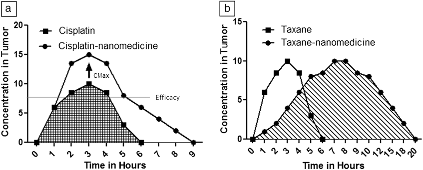
In a recent study, Dhar et al. generated a platinum (IV) complex, which had sufficient hydrophobicity for encapsulation in PLGA-b-PEG nanoparticles. However, the prodrug (a drug precursor) had to be intracellularly processed into active cisplatin. Reference Dhar, Gu, Langer, Farokhzad and Lippard37 Alternative strategies based on the conjugation of platinum to polymers (for example, a polyamidoamine dendrimer-platinum complex) produced a 200–550 fold reduction in cytotoxicity compared with free cisplatin. These results are due to stable linkages that are formed between the polymer and Pt. Reference Haxton and Burt38 Similarly, AP5280, a N-(2-hydroxypropyl) methacrylamide copolymer-bound platinum exhibited minimal nephrotoxicity in clinical studies Reference Rademaker-Lakhai, Terret, Howell, Baud, de Boer, Pluim, Beijnen, Schellens and Droz39 but was less potent than carboplatin, as the platinum was stably coordinated to an aminomalonic acid chelating agent, which prevented the aquation of cisplatin. Reference Lin, Zhang, Rice, Stewart, Nowotnik and Howell40 These studies highlight the fact that increased nanoparticle-mediated tumor delivery of a drug may not always translate to increased efficacy, and in the case of cisplatin, it is likely that intratumoral maximal concentration (CMax) of the active moiety (cisplatin gets aquated, i.e., chlorides are replaced by water molecules to generate the active form) has a bigger role to play than an increased area under the curve (AUC) of an intratumoral concentration-time curve.
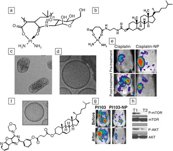
Indeed, in a recent study, we engineered a novel platinum analogue that could self-assemble into a supramolecular nanostructure, resulting in high loading efficiencies. In previous studies, loading efficiencies were low because the drug was encapsulated in nanoparticles. Reference Sengupta, Basu, Soni, Pandey, Roy, Oh, Chin, Paraskar, Sarangi, Connor, Sabbisetti, Kopparam, Kulkarni, Muto, Amarasiriwardena, Jayawardene, Lupoli, Dinulescu, Bonventre, Mashelkar and Sengupta41 Furthermore, we introduced a novel leaving group chemistry, specifically introducing a monocarboxylato and an O→Pt coordination bond, based on structure activity relationships, which resulted in faster aquation and drug release ( Figure 1 ). Reference Paraskar, Soni, Chin, Chaudhuri, Muto, Berkowitz, Handlogten, Alves, Bilgicer, Dinulescu, Mashelkar and Sengupta42 Together with preferential accumulation in the tumor and bypassing the kidney, this resulted in an increased efficacy with amelioration of nephrotoxicity. Thus, drugs that depend on CMax and possess physicochemical properties that limit efficient loading and release kinetics will likely require structure activity relationship (SAR)-based modification to achieve greater efficacy if formulated in nanoparticles ( Figure 2 ).
Using structure activity relationships to engineer nanoparticles. (a) A leaving group that could coordinate Pt was designed using a polyisobutylene-maleic acid polymer (n = 40) that we derivatized with glucosamine. At the optimal Pt to polymer ratio of 15:1, it self-assembled into a nanoparticle. (b) As polymers can be limited by polydispersity, we created the same Pt coordination environment using a cholesterol succinic acid conjugate. The coordination with Pt results in analogues of cisplatin/carboplatin, which can supramolecularly assemble into lipidic nanoparticles with phosphatidylcholine and poly(ethylene glycol)-distearoylphosphatidylethanolamine. (c) High-resolution transmission electron microscope image shows typical nanoparticles that are formed. (d) Cryo-transmission electron microscope image of a typical supramolecular nanoparticle. These molecules optimize the aquation kinetics and therefore increase the antitumor efficacy of the active molecule, as seen in (e) an ovarian cancer mouse model, where the animals were treated with 3 mg/kg Pt dose equivalent of cisplatin or cisplatin-nanoparticles (NPs). (f) The cholesterol backbone was also used to engineer a novel analogue of the PI3K (phosphatidylinositol 3 kinase) inhibitor, PI103, which could self-assemble into a nanoparticle (see inset). Note that the particle sizes are 100 nm for (c), (d), and (f). (g–h) The PI3K-inhibiting nanoparticles exert greater anti-tumor efficacy in a PI3K-driven ovarian cancer model, as it can exert a sustained inhibition of signaling molecules downstream of PI3K, shown in the Western blot (h). T1 is a tumor treated with PI103, while T2 is a tumor from animals treated with PI103-supramolecular nanoparticle (PI103-SNP). Note: P-mTOR, phospho-mammalian target of rapamycin; P-AKT, phosphor-AKT, an antibody. Figures adapted from References 41, 42, and 58.

Selecting the right drug for designing a nanoparticle. (a) If the mechanism of action depends on a minimal intratumoral concentration for efficacy, and the parent molecule currently achieves an intratumoral CMax (maximal concentration) concentration that exceeds this “efficacy” concentration, then a further increase in intratumoral concentration achieved using a nanoparticle may not translate into greater efficacy. In such a case, the molecule may need to be optimized to tailor increased efficacy. (b) On the other hand, if a molecule exerts activity based on the area under the curve, then a simple increase in intratumoral concentration will translate into greater antitumor outcome.

In contrast to platinum-based chemotherapies, low but sustained concentration of paclitaxel or docetaxel, also called metronomic dosing, has been shown to improve antitumor efficacy through an antiangiogenic effect (i.e., some drugs exert the anti-tumor effect by inhibiting the blood supply to the tumor at a lower but sustained dose), Reference Ng, Sparreboom, Shaked, Lee, Man, Desai, Soon-Shiong, Figg and Kerbel43 indicating sustained intratumoral concentrations might be clinically effective. Here, increased intratumoral AUC (in a concentration time plot) as a result of delivery using a nanoparticle can potentially translate into a greater outcome by not only exerting a cytotoxic effect but also a metronomic activity (Figure 2). Indeed, taxanes are one of the most widely used anticancer drugs, Reference Bedard, Di Leo and Piccart-Gebhart44 but are extremely hydrophobic due to their structure (a diterpenoid centered around a bulky and fused taxane ring with multiple hydrophobic substitutions), and the delivery vehicles that can be used are associated with adverse effects. Reference Zhang, Mei and Feng45,Reference Hennenfent and Govindan46
While formulating taxanes in nanoparticles can overcome these adverse effects, significant challenges arising from physicochemical properties of paclitaxel, including bulkiness, structural complexity, asymmetry, non-flexible structure, and tendency to interact with each other to form crystals in water, have limited the translation of such formulations even after two decades of research. Reference Koudelka and Turanek47 While nanoparticles with taxanes have been synthesized, they are all at low mol% (typically <5 mol%) drug concentrations, and this poses a significant hurdle for clinical translation. In a recent study, we used an all-atomistic molecular dynamics simulation approach to reveal that at high mol% (<20 mol%), taxanes form highly mobile fluidic domains that are mostly oriented near the lipid head group–water interface, thus increasing ripple formation in the lipid bilayer on a very small length scale that is indicative of pre-transition between fluid and gel phases.
A likely approach for formulating paclitaxel in a nanoparticle would be to increase its hydrophobicity, counter-intuitive to current approaches that are focused on making more hydrophilic variants of taxanes. These studies indicate that a fundamental understanding of the drug and an introduction of necessary modifications is critical for future translation, as simple entrapment approaches are unlikely to work (in the case of taxanes, due to the lack of stability at high mol% required for reaching therapeutic doses). Additionally, such approaches, where the active molecules are modified to self-assemble into nanoparticles through supramolecular interactions, can offer the advantage of ease of scaling up and characterization, homogeneity, and high loading efficiencies (compared with drug-encapsulated polymeric nanoparticles), which are all critical hurdles for translation of nanomedicines.
Learning from antibody drug conjugates
Previous strategies can facilitate the translation of nanomedicines that are built around existing clinically used molecules; however, a significant learning experience in the design of clinically effective nanomedicines can also come from studying the evolution of antibody drug conjugates (ADCs), an analogous platform, wherein early work in the 1980s translated into the first approved drug in 2000. Reference Teicher and Chari48 Both platforms have similar goals (i.e., targeted delivery of chemotherapy payloads to tumor cells). However, while nanomedicine research has focused on the delivery of existing therapeutics, ADCs were built around new, highly potent cytotoxic agents. For example, calicheamycin, maytansinoids, and auristatins, which are commonly used as payloads in ADCs, exhibit IC50 values (i.e., concentration at which 50% of the cells die) in the range of 10–9 M to 10–11 M. Reference Gref, Lück, Quellec, Marchand, Dellacherie, Harnisch, Blunk and Müller16 In comparison, doxorubicin, which has been widely used as the cytotoxic payload in nanoparticles, exhibits an IC50 in the high nanomolar range. The advantage of using such potent cytotoxics together with the use of linkers that allow the cleaved metabolites to retain activity translates into significant efficacy. Reference Beck, Haeuw, Wurch, Goetsch, Bailly and Corvaïa49 Indeed, the linker chemistry is crucial. The linkers should not only confer stability in blood plasma (to avoid premature release of the drug), but also become chemically labile once internalized to release the drug in an active form. From a technical viewpoint, this knowledge could be transferred to the design of nanomedicines. For example, camptothecin—a topoisomerase I inhibitor—is a highly potent cytotoxic, with an IC50 significantly lower than that of irinotecan, the current gold standard drug in the same class. As a result, it is likely that a camptothecin-cyclodextrin conjugate-based nanoparticle can confer enhanced therapeutic efficacy as compared to irinotecan. Reference Cheng, Khin and Davis50 The use of a cleavable ester, disulfide, or non-cleavable thio-ether linkages can further optimize these products.
Interestingly, nanoparticles can offer unique advantages over ADCs. Specifically, the use of multivalent building blocks such as cyclodextrins offers the flexibility to add both cytotoxic molecules as well as targeting antibodies. Similarly, employing a supramolecular nanochemistry approach, where the targeting ligand and the payload can self-assemble into nanoparticles when mixed at the desired ratio, can promote precisely controlled stoichiometric loading of the targeting ligands and drugs. Both of these approaches can result in nanoparticles composed of a higher number of cytotoxic molecules per targeting antibody as compared with four to six payloads per antibody in the case of an ADC. Interestingly, controlling the density of targeting ligands can also increase the avidity of the system to significantly higher values than the affinity value of a single ligand. However, it is critical that the targeting ligands are displayed in the correct orientation.
In a recent study, a nanoparticle with a PEG brush surface exhibited a 200-fold decrease in clearance from the body versus a non-PEGylated nanoparticle and a 1.5-fold decrease versus a nanoparticle with a PEG mushroom surface (mushroom conformation is defined by low density, <4 mol% DSPE-PEG2000, while brush mode is defined as >8 mol% PEGylation). Reference Perry, Reuter, Kai, Herlihy, Jones, Luft, Napier, Bear and DeSimone51 This results from the fact that the mushroom conformation can bury a large fraction of the targeting ligand into the PEG coating and sterically hinder binding to the epitope. This can be optimized by changing the length of the PEG chains. Reference Stefanick, Ashley, Kiziltepe and Bilgicer17 Such optimizations can potentially result in synergistic efficacy outcomes, especially if the targeting antibody exerts an additional therapeutic function as a neutralizing antibody. Interestingly, regulatory experience with ADCs offer a roadmap for the translation of such nanomedicines.
Strategies for addressing emerging challenges
Nanomedicines also offer unique advantages that can address some of the critical challenges that are emerging in the treatment of cancer (see the Introductory, Yhee et al., and Pearson et al. articles in this issue). For example, it is increasingly becoming evident that tumor heterogeneity can be a significant limiting factor, resulting in chemotherapy failure. Indeed, emerging reports indicate that variability in adaptive resistance could exist within lineages from a single clone, Reference Kreso, O’Brien, van Galen, Gan, Notta, Brown, Ng, Ma, Wienholds, Dunant, Pollett, Gallinger, McPherson, Mullighan, Shibata and Dick52 an indication of heterogeneity within a tumor that can confer resistance. This could arise from intrinsic stem-like cells (as defined in the hierarchical theory), through random transitions between cell states or phenotypic outcomes of a random pathway or gene activation (the stochastic model). Reference Shackleton, Quintana, Fearon and Morrison53–Reference Marusyk and Polyak56 From the perspective of chemotherapy outcomes, this means that a cell may or may not respond to a targeted therapeutic even if the target is present. Similarly, the efficacy of an ADC may be limited due to tumor heterogeneity. This emerging challenge can offer a potential translational path for nanomedicines. For example, nanomedicines can target the tumor dynamically and deliver the payload to all cells despite their phenotypic status. Additionally, it may be possible to deliver two active agents, a cytotoxic and a targeted therapeutic, from the same multifunctional nanoparticle, which can act synergistically and prevent the phenotypic switching or acquisition of a resistant cell state.
Indeed, we observed that cisplatin as well as cisplatin nanoparticles can induce an adaptive response through the activation of phosphatidylinositol 3 kinase signaling in tumor cells. Reference Pandey, Kulkarni, Roy, Goldman, Sarangi, Sengupta, Phipps, Kopparam, Oh, Basu, Kohandel and Sengupta57 Interestingly, a supramolecular nanoparticle engineered using structure activity relationship studies on an inhibitor of phosphatidylinositol 3 kinase (PI3K, an enzyme implicated in cancer), PI103, was found to exert a significantly superior antitumor outcome as compared with the parent molecule, Reference Kulkarni, Roy, Rao, Wyant, Mahmoud, Ramachandran, Sengupta, Goldman, Kotamraju, Basu, Mashelkar, Ruoslahti, Dinulescu and Sengupta58 which unlike the nanoparticle is limited by rapid clearance from the circulation due to glucuronidation. Additionally, the sustained inhibition of PI3K signaling by the nanoparticle prevented the feedback signaling loop through insulin-like growth factors that has typically limited the efficacy of PI3K inhibitors in the clinic (Figure 1). Interestingly, this is a classical example of how nanomedicines could be used to rescue shelved drugs. The possibility of generating multifunctional nanoparticles through supramolecular assembly of stoichiometrically mixed cisplatin- and PI3K-building blocks could mean that both drugs will be deployed to all cells that internalize the nanoparticles, therefore preventing an adaptive response and resulting in greater efficacy. Thus, mechanistically inspired nanomedicines can play a critical role in overcoming emerging challenges in cancer chemotherapy.
Although the future of nanomedicines for treatment of cancer lies in enabling combination chemotherapy, for successful translation, it may not be as trivial as delivering two agents. An emerging paradigm is that efficacy is governed by presenting two active agents in the right sequence. For example, in a recent study, Lee et al. demonstrated that pre-treatment, but not post-treatment or co-treatment, of a triple negative (estrogen, progesterone, and Her-2 negative) breast cancer with epidermal growth factor receptor inhibitors could enhance the apoptotic, or cell death, response to DNA-damaging cytotoxic agents. Reference Lee, Ye, Gardino, Heijink, Sorger, MacBeath and Yaffe59 Using mathematical modeling, in vitro, and in vivo validation, we observed that unlike the results in the Lee et al. study, post-treatment, and not pre-treatment, with a PI3K inhibitor synergized with cisplatin nanoparticles. These studies highlight the need to incorporate a temporal function into the design of nanomedicines for increasing efficacy because rational combination therapies are emerging as the norm for the management of cancer. In a previous study, we demonstrated that a nanoparticle model system enabling combination therapy with temporal release can exert a superior antitumor outcome. Reference Sengupta, Eavarone, Capila, Zhao, Watson, Kiziltepe and Sasisekharan60 Temporal sequencing of active agents in combination therapy can be achieved using distinct linker chemistries or using a layer-by-layer approach during fabrication of the nanoparticles.
Summary
It is becoming increasingly evident that successful translation of nanomedicines in cancer will require more than just a reduction in side effects. The benchmark has shifted toward clear efficacy outcome, which will require a serious analysis of the payload. It is unlikely that looking at nanomedicines as simple delivery platforms will confer that advantage, and therefore selecting the right molecule is critical. For this same reason, even targeted nanoparticles with the wrong molecule may fail in the clinic. However, the right molecule, for example highly potent molecules similar to those used in antibody drug conjugates, could offer a translational advantage, while existing low potency molecules will require significant tweaking based on SAR studies to generate analogues that engineer nanomedicines with increased efficacy. Finally, emerging challenges, such as tumor heterogeneity, adaptive resistance, and the need for sequential dosing of combination therapies, offer new opportunities that are unmet and can be addressed with nanomedicines, thus offering clear translational paths.
Acknowledgments
This work is supported by the US Department of Defense (DOD) Breast Cancer Research Program (BCRP) Era of Hope Scholar Award (W81XWH-07–1-0482), DOD Collaborative Innovator Grant (W81XWH-09–700), NIH Grant R01 (1R01CA135242–01A2), an IUSSTF Joint Center Grant, and an American Lung Association Discovery Grant 481 (LCD-259932-N).